Effect of experimental type 2 diabetes complicated by pyelonephritis on ltrastructural changes in the choroid, retina and nephrons
DOI:
https://doi.org/10.31288/oftalmolzh202233238Keywords:
ultrastructure, pyelonephritis, type 2 diabetes mellitus, choroid, retina, renal glomeruli and tubulesAbstract
Background: The clinical and morphological picture of diabetic microangiopathy is rather specific. Diabetic retinal ischemia can lead to irreversible damage to retinal neural elements and choroidal capillaries. Diabetic nephropathy can lead to progressive renal dysfunction and chronic renal failure. Choroidal and retinal capillaries are structurally and functionally similar to those of the intestinal mucosa and renal tissue.
Purpose: To assess vascular ultrastructural changes in the choroid, retina, and renal glomerular and tubular system in a rat model of pyelonephritis in the presence of type 2 diabetes mellitus.
Material and Methods: Samples were obtained from 95 Wistar rats divided into three groups: group 1 (or control group) of 30 intact animals; group 2 of 15 animals with type 2 diabetes mellitus induced by intraperitoneal streptozotocin 15.0 mg/kg for 5 consecutive days; and group 3 of 50 animals with acute pyelonephritis in the presence of type 2 diabetes mellitus (streptozotocin 35.0 mg/kg on 2 days spaced by a week). Acute pyelonephritis was induced by Escherichia coli administration (107 CFU/kg) rectally. The ultrastructure of rat choroidal, retinal and renal glomerular-and-tubular vessels was examined with electron microscopy (PEM-100-01 electron microscope).
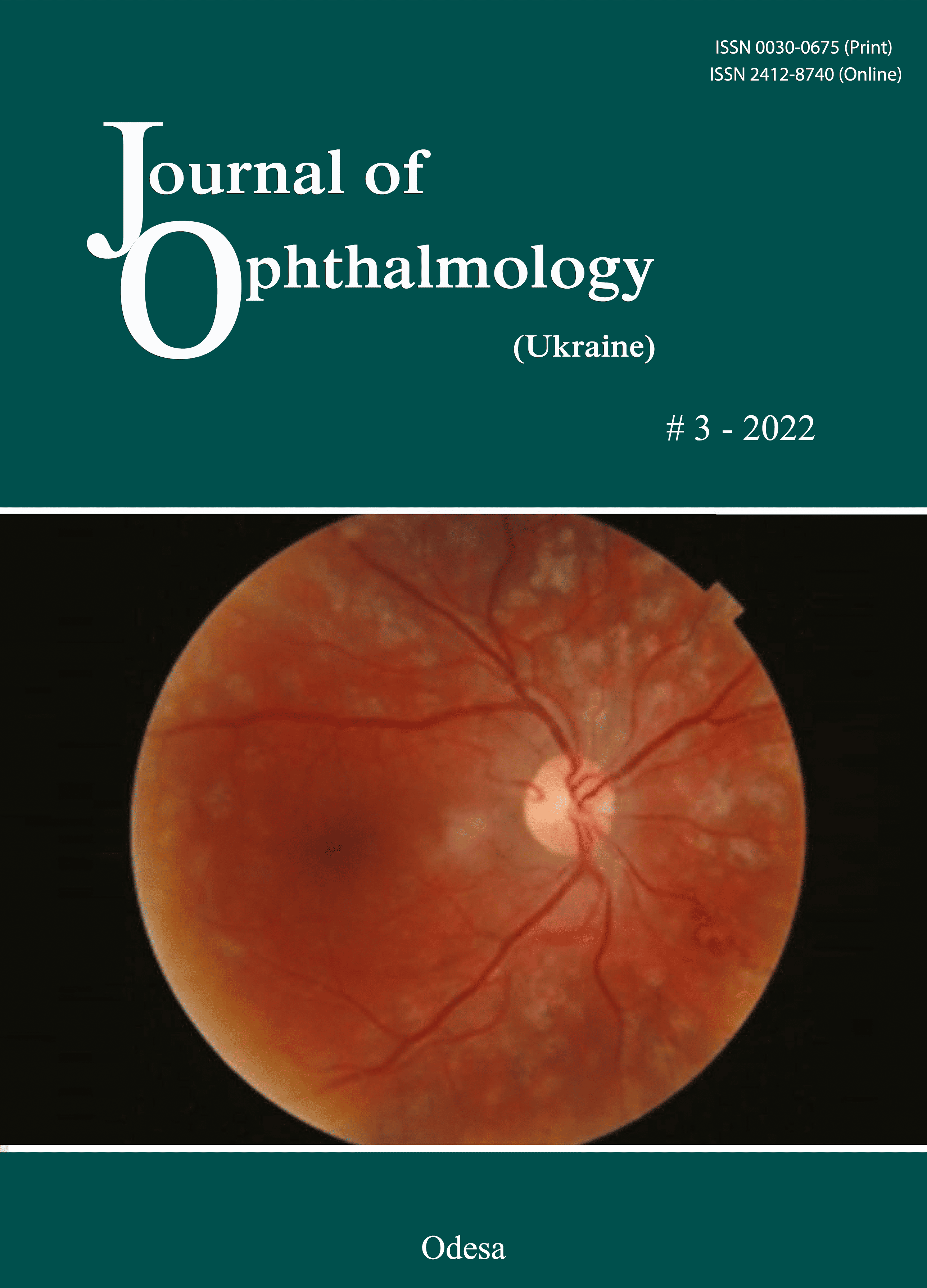
Results: In rats with induced type 2 diabetes mellitus, the most significant ocular vascular changes and renal vascular changes were found in endothelial cells. These changes included findings of vacuolar degeneration in some epithelial cells, basal membrane thickening and focal necrosis of individual epithelial cells. Vessel lumens appeared focally narrowed or expanded, with red blood cells forming clumps or sludge in lumens. Some capillaries were obliterated. These changes obviously caused secondary changes in the surrounding structures. Common ocular changes included focal destruction in retinal pigment epithelium cells, destruction of retinal photoreceptor inner segments and choroidal stromal edema. Common renal changes included destruction of the podocytes of the glomerular capillary network and homogenization of the basal membrane. Vascular ultrastructural changes in the renal glomerular system were more marked in rats with experimental type 2 diabetes mellitus and pyelonephritis than in those with type 2 diabetes mellitus only.
Conclusion: Electron microscopy micrographs demonstrated the ultrastructural changes in the retinal and uveal vascular systems which were of a similar type to those in the renal glomerular-and-tubular system in rats with experimental pyelonephritis in the presence of type 2 diabetes mellitus.
References
1.Ighodaro OM, Adeosun АМ. Vascular complications in diabetes mellitus. Glob J Endocrinol Metab. 2017;1(2):1-3. https://doi.org/10.31031/GJEM.2017.01.000506
2.Gross JL, de Azevedo MJ, Silveiro SP, et al. Diabetic nephropathy: diagnosis, prevention, and treatment. Diabetes Care. 2005 Jan;28(1):164-76. https://doi.org/10.2337/diacare.28.1.164
3.Maltsev EV, Zborovska OV, Dorokhova OE. [Fundamental aspects of the development and treatment of diabetic retinopathy]. Odesa: Astroprint; 2018. Russian.
4.Maltsev EV, Zborovska OV, Dorokhova ОE. [State of the retina and choroid in the rabbit with dithizone-induced diabetes. Report 1. Structural changes]. Oftalmol Zh. 2011;6: Russian.
5.Antonetti DA, Klein R, Gardner TW. Diabetic retinopathy. N Engl J Med. 2012;366(13):1227-1239. https://doi.org/10.1056/NEJMra1005073
6.Keen H, Lee ET, Russell D, et al. The appearance of retinopathy and progression to proliferative retinopathy: The WHO multinational study of vascular disease in diabetes. Diabetologia. 2001 Sep;44 Suppl 2:S22-30. https://doi.org/10.1007/PL00002935
7.Oganezova ZhG, Egorov EA. [The use of angioprotectors in the treatment of diabetic angiopathy: focus on calcium dobesilate]. RMJ Klinicheskaia oftalmologiia. 2015;4:201-4. Russian.
8.Brownlee M. The pathobiology of diabetic complications: a unifying mechanism. Diabetes. 2005 Jun;54(6):1615-25. https://doi.org/10.2337/diabetes.54.6.1615
9.Bhavsar AR, Emerson GG, Emerson MV, Browning DJ. Diabetic Retinopathy. Epidemiology of Diabetic Retinopathy. In: Browning DJ, editor. Diabetic Retinopathy: Evidence-Based Management. New York: Springer; 2010. https://doi.org/10.1007/978-0-387-85900-2_3
10.Gusev YuA, Kapkova SG, Varkentina IV, Tret'yak EB. [Calcium dobesilate in the treatment of diabetic retinopathy: the most important thing is not to lose time]. RMJ Klinicheskaia oftalmologiia. 2016;1:50-4. Russian.
11.Cao J, McLeod S, Merges CA, Lutty GA. Choriocapillaris degeneration and related pathologic changes in human diabetic eyes. Arch Ophthalmol. 1998 May;116(5):589-97. https://doi.org/10.1001/archopht.116.5.589
12.Lutty GA, Cao J, McLeod DS. Relationship of polymorphonuclear leukocytes to capillary dropout in the human diabetic choroids. Am J Pathol. 1997 Sep;151(3):707-14.
13.Vit VV. [The structure of the human visual system]. Odesa: Astroprint; 2003. Russian.
14.Vit VV. [Pathology of the eye, ocular adnexa and orbit]. Odesa: Astroprint; 2019. Russian.
15.Reynolds ES. The use of lead citrate at high pH as an electron-opaque stain in electron microscopy. 1963 Apr;17(1):208-12. https://doi.org/10.1083/jcb.17.1.208
16.Borysov SO, Kostyev FI, Borysov OV, Molchanyuk NI. [Electron microscopic diagnostics of apoptosis processes under simulation conditions in the experiment of acute pyelonephritis and concomitant diabetes mellitus type I and II]. Reports of Morphology. 2018;24(1):39-46. https://doi.org/10.31393/morphology-journal-2018-24(1)-07
17.Hrytsiuk NI. [Changes in tortuous canaliculi after NADP injection in rats with streptozotocin-induced diabetes]. Mizhnarodnyi endokrynologichnyi zhurnal. 2017;13(8):624-9. Ukrainian.
18.Borysov SO, Kostiev FI, Borysov OV, Artiomov OV. [Comparison of renal morphological changes in rat models of pyelonephritis and concomitant type 1 or 2 diabetes mellitus]. Urologiia. 2019. 23(3):205-10.
Downloads
Published
How to Cite
Issue
Section
License
Copyright (c) 2025 С. О. Борисов, Ф. І. Костєв, О. В. Борисов, Ю. М. Дехтяр, В. В. Віт, Н. І. Молчанюк, І. М. Михейцева, С. Г. Коломійчук, Ахмед Амаієд

This work is licensed under a Creative Commons Attribution 4.0 International License.
This work is licensed under a Creative Commons Attribution 4.0 International (CC BY 4.0) that allows users to read, download, copy, distribute, print, search, or link to the full texts of the articles, or use them for any other lawful purpose, without asking prior permission from the publisher or the author as long as they cite the source.
COPYRIGHT NOTICE
Authors who publish in this journal agree to the following terms:
- Authors hold copyright immediately after publication of their works and retain publishing rights without any restrictions.
- The copyright commencement date complies the publication date of the issue, where the article is included in.
DEPOSIT POLICY
- Authors are permitted and encouraged to post their work online (e.g., in institutional repositories or on their website) during the editorial process, as it can lead to productive exchanges, as well as earlier and greater citation of published work.
- Authors are able to enter into separate, additional contractual arrangements for the non-exclusive distribution of the journal's published version of the work with an acknowledgement of its initial publication in this journal.
- Post-print (post-refereeing manuscript version) and publisher's PDF-version self-archiving is allowed.
- Archiving the pre-print (pre-refereeing manuscript version) not allowed.


