Relationship between changes in retinal brain-derived neurotrophic factor (BDNF) concentration and morphological changes in retinal neurons in rats with induced diabetes and axial myopia
DOI:
https://doi.org/10.31288/oftalmolzh202434044Keywords:
diabetic retinopathy, myopia, rats, retina, BDNF, retinal neuronal cells, structural changes, ganglion cellsAbstract
Background: Myopia significantly decreases the frequency and severity of diabetic retinopathy (DR). The proliferative form of this diabetic complication in the retina is known to be very uncommon in diabetic myopes. The mechanisms of this phenomena are, however, still unclear. Studies on these mechanisms and the clarification of the structural changes in retinal neurons in animal models of both diabetes and myopia are critical to the research of the pathogenesis and further identification of therapeutic and preventive targets for these pathological conditions.
Purpose: To examine changes in the retinal brain-derived neurotrophic factor (BDNF) concentration and the relationship of the latter with the structure of retinal neuronal cells in rats with induced both diabetes and axial myopia.
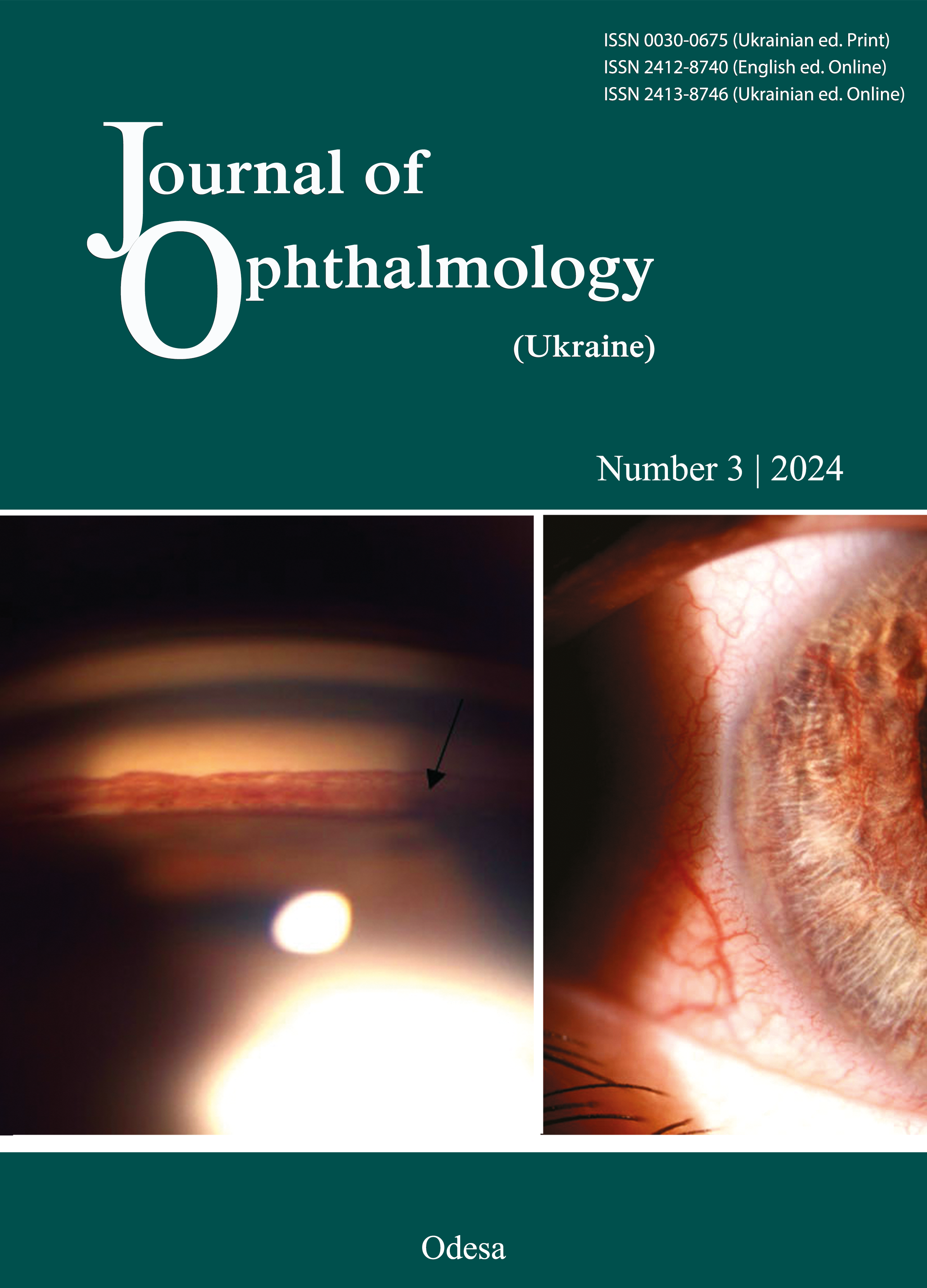
Material and Methods: Rats (age, 2 to 10 weeks) were assigned to four groups: group 1 (myopia only, n = 15), group 2 (diabetes only, n = 15), group 3 (myopia plus diabetes, n = 15), and group 4 (healthy controls, n = 10). Axial myopia was produced in two-month-old animals by surgically fusing the eyelids of both eyes. Streptozotocin (STZ) (15 mg/kg body weight, intraperitoneally consequently for 5 days) was used to induce diabetes. Diabetes was induced in group 3 at 3 weeks after the initiation of the experiment. At 2 months, all rats were euthanized under anesthesia, and their eyes were enucleated. To perform a histomorphological study, serial retinal sections were made and stained with hematoxylin and eosin, and microscopy was performed and images were collected and evaluated on a light microscope Jenamed 2. Rat BDNF enzyme-linked immunosorbent kits (Elabscience, Houston, TX) were used to determine BDNF concentrations in retinal supernatant and plasma.
Results: Rats with both diabetes and myopia exhibited smaller reductions in plasma and, especially, retinal BDNF concentrations compared to rats with diabetes only. Retinal BDNF concentrations in rats with both diabetes and myopia were 36.1% higher than in rats with diabetes only. Unlike rats with STZ-induced diabetes only, those with STZ-induced diabetes in the presence of experimental myopia exhibited a rather high neuronal cell density in the retinal ganglion cell layer. No noticeable change in the cell density in the inner nuclear layer and photoreceptor layer was observed in the latter animals.
Conclusion: Axial length elongation secondary to experimental myopia in animals facilitates the protection against diabetic changes in the retina, which was confirmed at the molecular and morphological levels, with BDNF being a possible component of this protective mechanism.
References
Lim LS, Lamoureux E, Saw SM, Tay WT, Mitchell P, Wong TY. Are myopic eyes less likely to have diabetic retinopathy? Ophthalmology. 2010;117(3):524-30.
https://doi.org/10.1016/j.ophtha.2009.07.044
Man RE, Sasongko MB, Sanmugasundram S, et al. Longer axial length is protective of diabetic retinopathy and macular edema. Ophthalmology. 2012;119(9):1754-9. https://doi.org/10.1016/j.ophtha.2012.03.021
Wang Q, Wang YX, Wu SL, et al. Ocular Axial Length and Diabetic Retinopathy: The Kailuan Eye Study. Invest Ophthalmol Vis Sci. 2019; 60(10):3689-95. https://doi.org/10.1167/iovs.19-27531
Lin Z, Li D, Zhai G, et al. High myopia is protective against diabetic retinopathy via thinning retinal vein: A report from Fushun Diabetic Retinopathy Cohort Study (FS-DIRECT). Diab Vasc Dis Res Jul-Aug. 2020;17(4):1479164120940988. https://doi.org/10.1177/1479164120940988
Weijung Ten, Ying Yuan, Wei Zhang, Yue Wu & Bilian Ke. High myopia is protective against diabetic retinopathy in the participants of the National Health and Nutrition Examination Survey BMC. Ophthalmology 2023; 23:468. https://doi.org/10.1186/s12886-023-03191-x
Mikheytseva I, Molchanuk N, Amayed A, Kolomiichuk S, Siroshtanenko T. [Features of ultrastructural changes in the neurosensory elements of the retina of rats in the modeling of diabetic retinopathy on the background of axial myopia]. Fiziol Zh. 2024;1:31-36. Ukrainian. https://doi.org/10.15407/fz70.01.031
Tuwar MN ,Wei-Hung Chen, Chiwaya AM et al. Brain-derived neurotrophic factor (BDNF) and translocator protein (TSPO) as diagnostic biomarkers for acute ischemic stroke. Diagnostics. 2023; 13(13): 2298. https://doi.org/10.3390/diagnostics13132298
Yun-Zheng Le, Bei Xu, Ana J Chucair-Elliott et al. VEGF Mediates Retinal Müller Cell Viability and Neuroprotection through BDNF in Diabetes. Biomolecules. 2021;11(5):712. https://doi.org/10.3390/biom11050712
Akamatsu S, Fujii S, Escaño MF Altered expression of genes in experimentally induced myopic chick eyes. Jap. J. Ophthalmology. 2001, 45 (2), 137-143. https://doi.org/10.1016/S0021-5155(00)00360-9
Mikheytseva IN, Abdulhadi Mohammad, Putienko AA, et al. Modelling form deprivation myopia in experiment. Journal of Ophthalmology (Ukraine). 2018; 2 (481):50-55. https://doi.org/10.31288/oftalmolzh/2018/2/5055
Simo R, Stitt AW, Gardner TW. Neurodegeneration in diabetic retinopathy: does it really matter? Diabetologia. 2018; 61 (9): 1902-1912. https://doi.org/10.1007/s00125-018-4692-1
Duh EJ, Sun JK, Stitt AW. Diabetic retinopathy: current understanding, mechanisms, and treatment strategies JCI Insight. 2017; 2: (14). https://doi.org/10.1172/jci.insight.93751
Rakieten N, Rakieten ML, Nadkarni MV. Studies on the diabetogenic action of streptozotocin (NSC-37917). Cancer Chemother Rep. 1963;29:91-98.
Rungger-Brändle E, Dosso AA, Leuenberger PM. Glial reactivity, an early feature of diabetic retinopathy. Invest Ophthalmol Vis Sci Assoc Res Vis Ophthalmol. 2000; 41(7):1971-1980.
Olivares AM, Althoff K, Chen GF et al. Animal Models of Diabetic Retinopathy. Curr Diab Rep. 2017; 17: 93. https://doi.org/10.1007/s11892-017-0913-0
Guo M, Liu H, Li SS et al. Low serum brain-derived neurotrophic factor but not brain-derived neurotrophic factor gene val66met polymorphism is associated with diabetic retinopathy in Chinese type 2 diabetic patients Retina. 2017; 37(2):350-358. https://doi.org/10.1097/IAE.0000000000001132
Ola MS, Nawaz MI, El-Asrar AA et al Reduced levels of brain derived neurotrophic factor (BDNF) in the serum of diabetic retinopathy patients and in the retina of diabetic rats. Cell Mol Neurobiol. 2013;33(3):359-67. https://doi.org/10.1007/s10571-012-9901-8
Downloads
Published
How to Cite
Issue
Section
License
Copyright (c) 2024 Mikheytseva I. M., Amaied Ahmed, Artiomov O.V., Kolomiichuk S. G., Kuznetsov M. K.

This work is licensed under a Creative Commons Attribution 4.0 International License.
This work is licensed under a Creative Commons Attribution 4.0 International (CC BY 4.0) that allows users to read, download, copy, distribute, print, search, or link to the full texts of the articles, or use them for any other lawful purpose, without asking prior permission from the publisher or the author as long as they cite the source.
COPYRIGHT NOTICE
Authors who publish in this journal agree to the following terms:
- Authors hold copyright immediately after publication of their works and retain publishing rights without any restrictions.
- The copyright commencement date complies the publication date of the issue, where the article is included in.
DEPOSIT POLICY
- Authors are permitted and encouraged to post their work online (e.g., in institutional repositories or on their website) during the editorial process, as it can lead to productive exchanges, as well as earlier and greater citation of published work.
- Authors are able to enter into separate, additional contractual arrangements for the non-exclusive distribution of the journal's published version of the work with an acknowledgement of its initial publication in this journal.
- Post-print (post-refereeing manuscript version) and publisher's PDF-version self-archiving is allowed.
- Archiving the pre-print (pre-refereeing manuscript version) not allowed.


