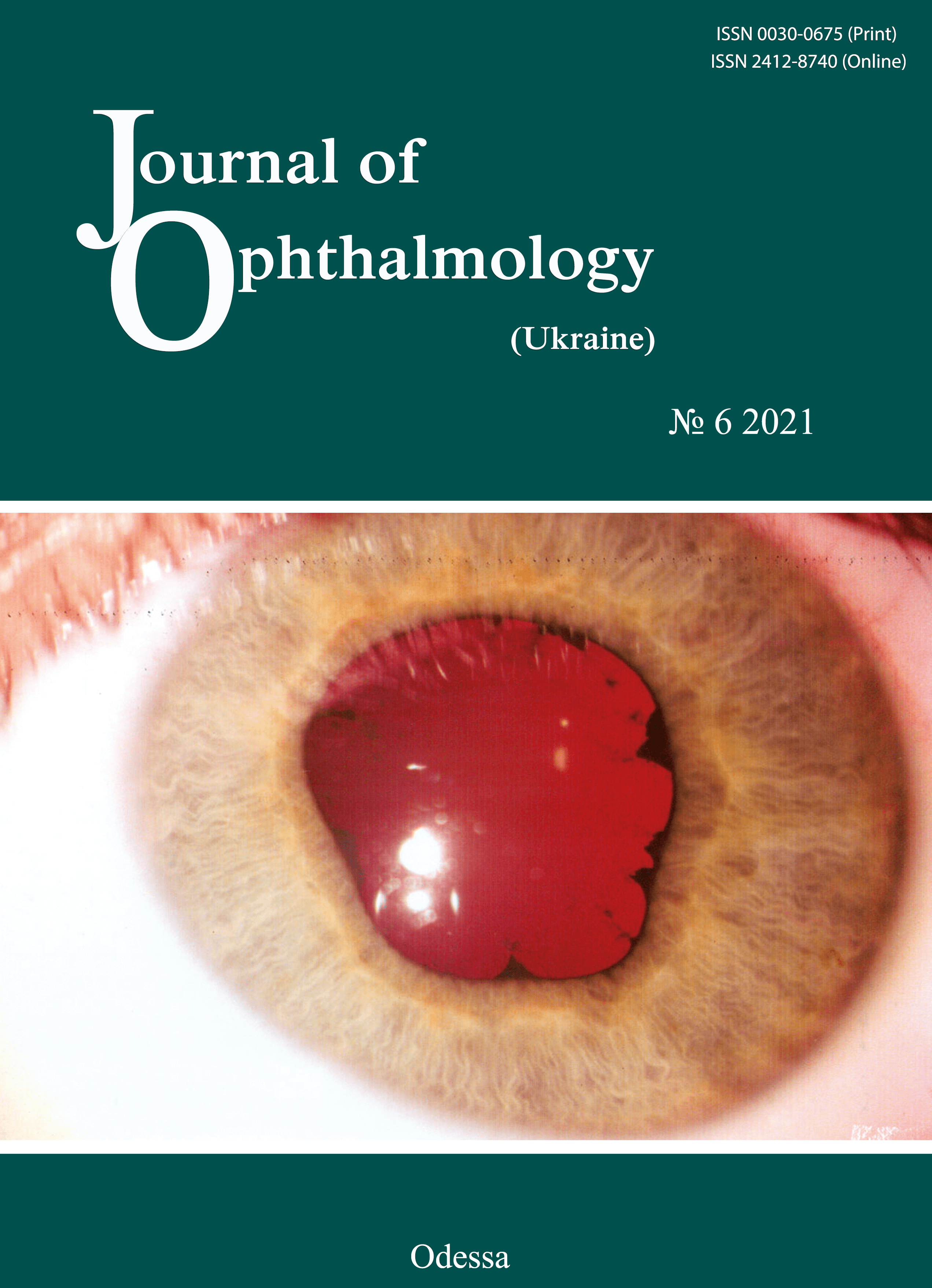
Ultrastructural features of epiretinal membranes after intravitreal injection of various doses of aflibercept in patients with proliferative diabetic retinopathy
DOI:
https://doi.org/10.31288/oftalmolzh202162530Keywords:
proliferative diabetic retinopathy, epiretinal membrane, afliberceptAbstract
Background: Proliferative diabetic retinopathy (PDR) can cause a loss of vision due to complications like vitreous hemorrhage and tractional retinal detachment, which require vitrectomy. The source of bleeding is usually in pathologic new blood vessels in the fibrovascular epiretinal membrane (ERM). Anti-vascular endothelial growth factor agents are widely used for prevention of intraoperative and postoperative hemorrhagic complications in vitrectomy for PDR.
Purpose: To examine ultrastructural features of ERM after intravitreal injection of various doses of aflibercept in patients with PDR.
Material and Methods: Five patients (5 eyes) were included in the study. ERM specimens with neovascular vessels were taken intraoperatively from the five patients for histological microscopy studies. Specimen No. 1 was taken from the ERM (control ERM) of the patient who did not receive preoperative injection of aflibercept. In addition, specimen No. 2 was taken on day 3 after intravitreal injection of 1.0 mg aflibercept; specimen No. 3, on day 5 after intravitreal injection of 1.0 mg aflibercept; specimen No. 4, on day 3 after intravitreal injection of 2.0 mg aflibercept; and specimen No. 5, on day 5 after intravitreal injection of 2.0 mg aflibercept.
Results: We found that aflibercept had a positive effect on obliteration of newly formed vessels and capillaries. There was a decrease in the size of the lumen of ERM microvessels with an increase in the injected dose and time after injection. In addition, endothelial cells in the newly formed vessels and fibroblasts exhibited active protein synthesis metabolism on day 3 after intravitreal injection of 1.0 mg aflibercept or 2.0 mg aflibercept.
References
1.Shkvorchenko DO, Levina LV. [Retinotomy and retinectomy in the management of proliferative diabetic retinopathy complicated by anterior proliferative vitreoretinopathy]. In: [Proceedings of the National Russian Conference on Modern Technologies for the Treatment of Vitreoretinal Pathology]. Moscow; 2006. p. 205-10. Russian.
2.Shibuya M. Vascular endothelial growth factor-dependent and -independent regulation of angiogenesis. BMB Rep. 2008 Apr 30;41(4):278-86. https://doi.org/10.5483/BMBRep.2008.41.4.278
3.Hendrick AM, Gibson MV, Kulshresthra A. Diabetic Retinopathy. Prim Care. 2015 Sep;42(3):451-64.https://doi.org/10.1016/j.pop.2015.05.005
4.Sawa H, Ikeda T, Matsumoto Y. Neovascularization from scleral wound as cause of vitreous rebleeding after vitrectomy for proliferative diabetic retinopathy. Jpn J Ophthalmol. Mar - Apr 2000;44(2):154-60.https://doi.org/10.1016/S0021-5155(99)00194-X
5.Adamis AP, Shima DT. The role of vascular endothelial growth factor in ocular health and disease. Retina. Feb-Mar 2005;25(2):111-8.https://doi.org/10.1097/00006982-200502000-00001
6.Aleman I, Castillo Velazquez J, Rush SW. Ziv-aflibercept versus bevacizumab administration prior to diabetic vitrectomy: a randomised and controlled trial. Br J Ophthalmol. 2019 Dec;103(12):1740-6.https://doi.org/10.1136/bjophthalmol-2018-313313
7.Velazquez JC, Aleman I, Rush SW. Bevacizumab before Diabetic Vitrectomy: A Clinical Trial Assessing 3 Dosing Amounts. Ophthalmol Retina. 2018 Oct; 2(10):1010-20.https://doi.org/10.1016/j.oret.2018.04.014
8.Antoszyk AN, Glassman AR, Beaulieu WT, et al. Effect of Intravitreous Aflibercept vs Vitrectomy With Panretinal Photocoagulation on Visual Acuity in Patients With Vitreous Hemorrhage From Proliferative Diabetic Retinopathy: A Randomized Clinical Trial. JAMA. 2020 Dec 15;324(23):2383-95.https://doi.org/10.1001/jama.2020.23027
9.Abd Elhamid AH, Mohamed AAEA., Khattab AM. Intravitreal Aflibercept injection with panretinal photocoagulation versus early vitrectomy for diabetic vitreous hemorrhage: randomized clinical trial. BMC Ophthalmol. 2020 Apr 6;20(1):130.https://doi.org/10.1186/s12886-020-01401-4
10.Ponomarchuk Vira S, Vit VV, Umanets MM. Morphological and ophthalmoscopic features of epiretinal membranes after intravitreal injection of various doses of aflibercept in patients with proliferative diabetic retinopathy. J Ophthalmol (Ukraine). 2021;5:3-9.https://doi.org/10.31288/oftalmolzh2021539
11.Kohno R, Hata Y, Mochisuki Y, et al. Histopathology of neovascular tissue from eyes with proliferative diabetic retinopathy after intravitreal bevacizumab injection. Am J Ophthalmol. 2010 Aug;150(2):223-229.e1.https://doi.org/10.1016/j.ajo.2010.03.016
12.Dumbrova NE, Korol AR, Nasinnik IO, et al. [Structural changes in the choroid and retina of the rabbit after intravitreal injections of selective and non-selective anti-VEGF agents]. Oftalmol Zh. 2012;3:68-72. Russian. https://doi.org/10.31288/oftalmolzh201236872
13.Vidinova C, Vidinov N. [The effect of bevacizumab on the ultrastructure of choroidal neovascular membranes in patients with age-related macular degeneration (AMD)]. Klin Monbl Augenheilkd. 2009 Jun;226(6):491-5.https://doi.org/10.1055/s-0028-1109429
14.Vidinova C, Vidinov N. [Comparative ultrastructural analysis of the proliferative tissue in PDR patients with or without vitreous haemorrhage: from ultrastructural approach to clinical practice]. Klin Monbl Augenheilkd. 2007 Mar;224(3):185-9.https://doi.org/10.1055/s-2007-962958
15.Ponomarchuk Vira S, Velychko LM, Umanets MM. Vitreous VEGF levels among patients with proliferative diabetic retinopathy depending on the general clinical status and ocular status. J Ophthalmol (Ukraine). 2021;4:19-25.https://doi.org/10.31288/oftalmolzh202141925
16.Reynolds ES. The use of lead citrate at high pH as an electron-opaque stain in electron microscopy. J Cell Biol. 1963 Apr;17(1):208-12.https://doi.org/10.1083/jcb.17.1.208
17.Julien S, Heiduschka P, Hofmeister S, Schraermeyer U. Immunohistochemical localisation of intravitreally injected bevacizumab at the posterior pole of the primate eye: implication for the treatment of retinal vein occlusion. Br J Ophthalmol. 2008 Oct;92(10):1424-8.https://doi.org/10.1136/bjo.2008.141317
18.Heiduschka P, Fietz H, Hofmeister S, et al. Penetration of bevacizumab through the retina after intravitreal injection in the monkey. Invest Ophthalmol Vis Sci. 2007 Jun;48(6):2814-23.https://doi.org/10.1167/iovs.06-1171
19.Peters S, Heiduschka P, Julien S, et al. Ultrastructural findings in the primate eye after intravitreal injection of bevacizumab. Am J Ophthalmol. 2007 Jun;143(6):995-1002.https://doi.org/10.1016/j.ajo.2007.03.007
Downloads
Published
How to Cite
Issue
Section
License
Copyright (c) 2025 Вера С. Пономарчук , Н. И. Молчанюк , Н. Н. Уманец

This work is licensed under a Creative Commons Attribution 4.0 International License.
This work is licensed under a Creative Commons Attribution 4.0 International (CC BY 4.0) that allows users to read, download, copy, distribute, print, search, or link to the full texts of the articles, or use them for any other lawful purpose, without asking prior permission from the publisher or the author as long as they cite the source.
COPYRIGHT NOTICE
Authors who publish in this journal agree to the following terms:
- Authors hold copyright immediately after publication of their works and retain publishing rights without any restrictions.
- The copyright commencement date complies the publication date of the issue, where the article is included in.
DEPOSIT POLICY
- Authors are permitted and encouraged to post their work online (e.g., in institutional repositories or on their website) during the editorial process, as it can lead to productive exchanges, as well as earlier and greater citation of published work.
- Authors are able to enter into separate, additional contractual arrangements for the non-exclusive distribution of the journal's published version of the work with an acknowledgement of its initial publication in this journal.
- Post-print (post-refereeing manuscript version) and publisher's PDF-version self-archiving is allowed.
- Archiving the pre-print (pre-refereeing manuscript version) not allowed.


