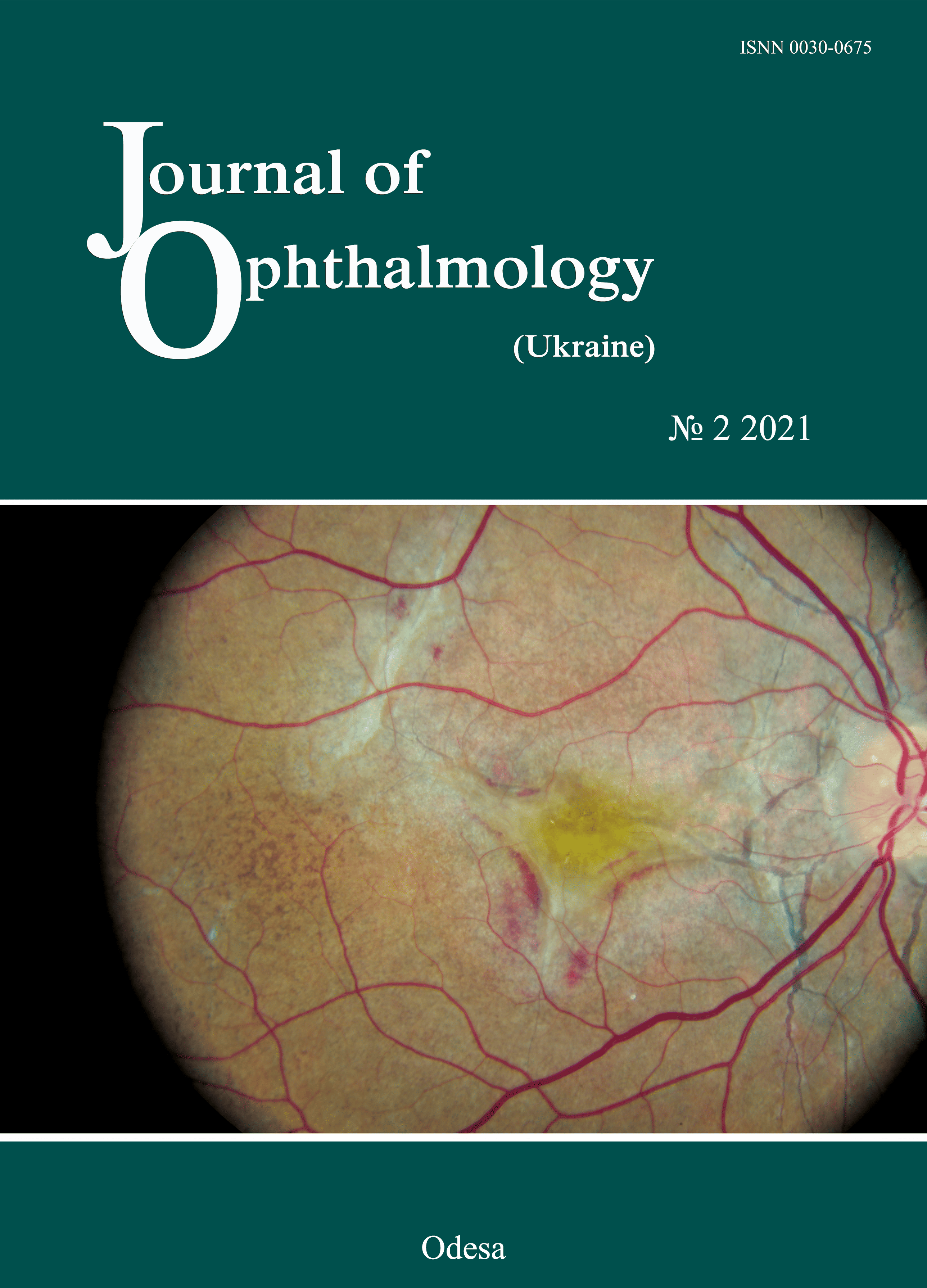
Ангіоїдні смужки сітківки
DOI:
https://doi.org/10.31288/oftalmolzh202124654Ключові слова:
ангіоїдні смужки сітківки, субретинальна неоваскулярна мембрана, інтравітреальні введенняАнотація
В статті представлено огляд сучасної літератури щодо етіології, патогенезу, клінічної картини та лікування ангіоїдних смужок сітківки.
Ангіоїдні смужки сітківки – це захворювання очного дна, яке характеризується наявністю тріщин в потовщеному, кальцифікованому і патологічно крихкому еластичному і колагеновому шарах мембрани Бруха, які в 50 % випадків поєднуються з системними захворюваннями: еластичною псевдоксантомою, хворобою Педжета та ін. Для діагностики, оцінки та моніторингу ангіоїдних смужок використовуються різні методи діагностики: візометрія, рефрактометрія, вимірювання внутрішньоочного тиску, визначення об'єму рухів очного яблука, біомікроскопія переднього відрізку очей, офтальмоскопія, флуоресцентна ангіографія сітківки, оптична когерентна томографія сітківки. Основним ускладненням ангіоїдних смужок сітківки, що спричиняє пониження зоpу, є субретинальна неоваскулярна мембрана (CНМ). На сьогоднішній день найефективнішим методом лікування СНМ є інтравітреальні введення інгібіторів факторів росту ендотелію судин.
Посилання
1.Doyne RW. Choroidal and retinal changes. The results of blows on the eyes. Trans Ophthalmol Soc UK. 1889;9:128.
2.Knapp H. On the formation of dark angioid streaks as unusual metamorphosis of retinal hemorrhage. Arch Ophthalmol. 1892;26:289-92.
3.Kofler A. Beitrage zur Kenntnis der Angioid streaks. Arch Augenheilkd. 1917;82:134-49.
4.Bock Z. KIinik und Anatomie der gefassahnlichen Streifen im Augenhintergrund. Z Augenheilkd. 1938;95:1-50. https://doi.org/10.1159/000313941
5.Hagedoorn A. Angioid streaks. Arch Ophthalmol. 1939;21:746-4. 935-65.https://doi.org/10.1001/archopht.1939.00860060045002
6.Chassaing N, Martin L, Calvas P, Le Bert M, Hovnanian A. Pseudoxanthoma elasticum: a clinical, pathophysiological and genetic update including 11 novel ABCC6 mutations. J Med Genet. 2005;42:881-92.https://doi.org/10.1136/jmg.2004.030171
7.Tripathy K, Quint JM. Angioid Streaks (Knapp Streaks). StatPearls [Internet]. Treasure Island (FL): StatPearls Publishing; 2020 June 01.
8.Chatziralli I, Saitakis G, Dimitriou E, Chatzirallis A, Stoungioti S, Theodossiadis G, Theodossiadis P. ANGIOID STREAKS: A Comprehensive Review From Pathophysiology to Treatment. Retina (Philadelphia, Pa.). 2019 Jan;39(1):1-11.https://doi.org/10.1097/IAE.0000000000002327
9.Georgalas I, Papaconstantinou D, Koutsandrea C, Kalantzis G, Karagiannis D, Georgopoulos G, Ladas I. Angioid streaks, clinical course, complications, and current therapeutic management. Ther Clin Risk Manag. 2009 Feb;5(1):81-9.https://doi.org/10.2147/TCRM.S4682
10.Singman E, Doyle J. Angioid streaks are not a common feature of Ehlers Danlos Syndrome. JAMA Ophthalmology. 2018.137(3).https://doi.org/10.1001/jamaophthalmol.2018.5995
11.Brady AF, Demirdas S, Fournel-Gigleux S, et al. The Ehlers-Danlos syndromes, rare types. Am J Med Genet C Semin Med Genet. 2017;175(1):70-115.https://doi.org/10.1002/ajmg.c.31550
12.Uitto J, Jiang Q, Varadi A, Bercovitch LG, Terry SF. Pseudoxanthoma Elasticum: diagnostic features, classification, and treatment options. Expert Opin Orphan Drugs. 2014;2:567-77.https://doi.org/10.1517/21678707.2014.908702
13.Bercovitch L, Terry P. Pseudoxanthoma elasticum 2004. J Am Acad Dermatol. 2004;51:S13-S14. https://doi.org/10.1016/j.jaad.2004.01.015
14.Finger RP, Charbel Issa P, Ladewig MS, Gotting C, Szliska C, Scholl HP, Holz FG. Pseudoxanthoma elasticum: genetics, clinical manifestations and therapeutic approaches. Surv Ophthalmol. 2009;54:272-285. doi: 10.1016/j.survophthal.2008.12.006. https://doi.org/10.1016/j.survophthal.2008.12.006
15.Mizutani Y, Nakayama T, Asai S, et al. ABCC6 mutation in patients with angioid streaks. Int J Biomed Sci. 2006;2:7-12. https://doi.org/10.59566/IJBS.2006.2009
16.Sato N, Nakayama T, Mizutani Y, Yuzawa M. Novel mutations of ABCC6 gene in Japanese patients with Angioid streaks. Biochem Biophys Res Commun. 2009;380:548-53. https://doi.org/10.1016/j.bbrc.2009.01.117
17.Katagiri S, Negishi Y, Mizobuchi K, et al. ABCC6 gene analysis in 20 Japanese patients with angioid streaks revealing four frequent and two novel variants and pseudodominantin heritance. J Ophthalmol. 2017;2017:1079687.https://doi.org/10.1155/2017/1079687
18.Booij JC, Baas DC, Beisekeeva J et al. The dynamic nature of Bruch's membrane. Prog Retin Eye Res. 2010;29:1-18. https://doi.org/10.1016/j.preteyeres.2009.08.003
19.19 Vit VV. [Pathology of the eye, ocular adnexa and orbit]. Vol. 2. Odesa:Astroprint; 2019. Russian.
20.Keith CG. Angioid streaks and pseudoxanthoma elasticum. Br J Ophthalmol. 1956;40:480-6. https://doi.org/10.1136/bjo.40.8.480
21.Martinez-Serrano MG, Rodriguez-Reyes A, Guerrero-Naranjo JL, et al. Long-term follow-up of patients with choroidal neovascularization due to angioid streaks. Clin Ophthalmol. 2016;11:23-30.https://doi.org/10.2147/OPTH.S118016
22.Myung JS, Bhatnagar P, Spaide RF et al. Long-term outcomes of intravitreal antivascular endothelial growth factor therapy for the management of choroidal neovascularization in pseudoxanthoma elasticum. Retina. 2010;30:748-55. https://doi.org/10.1097/IAE.0b013e3181c596b1
23.Gal-Or O, Balaratnasingam C, Freund KB. Optical coherence tomography angiography findings of choroidal neovascularization in pseudoxanthoma elasticum. Int J Retina Vitreous. 2015;1:11. https://doi.org/10.1186/s40942-015-0011-x
24.Abusamak M, Lee AG, Berry S, Sadaka A. Angioid streaks. Medscape [Internet]. 2019 Jul 22. Available from: https://emedicine.medscape.com/article/1190444-overview#a2.
25.Ellabban AA, Tsujikawa A, Matsumoto A, Ogino K, Hangai M, Ooto S, Yamashiro K, Akiba M, Yoshimura N. Macular choroidal thickness and volume in eyes with angioid streaks measured by swept source optical coherence tomography. Am J Ophthalmol. 2012 Jun;153(6):1133-43.e1. https://doi.org/10.1016/j.ajo.2011.12.013
26.Roach ES, Islam MP. Pseudoxanthoma elasticum. Handb Clin Neurol. 2015;132:215-21. https://doi.org/10.1016/B978-0-444-62702-5.00015-9
27.Gandorfer A, Ulbig M, Bechmann M, et al. Retinal telangiectasis and angioid streaks. Br J Ophthalmol. 2000;84:1327-8. https://doi.org/10.1136/bjo.84.11.1318j
28.Baillif-Gostoli S, Quaranta-El Maftouhi M, Mauget-FaysseM. Polypoidal choroidal vasculopathy in a patient with angioid streaks secondary to pseudoxanthoma elasticum. Graefes Arch Clin Exp Ophthalmol. 2010;248:1845-8. https://doi.org/10.1007/s00417-010-1328-7
29.Cebeci Z, Bayraktar S, Oray M, Kir N. Silent polypoidalchoroidal vasculopathy in a patient with angioid streaks. Arq Bras Oftalmol. 2016;79:200-1.https://doi.org/10.5935/0004-2749.20160058
30.Shields JA, Federman JL, Tomer TL, Annesley WH., Jr Angioid streaks. I. Ophthalmoscopic variations and diagnostic problems. Br J Ophthalmol. 1975;59:257-66.https://doi.org/10.1136/bjo.59.5.257
31.Mansour AM, Ansari NH, Shields JA, Annesley WH, Jr, Cronin CM, Stock EL. Evolution of angioid streaks. Ophthalmologica. 1993;207:57-61.https://doi.org/10.1159/000310407
32.Mansour AM, Shields JA, Annesley WH, Jr, el-Baba F, Tasman W, Tomer TL. Macular degeneration in angioid streaks. Ophthalmologica. 1988;197:36-41.https://doi.org/10.1159/000309915
33.Al-Rashaed S, Arevalo JF. Long-term follow-up of choroidal neovascularization secondary to angioid streaks: case series and literature review. Clin Ophthalmol. 2012;6:1029-34.https://doi.org/10.2147/OPTH.S30550
34.Savko VV, Naritsyna NI, Chechin PP, Konovalova NV, Serebrina TM, Novik AIa. [Grönblad-Strandberg syndrome: clinical features, diagnosis and treatment]. Oftalmol Zh. 2009;4:82-5. Russian.https://doi.org/10.31288/oftalmolzh200948285
35.Smith JL, Gass JD, Justice JJr. Fluorescein fundus photography of angioid streaks. Br J Ophthalmol. 1964;48:517-21.https://doi.org/10.1136/bjo.48.10.517
36.Sawa M, Ober MD, Freund KB, Spaide RF. Fundus autofluorescence in patients with pseudoxanthoma elasticum. Ophthalmology. 2006113820e1-2.
37.Finger RP, Charbel Issa P, Ladewig M, et al. Fundus autofluorescence in Pseudoxanthoma elasticum. Retina. 2009;29:1496-1505.https://doi.org/10.1097/IAE.0b013e3181aade47
38.De Zaeytijd J, Vanakker OM, Coucke PJ, et al. Added value of infrared, red-free and autofluorescence fundus imaging in pseudoxanthoma elasticum. Br J Ophthalmol. 2010;94:479-86.https://doi.org/10.1136/bjo.2009.162644
39.Lafaut BA, Leys AM, Scassellati-Sforzolini B et al. Comparison of fluorescein and indocyanine green angiography in angioid streaks. Graefes Arch Clin Exp Ophthalmol.1998;236:346-53.https://doi.org/10.1007/s004170050089
40.Peretyagina DO, Ulyanova NA. SS-OCT-derived morphometric changes in the choroid in patients with age-related macular degeneration. Journal of Ophthalmology (Ukraine). 2019;6:63-9. https://doi.org/10.31288/oftalmolzh201966369
41.Iegorova KS, Znamenska MA, Guk MO, Mumliev AO. Early signs of primary compressive optic atrophy evidenced by OCT in patients with basal brain tumors. Journal of Ophthalmology (Ukraine). 2020;1:35-9.https://doi.org/10.31288/oftalmolzh202013539
42.Marchese A, Parravano M, Rabiolo A, Carnevali A, Corbelli E, Cicinelli MV, et al. Optical coherence tomography analysis of evolution of Bruch's membrane features in angioid streaks. Eye (Lond). 2017 Nov; 31(11): 1600-5.https://doi.org/10.1038/eye.2017.112
43.Chapron T, Mimoun G, Miere A, Srour M, Ameen El A, Semoun O, Souied EN. Optical coherence tomography angiography features of choroidal neovascularization secondary to angioid streaks. Eye (Lond). 2019 Mar; 33(3): 385-91.https://doi.org/10.1038/s41433-018-0213-1
44.Orly Gal-Or, Chandrakumar Balaratnasingam, Bailey Freund K. Optical coherence tomography angiography findings of choroidal neovascularization in pseudoxanthoma elasticum. Int J Retina Vitreous. 2015; 1:11.https://doi.org/10.1186/s40942-015-0011-x
45.Mansour AM; Ansari NH; Shields JA; Annesley WH; Cronin CM; Stock E. Evolution of angioid streaks. Ophthalmologica. 1993; 207(2):57-61.https://doi.org/10.1159/000310407
46.Clarkson JG, Altman RD. Angioid streaks. Surv Ophthalmol. 1982;26:235-46.https://doi.org/10.1016/0039-6257(82)90158-8
47.Singerman LJ, Hatem G. Laser treatment of choroidal neovascular membranes in angioid streaks. Retina. 1981;1:75-83. https://doi.org/10.1097/00006982-198101020-00001
48.Meislik J, Neldner K, Reeve EB, Ellis PP. Laser treatment in maculopathy of pseudoxanthoma elasticum. Can J Ophthalmol. 1978;13:210-2.
49.Deutman AF, Kovacs B. Argon laser treatment in complications of angioid streaks. Am J Ophthalmol. 1979;88:12-7. https://doi.org/10.1016/0002-9394(79)90744-X
50.Gelisken O, Hendrikse F, Deutman AF. A long-term follow-up study of laser coagulation of neovascular membranes in angioid streaks. Am J Ophthalmol. 1988;105:299-303.https://doi.org/10.1016/0002-9394(88)90014-1
51.Aras C, Baserer T, Yolar M, et al. Two cases of choroidal neovascularization treated with transpupillary thermotherapy in angioid streaks. Retina. 2004;24:801-803.https://doi.org/10.1097/00006982-200410000-00020
52.Machemer R, Steinhorst UH. Retinal separation, retinotomy, and macular relocation: II. A surgical approach for age-related macular degeneration. Graefes Arch Clin Exp Ophthalmol. 1993;231:635-41.https://doi.org/10.1007/BF00921957
53.Roth DB, Estafanous M, Lewis H. Macular translocation for subfoveal choroidal neovascularization in angioid streaks. Am J Ophthalmol. 2001;131:390-2.https://doi.org/10.1016/S0002-9394(00)00809-6
54.Fujii GY, Humayun MS, Pieramici DJ, Schachat AP, Au Eong KG, E de Juan Jr. Initial experience of inferior limited macular translocation for subfoveal choroidal neovascularization resulting from causes other than age-related macular degeneration. Am J Ophthalmol. 2001;131:90-100.https://doi.org/10.1016/S0002-9394(00)00769-8
55.S ickenberg M, Schmidt-Erfurth U, Miller JW, et al. A preliminary study of photodynamic therapy using verteporfin for choroidal neovascularization in pathologic myopia, ocular histoplasmosis syndrome, angioid streaks, and idiopathic causes. Arch Ophthalmol. 2000;118:327-36.https://doi.org/10.1001/archopht.118.3.327
56.Karacorlu M, Karacorlu S, Ozdemir H, Mat C. Photodynamic therapy with verteporfin for choroidal neovascularization in patients with angioid streaks. Am J Ophthalmol. 2002;134:360-6.https://doi.org/10.1016/S0002-9394(02)01626-4
57.Shaikh S, Ruby AJ, Williams GA. Photodynamic therapy using verteporfin for choroidal neovascularization in angioid streaks. Am J Ophthalmol. 2003;135:1-6.https://doi.org/10.1016/S0002-9394(02)01835-4
58.Menchini U, Virgili G, Introini U, et al. Outcome of choroidal neovascularization in angioid streaks after photodynamic therapy. Retina. 2004;24:763-71.https://doi.org/10.1097/00006982-200410000-00013
59.Arias L, Pujol O, Rubio M, Caminal J. Long-term results of photo-dynamic therapy for the treatment of choroidal neovascularization secondary to angioid streaks. Graefes Arch Clin Exp Ophthalmol. 2006 Jun;244(6):753-7.https://doi.org/10.1007/s00417-005-0131-3
60.Ladas ID, Georgalas I, Rouvas AA, Gotsis S, Karagiannis DA, Moschos M. Photodynamic therapy with verteporfin of choroidal neovascularization in angioid streaks: conventional versus early retreatment. Eur J Ophthalmol. 2005;15:69-73. https://doi.org/10.1177/112067210501500111
61.Al-Zamil WM, Yassin SA. Recent developments in age-related macular degeneration: a review. Clin Interv Aging. 2017;12:1313-1330. https://doi.org/10.2147/CIA.S143508
62.Korol AR, Zadorozhnyy OS, Naumenko VO, Kustryn TB, Pasyechnikova NV. Intravitreal aflibercept for the treatment of choroidal neovascularization associated with pathologic myopia: a pilot study. Clin Ophthalmol. 2016;10:2223-9. https://doi.org/10.2147/OPTH.S117791
63.Pasyechnikova NV, Naumenko VO, Korol AR, Zadorozhnyy OS, Kustryn TB, Henrich PB. Intravitreal ranibizumab for the treatment of choroidal neovascularizations associated with pathologic myopia: a prospective study. Ophthalmologica. 2015;233(1):2-7. https://doi.org/10.1159/000369397
64.Wecke T, Knop C, Schreiber W, Behrens-Baumann W. Intraocular injections of bevacizumab in rare indications - two cases. Ophthalmologe. 2009 May; 106(5):435-42. doi: 10.1007/s00347-008-1782-3. https://doi.org/10.1007/s00347-008-1782-3
65.Chang LK, Spaide RF, Brue C, Freund KB, Klancnik JM, Jr, Slakter JS. Bevacizumab treatment for subfoveal choroidal neovascularization from causes other than age-related macular degeneration. Arch Ophthalmol. 2008;126:941-5. https://doi.org/10.1001/archopht.126.7.941
66.Donati MC, Virgili G, Bini A, et al. Intravitreal bevacizumab (Avastin) for choroidal neovascularization in angioid streaks: A case series. Ophthalmologica. 2008;223:24-7. https://doi.org/10.1159/000161879
67.Tilleul J, Mimoun G, Querques G, Puche N, Zerbib J, Lalloum F, Srour M, Souied EH. Intravitreal ranibizumab for choroidal neovascularization in angioid streaks: Four-Year Follow-up. Retina. 2016; 36(3):483-91. https://doi.org/10.1097/IAE.0000000000000745
68.Martinez-Serrano MG, Rodriguez-Reyes A, Guerrero-Naranjo JL, Salcedo-Villanueva G, Fromow-Guerra J, García-Aguirre G, et al. Long-term follow-up of patients with choroidal neovascularization due to angioid streaks. Clin Ophthalmol. 2017; 11:23-30. https://doi.org/10.2147/OPTH.S118016
69.Ebran JM, Mimoun G, Cohen SY, Grenet T, Donati A, Jean-Pastor MJ, Ponthieux A, Bouchet C. Treatment with ranibizumab for choroidal neovascularization secondary to a pseudoxanthoma elasticum: Results of the French observational study PiXEL. J Fr Ophtalmol. 2016; 39(4):370-5.
70.Mimoun G, Tilleul J, Leys A, et al. Intravitreal ranibizumab for choroidal neovascularization in angioid streaks. Am J Ophthalmol. 2010;150:692-700. https://doi.org/10.1016/j.ajo.2010.06.004
71.Makri OE, Tsapardoni FN, Plotas P, Pallikari A, Georgakopoulos CD. Intravitreal aflibercept for choroidal neovascularization secondary to angioid streaks in an on-responderto intravitreal ranibizumab. Int Med Case Rep J. 2018; 11:229-31. https://doi.org/10.2147/IMCRJ.S166473
72.Esen E, Sizmaz S, Demircan N. Intravitreal aflibercept for management of subfoveal choroidal neovascularization secondary to angioid streaks. Indian J Ophthalmol. 2015; 63(7):616-8. https://doi.org/10.4103/0301-4738.167121
73.Gliem M, Birtel J, Herrman P, Fimmers R, Berger M, Coch C, Wingen A, Holz FG, Issa PC. Aflibercept for choroidal neovascularizations secondary to pseudoxanthoma elasticum: a prospective study. Graefes Arch Clin Exp Ophthalmol. 2020; 258:311-8. https://doi.org/10.1007/s00417-019-04551-4
74.Parodi MB, Cicinelli MV, Marchese A, Giuffre C, Francesco V, Staurenghi G, Varano M, Bandello F. Intravitreal aflibercept for management of choroidal neovascularization secondary to angioid streaks: The Italian EYLEA-STRIE study. Eur J Ophtalmol. 2020 Jun 2;1120672120928305. https://doi.org/10.1177/1120672120928305
75.Savastano MC, Minnella AM, Zinzanella G, et al. Successful long-term management of choroidal neovascularization secondary to angioid streaks in a patient with pseudoxanthoma elasticum: a case report. J Med Case Rep. 2014;8:458. https://doi.org/10.1186/1752-1947-8-458
##submission.downloads##
Опубліковано
Як цитувати
Номер
Розділ
Ліцензія
Авторське право (c) 2025 А. Р. Король, В. В. Ростель

Ця робота ліцензується відповідно до Creative Commons Attribution 4.0 International License.
Ця робота ліцензується відповідно до ліцензії Creative Commons Attribution 4.0 International (CC BY). Ця ліцензія дозволяє повторно використовувати, поширювати, переробляти, адаптувати та будувати на основі матеріалу на будь-якому носії або в будь-якому форматі за умови обов'язкового посилання на авторів робіт і первинну публікацію у цьому журналі. Ліцензія дозволяє комерційне використання.
ПОЛОЖЕННЯ ПРО АВТОРСЬКІ ПРАВА
Автори, які подають матеріали до цього журналу, погоджуються з наступними положеннями:
- Автори отримують право на авторство своєї роботи одразу після її публікації та назавжди зберігають це право за собою без жодних обмежень.
- Дата початку дії авторського права на статтю відповідає даті публікації випуску, до якого вона включена.
ПОЛІТИКА ДЕПОНУВАННЯ
- Редакція журналу заохочує розміщення авторами рукопису статті в мережі Інтернет (наприклад, у сховищах установ або на особистих веб-сайтах), оскільки це сприяє виникненню продуктивної наукової дискусії та позитивно позначається на оперативності і динаміці цитування.
- Автори мають право укладати самостійні додаткові угоди щодо неексклюзивного розповсюдження статті у тому вигляді, в якому вона була опублікована цим журналом за умови збереження посилання на первинну публікацію у цьому журналі.
- Дозволяється самоархівування постпринтів (версій рукописів, схвалених до друку в процесі рецензування) під час їх редакційного опрацювання або опублікованих видавцем PDF-версій.
- Самоархівування препринтів (версій рукописів до рецензування) не дозволяється.


