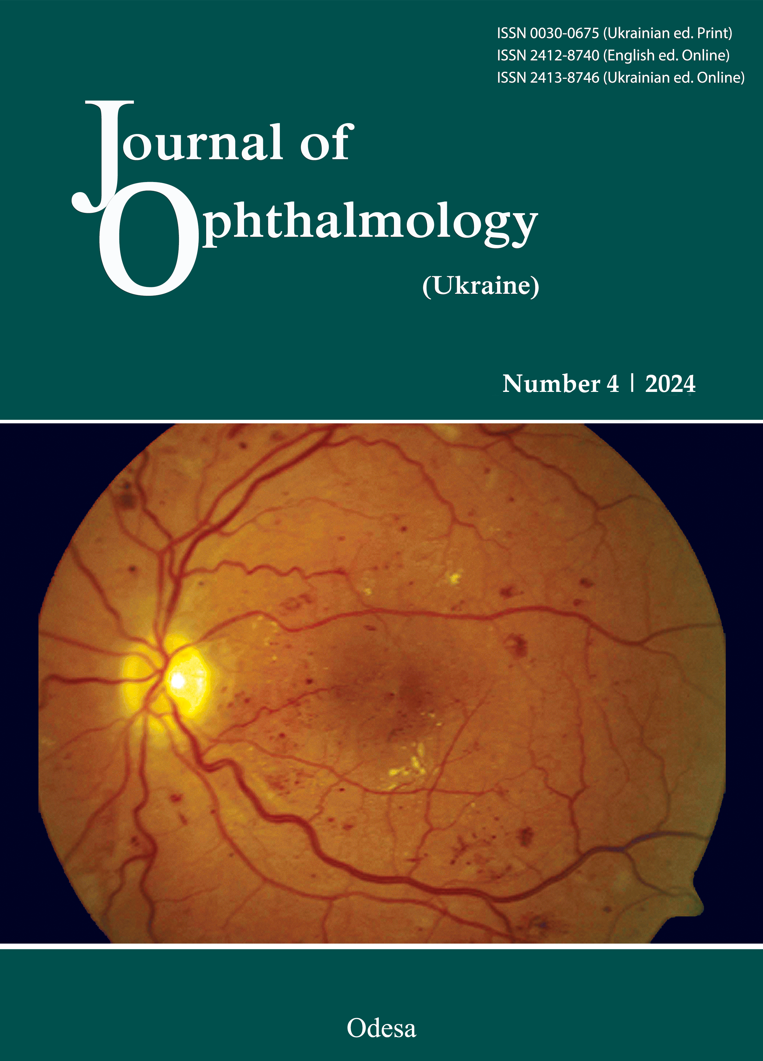
Clinical outcomes of a treat-and-extend regimen with intravitreal aflibercept injections in patients with choroidal neovascularization secondary to chronic central serous chorioretinopathy
DOI:
https://doi.org/10.31288/oftalmolzh202444651Keywords:
chronic central serous chorioretinopathy, occult type 1 choroidal neovascularization, intravitreal aflibercept, treat-and-extend, optical coherence tomographyAbstract
Purpose: To evaluate 12-month clinical outcomes of a treat-and-extend regimen with intravitreal aflibercept injections in patients with occult (type 1) choroidal neovascularization (CNV) secondary to chronic central serous chorioretinopathy (CSC).
Methods: This was a prospective observational single-center study involving 24 patients (24 eyes) with occult (type 1) CNV secondary to chronic CSC. All patients received three initial loading doses of intravitreal 2 mg (0.05 ml) aflibercept at 4-weekly intervals, followed by a treat-and-extend protocol. The primary outcome was best-corrected visual acuity (BCVA) at 12 months. Statistical analyses were conducted and graphs were created using Statistica 10.0 software.
Results: Mean BCVA increased significantly from 0.44 ± 0.35 at baseline to 0.58 ± 0.3 at month 12 (р = 0.01). At month 12, complete resolution of SRF was observed in 18 eyes (75%). The mean number of intravitreal aflibercept injections over 12 months was 7.5 ± 1.4.
Conclusion: Treat-and-extend intravitreal aflibercept is an effective and safe approach for managing patients with occult (type 1) CNV secondary to chronic CSC.
References
Spaide RF, Campeas L, Haas A, et al. Central serous chorioretinopathy in younger and older adults. Ophthalmology. 1996;103(12):2070-2080. https://doi.org/10.1016/S0161-6420(96)30386-2
Hage R, Mrejen S, Krivosic V, et al. Flat irregular retinal pigment epithelium detachments in chronic central serous chorioretinopathy and choroidal neovascularization. Am J Ophthalmol. 2015;159(5):890-903. https://doi.org/10.1016/j.ajo.2015.02.002
Lafaut BA, Salati C, Priem H, De Laey JJ. Indocyanine green angiography is of value for the diagnosis of chronic central serous chorioretinopathy in elderly patients. Graefes Arch Clin Exp Ophthalmol. 1998;236(7):513-521. https://doi.org/10.1007/s004170050114
Spaide RF, Hall L, Haas A, et al. Indocyanine green videoangiography of older patients with central serous chorioretinopathy. Retina. 1996;16(3):203-213. https://doi.org/10.1097/00006982-199616030-00004
Fung AT, Yannuzzi LA, Freund KB. Type 1 (sub-retinal pigment epithelial) neovascularization in central serous chorioretinopathy masquerading as neovascular age-related macular degeneration. Retina. 2012;32(9):1829-1837. https://doi.org/10.1097/IAE.0b013e3182680a66
Zhou X, Komuku Y, Araki T, et al. Risk factors and characteristics of central serous chorioretinopathy with later development of macular neovascularisation detected on OCT angiography: a retrospective multicentre observational study. BMJ Open Ophthalmol. 2022;7(1):e000976. https://doi.org/10.1136/bmjophth-2022-000976
Loo RH, Scott IU, Flynn HW Jr, et al. Factors associated with reduced visual acuity during long-term follow-up of patients with idiopathic central serous chorioretinopathy. Retina. 2002;22(1):19-24. https://doi.org/10.1097/00006982-200202000-00004
Sulzbacher F, Schuёtze C, Burgmuёller M, et al. Clinical evaluation of neovascular and non-neovascular chronic central serous chorioretinopathy (CSC) diagnosed by swept source optical coherence tomography angiography (SS OCTA). Graefes Arch Clin Exp Ophthalmol. 2019; 257(8):1581-1590. https://doi.org/10.1007/s00417-019-04297-z
Shiragami C, Takasago Y, Osaka R, et al. Clinical Features of Central Serous Chorioretinopathy With Type 1 Choroidal Neovascularization. Am J Ophthalmol. 2018;193:80-86. https://doi.org/10.1016/j.ajo.2018.06.009
Mrejen S, Balaratnasingam C, Kaden TR, et al. Long-term visual outcomes and causes of vision loss in chronic central serous chorioretinopathy. Ophthalmology. 2019;126(4):576-588. https://doi.org/10.1016/j.ophtha.2018.12.048
Bousquet E, Bonnin S, Mrejen S, et al. Optical coherence tomography angiography of flat irregular pigment epithelium detachment in chronic central serous chorioretinopathy. Retina. 2018;38:629-638. https://doi.org/10.1097/IAE.0000000000001580
Quaranta-El Maftouhi M, El Maftouhi A, Eandi CM. Chronic central serous chorioretinopathy imaged by optical coherence tomographic angiography. Am J Ophthalmol. 2015;160:581-587. https://doi.org/10.1016/j.ajo.2015.06.016
Wu JS, Chen SN. Optical Coherence Tomography Angiography for Diagnosis of Choroidal Neovascularization in Chronic Central Serous Chorioretinopathy after Photodynamic Therapy. Sci Rep. 2019;9:9040. https://doi.org/10.1038/s41598-019-45080-8
Yang C, Chen K, Lee S, Lee F. Photodynamic therapy in the treatment of choroidal neovascularization complicating central serous chorioretinopathy. J Chin Med Assoc. 2009;72:501-505. https://doi.org/10.1016/S1726-4901(09)70417-4
Ergun E, Tittl M, Stur M. Photodynamic therapy with verteporfin in subfoveal choroidal neovascularization secondary to central serous chorioretinopathy. Arch Ophthalmol. 2004;122:37-41. https://doi.org/10.1001/archopht.122.1.37
Chan WM, Lam DSC, Lai TYY, et al. Treatment of choroidal neovascularization in central serous chorioretinopathy by photodynamic therapy with verteporfin. Am J Ophthalmol. 2003;136:836-845. https://doi.org/10.1016/S0002-9394(03)00462-8
Hu YC, Chen YL, Chen YC, Chen SN. 3-year follow-up of half-dose verteporfin photodynamic therapy for central serous chorioretinopathy with OCT-angiography detected choroidal neovascularization. Sci Rep. 2021;11:13286. https://doi.org/10.1038/s41598-021-92693-z
Peiretti E, Caminiti G, Serra R, Querques L, Pertile R, Querques G. Anti-vascular endothelial growth factor therapy versus photodynamic therapy in the treatment of choroidal neovascularization secondary to central serous chorioretinopathy. Retina. 2018;38(8):1526-1532. https://doi.org/10.1097/IAE.0000000000001750
Lai TYY, Staurenghi G, Lanzetta P, et al. Efficacy and safety of ranibizumab for the treatment of choroidal neovascularization due to uncommon cause: twelve-month results of the MINERVA study. Retina. 2018;38(8):1464-1477. https://doi.org/10.1097/IAE.0000000000001744
Jung BJ, Kim JY, Lee JH, et al. Intravitreal aflibercept and ranibizumab for pachychoroid neovasculopathy. Sci Rep. 2019;9(1):2055. https://doi.org/10.1038/s41598-019-38504-y
Romdhane K, Zola M, Matet A, et al. Predictors of treatment response to intravitreal anti-vascular endothelial growth factor (anti-VEGF) therapy for choroidal neovascularisation secondary to chronic central serous chorioretinopathy. Br J Ophthalmol. 2020;104(7):910-916. https://doi.org/10.1136/bjophthalmol-2019-314625
Schworm B, Luft N, Keidel LF, et al. Response of neovascular central serous chorioretinopathy to an extended upload of anti-VEGF agents. Graefes Arch Clin Exp Ophthalmol. 2020;258:1013-1021. https://doi.org/10.1007/s00417-020-04623-w
Matsumoto H, Hiroe T, Morimoto M, et al. Efficacy of treat-and-extend regimen with aflibercept for pachychoroid neovasculopathy and Type 1 neovascular age-related macular degeneration. Jpn J Ophthalmol. 2018;62(2):144-150. https://doi.org/10.1007/s10384-018-0562-0
Avery RL, Bakri SJ, Blumenkranz MS, et al. Intravitreal injection technique and monitoring: updated guidelines of an expert panel. Retina. 2014;34(12):1-18. https://doi.org/10.1097/IAE.0000000000000399
Yeo JH, Oh R, Kim YJ, et al. Choroidal Neovascularization Secondary to Central Serous Chorioretinopathy: OCT Angiography Findings and Risk Factors. J Ophthalmol. 2020:7217906. https://doi.org/10.1155/2020/7217906
Chhablani J, Kozak I, Pichi F, et al. Outcomes of treatment of choroidal neovascularization associated with central serous chorioretinopathy with intravitreal antiangiogenic agents. Retina. 2015;35(12):2489-2497. https://doi.org/10.1097/IAE.0000000000000655
Lee GI, Kim AY, Kang SW, et al. Risk Factors and Outcomes of Choroidal Neovascularization Secondary to Central Serous Chorioretinopathy. Sci Rep. 2019;9:3927. https://doi.org/10.1038/s41598-019-40406-y
Smretschnig E, Hagen S, Glittenberg C, et al. Intravitreal anti-vascular endothelial growth factor combined with half-fluence photodynamic therapy for choroidal neovascularization in chronic central serous chorioretinopathy. Eye. 2016;30:805-811. https://doi.org/10.1038/eye.2016.41
Konstantinidis L, Mantel I, Zografos L, Ambresin A. Intravitreal ranibizumab in the treatment of choroidal neovascularization associated with idiopathic central serous chorioretinopathy. Eur J Ophthalmol. 2010;20:955-958. https://doi.org/10.1177/112067211002000524
Pang CE, Freund KB. Pachychoroid neovasculopathy. Retina. 2015;35:1-9. https://doi.org/10.1097/IAE.0000000000000331
Mao J, Zhang C, Liu C, et al. The Efficacy of Intravitreal Conbercept for Chronic Central Serous Chorioretinopathy. J Ophthalmol. 2019;2019:7409426. https://doi.org/10.1155/2019/7409426
Pitcher JD 3rd, Witkin AJ, DeCroos FC, Ho AC. A prospective pilot study of intravitreal aflibercept for the treatment of chronic central serous chorioretinopathy: the CONTAIN study. Br J Ophthalmol. 2015;99(6):848-852. https://doi.org/10.1136/bjophthalmol-2014-306018
Downloads
Published
How to Cite
Issue
Section
License
Copyright (c) 2024 Kustryn T. B., Zadorozhnyy O. S., Nasinnyk I. O., Nevska A. O., Pasyechnikova N. V., Korol A. R.

This work is licensed under a Creative Commons Attribution 4.0 International License.
This work is licensed under a Creative Commons Attribution 4.0 International (CC BY 4.0) that allows users to read, download, copy, distribute, print, search, or link to the full texts of the articles, or use them for any other lawful purpose, without asking prior permission from the publisher or the author as long as they cite the source.
COPYRIGHT NOTICE
Authors who publish in this journal agree to the following terms:
- Authors hold copyright immediately after publication of their works and retain publishing rights without any restrictions.
- The copyright commencement date complies the publication date of the issue, where the article is included in.
DEPOSIT POLICY
- Authors are permitted and encouraged to post their work online (e.g., in institutional repositories or on their website) during the editorial process, as it can lead to productive exchanges, as well as earlier and greater citation of published work.
- Authors are able to enter into separate, additional contractual arrangements for the non-exclusive distribution of the journal's published version of the work with an acknowledgement of its initial publication in this journal.
- Post-print (post-refereeing manuscript version) and publisher's PDF-version self-archiving is allowed.
- Archiving the pre-print (pre-refereeing manuscript version) not allowed.


