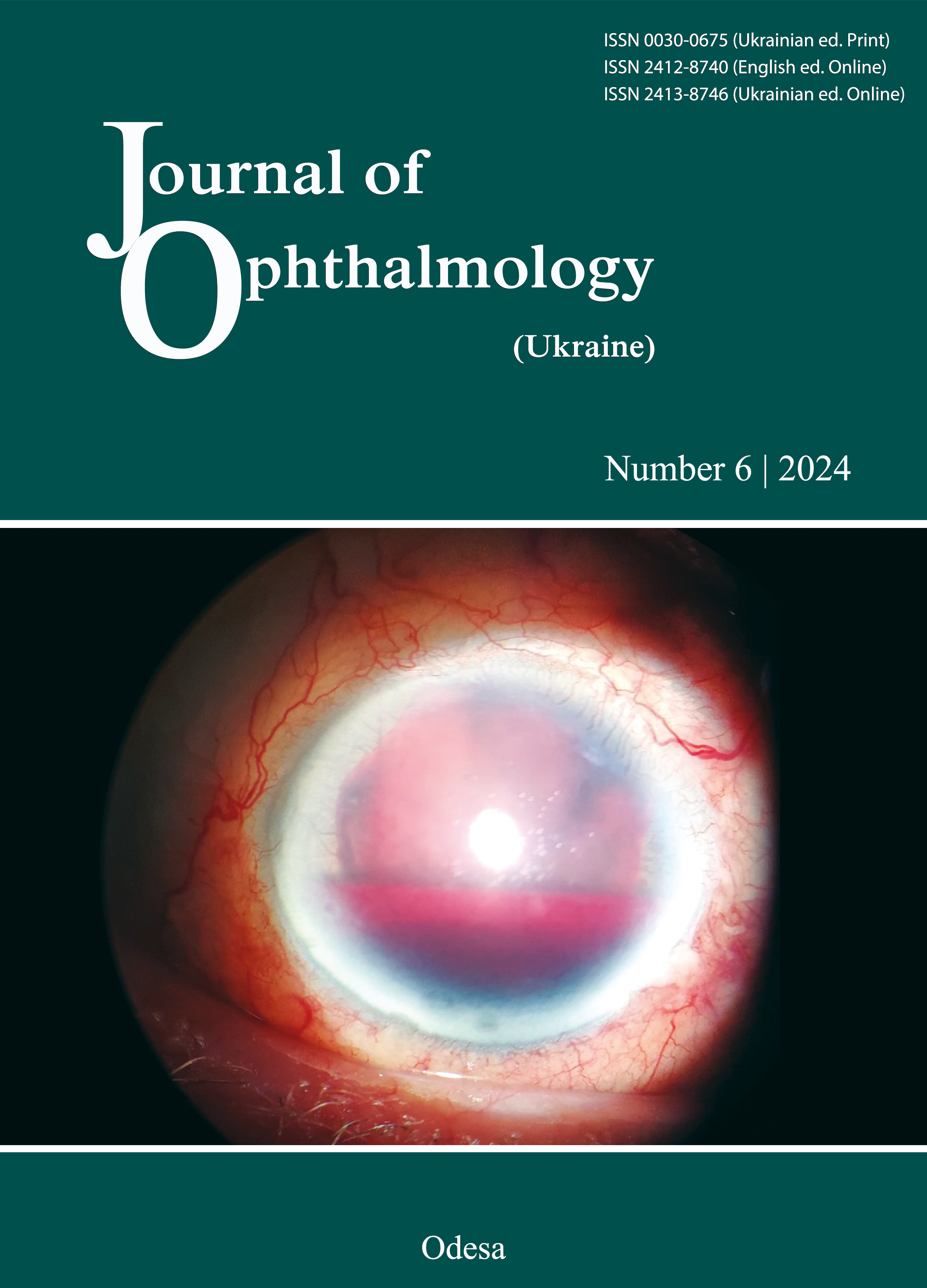
Vitrectomy with internal limiting membrane peeling for stellate nonhereditary idiopathic foveomacular retinoschisis: a case report
DOI:
https://doi.org/10.31288/oftalmolzh202466770Keywords:
stellate nonhereditary idiopathic foveomacular retinoschisis, vitrectomy, optical coherence tomography, foveomacular retinoschisis, internal limiting membraneAbstract
Purpose: To report a case of a rare disease, stellate nonhereditary idiopathic foveomacular retinoschisis (SNIFR), its clinical course and the results of vitrectomy with internal limiting membrane (ILM) peeling.
Material and Methods: Comprehensive ophthalmological examination (including visual acuity testing, biomicroscopy, ophthalmoscopy, tonometry, Goldmann kinetic perimetry and imaging with ocular ultrasound, optical coherence tomography (OCT), and fluorescein angiography (FA)) was performed before surgery and 1 month, 3 months and 12 months thereafter.
Results: The patient had no somatic comorbidity and complained only of a gradual deterioration in vision over two years. She experienced an improvement in vision after vitrectomy with ILM peeling. This treatment contributed to a substantial reduction in retinal thickness and the restoration of macular vitreoretinal interface.
Conclusion: This case indicates the efficacy and safety of surgical treatment for SNIFR, because this treatment contributed to the restoration of macular vitreoretinal interface and no recurrence was observed over a 1-year follow-up period.
References
Bloch E, Flores-Sánchez B, Georgiadis O, et al. An association between stellate nonhereditary idiopathic foveomacular retinoschisis, peripheral retinoschisis, and posterior hyaloid attachment. Retina Phila Pa. 2021;41(11):2361-9. https://doi.org/10.1097/IAE.0000000000003191
Yoshida-Uemura T, Katagiri S, Yokoi T, et al. Different foveal schisis patterns in each retinal layer in eyes with hereditary juvenile retinoschisis evaluated by en-face optical coherence tomography. Graefes Arch Clin Exp Ophthalmol. 2017;255:719-23. https://doi.org/10.1007/s00417-016-3552-2
Bloch E, Georgiadis O, Lukic M, da Cruz L. Optic disc pit maculopathy: new perspectives on the natural history. Am J Ophthalmol. 2019;207:159-69. https://doi.org/10.1016/j.ajo.2019.05.010
Steel DHW, Suleman J, Murphy DC, et al. Optic disc pit maculopathy: a two-year nationwide prospective population-based study. Ophthalmology. 2018;125:1757-64. https://doi.org/10.1016/j.ophtha.2018.05.009
Ober MD, Freund KB, Shah M, et al. Stellate nonhereditary idiopathic foveomacular retinoschisis. Ophthalmology. 2014;121:1406-13. https://doi.org/10.1016/j.ophtha.2014.02.002
Gass JD. Müller cell cone, an overlooked part of the anatomy of the fovea centralis; hypotheses concerning its role in the pathogenesis of macular hole and foveomacular retinoschisis. Arch Ophthalmol. 1999;6:821-3. https://doi.org/10.1001/archopht.117.6.821
Govetto A, Hubschman J-P, Sarraf D, et al. The role of Müller cells in tractional macular disorders: an optical coherence tomography study and physical model of mechanical force transmission. Br J Ophthalmol. 2019;104:466-72. https://doi.org/10.1136/bjophthalmol-2019-314245
Bringmann A, Unterlauft JD, Weidemann R, et al. Two different populations of Müller cells stabilize the structure of the fovea: an optical coherence tomography study. Int Ophthalmol. 2020;11:2931-2948. https://doi.org/10.1007/s10792-020-01477-3
Govetto A, Sarraf D, Hubschman JP, et al. Distinctive mechanisms and patterns of exudative versus tractional intraretinal cystoid spaces as seen with multimodal imaging. Am J Ophthalmol. 2020;212:43-56. https://doi.org/10.1016/j.ajo.2019.12.010
Rao P, Dedania VS, Drenser KA. Congenital X-linked retinoschisis: an updated clinical review. Asia Pac J Ophthalmol (Phila). 2018;7:169-75. https://doi.org/10.22608/APO.201803
Wu PC, Chen YJ, Chen YH, Chen CH, Shin SJ, Tsai CL, Kuo HK. Factors associated with foveoschisis and foveal detachment without macular hole in high myopia. Eye (Lond). 2009;23:356-61. https://doi.org/10.1038/sj.eye.6703038
Fragiotta S, Leong BC, Kaden TR, Bass SJ, Sherman J, Yannuzzi LA, Freund KB. A proposed mechanism influencing structural patterns in X-linked retinoschisis and stellate nonhereditary idiopathic foveomacular retinoschisis. Eye (Lond). 2019;33:724-8. https://doi.org/10.1038/s41433-018-0296-8
McBride M, Williamson JA. Foveal Retinoschisis: Case Report and Clinical Review. Clin Refract Optom. 2020;31:5. https://doi.org/10.57204/001c.36933
Moraes BR, Ferreira BF, Nogueira TM, Nakashima Y, Júnior HP, Souza EC. Vitrectomy for stellate nonhereditary idiopathic foveomacular retinoschisis associated with outer retinal layer defect. Retin Cases Brief Rep. 2022;16:289-292. https://doi.org/10.1097/ICB.0000000000000966
Schildroth KR, Mititelu M, Etheridge T, Holman I, Chang JS. Stellate nonhereditary idiopathic foveomacular retinoschisis: novel findings and optical coherence tomography angiography analysis. Retin Cases Brief Rep. 2023;17:165-169. https://doi.org/10.1097/ICB.0000000000001132
Ajlan RS, Hammamji KS. Stellate nonhereditary idiopathic foveomacular retinoschisis: response to topical dorzolamide therapy. Retin Cases Brief Rep. 2019;13:364-366. https://doi.org/10.1097/ICB.0000000000000599
Fine BS. Limiting membranes of the sensory retina and pigment epithelium. An electron microscopic study. Arch Ophthalmol. 1961 Dec;66:847-860. https://doi.org/10.1001/archopht.1961.00960010849012
Ho TC, Chen MS, Huang JS, et al. Foveola nonpeeling technique in internal limiting membrane peeling of myopic foveoschisis surgery. Retina. 2012;32:631-634. https://doi.org/10.1097/IAE.0b013e31824d0a4b
Shimada N, Sugamoto Y, Ogawa M, Takase H, Ohno-Matsui K. Fovea-sparing internal limiting membrane peeling for myopic traction maculopathy. Am J Ophthalmol. 2012;154:693-701. https://doi.org/10.1016/j.ajo.2012.04.013
Wakabayashi T, Oshima Y, Fujimoto H, Murakami Y, Sakaguchi H, Kusaka S, Tano Y. Foveal microstructure and visual acuity after retinal detachment repair: imaging analysis by Fourier-domain optical coherence tomography. Ophthalmology. 2009 Mar;116(3):519-528. https://doi.org/10.1016/j.ophtha.2008.10.001
Lois N, Burr J, Norrie J, Vale L, Cook J, McDonald A, etal. Internal limiting membrane peeling versus no peeling for idiopathic full-thickness macular hole: a pragmatic randomized controlled trial. Invest Ophthalmol Vis Sci. 2011 Mar 1;52(3):1586-92. https://doi.org/10.1167/iovs.10-6287
Downloads
Published
How to Cite
Issue
Section
License
Copyright (c) 2024 Pyrozhkova O. S., Umanets M. M.

This work is licensed under a Creative Commons Attribution 4.0 International License.
This work is licensed under a Creative Commons Attribution 4.0 International (CC BY 4.0) that allows users to read, download, copy, distribute, print, search, or link to the full texts of the articles, or use them for any other lawful purpose, without asking prior permission from the publisher or the author as long as they cite the source.
COPYRIGHT NOTICE
Authors who publish in this journal agree to the following terms:
- Authors hold copyright immediately after publication of their works and retain publishing rights without any restrictions.
- The copyright commencement date complies the publication date of the issue, where the article is included in.
DEPOSIT POLICY
- Authors are permitted and encouraged to post their work online (e.g., in institutional repositories or on their website) during the editorial process, as it can lead to productive exchanges, as well as earlier and greater citation of published work.
- Authors are able to enter into separate, additional contractual arrangements for the non-exclusive distribution of the journal's published version of the work with an acknowledgement of its initial publication in this journal.
- Post-print (post-refereeing manuscript version) and publisher's PDF-version self-archiving is allowed.
- Archiving the pre-print (pre-refereeing manuscript version) not allowed.


