Surgical approach to pediatric optic disc pit maculopathy: a case report
DOI:
https://doi.org/10.31288/oftalmolzh202535052Abstract
Purpose: To evaluate postoperative results of pars plana vitrectomy combined with an inverted internal limiting membrane-flap technique and intravitreal injection of viscoelastic material for optic disc pit maculopathy complicated by serous macular detachment in a child.
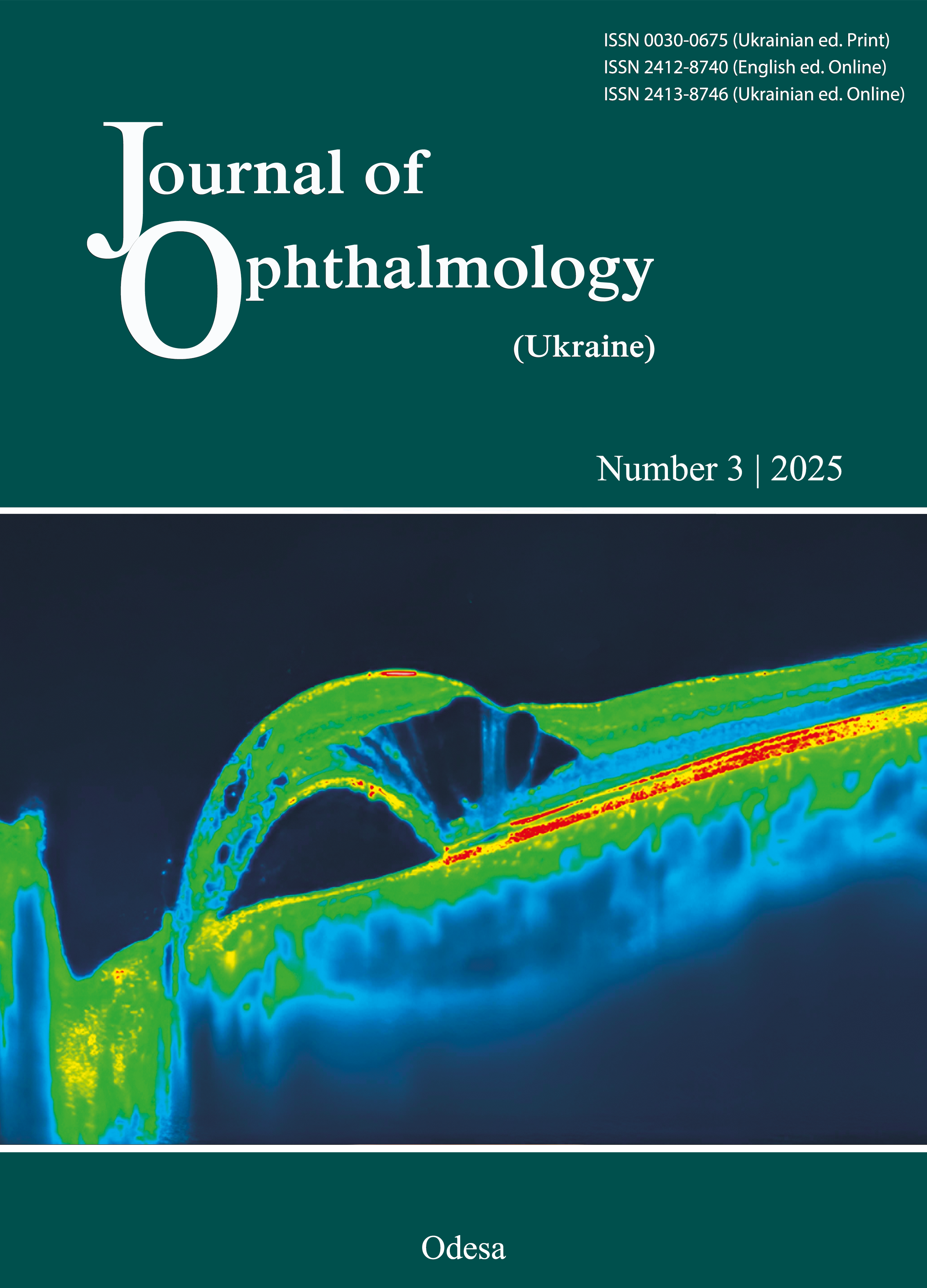
Observations: Ocular examination included best-corrected visual acuity (BCVA) testing, slit-lamp biomicroscopy, dilated fundus examination, intraocular pressure measurement, optical coherence tomography (OCT) and color fundus photography. BCVA in the left eye was 20/50 (0.4). Preoperative OCT findings showed distortion of retinal layers, serous macular detachment and a large schisis cavity in the left eye. Foveolar depression was not determined due to the height of intraretinal fluid and subretinal fluid extending towards the optic disc pit. Retinal thickness in the macular area was 507 μm.
Pars plana vitrectomy was performed in combination with an inverted internal limiting membrane internal limiting membrane-flap technique and intravitreal injection of viscoelastic material, followed by 15% C3F8 gas endotamponade.
A follow-up OCT examination in 3 months showed decreased subretinal fluid, residual edema, and restored foveolar depression. Retinal thickness in the macular area was 328 μm. BCVA of the left eye improved to 20/32 (0.63).
Conclusions: Pars plana vitrectomy with an inverted internal limiting membrane-flap technique for optic disc pit maculopathy allows to reduce an amount of intra- and subretinal fluid. A visco-associated flap fixation technique creates conditions for its stabilization, which ultimately contributes to improving anatomical and functional outcomes during surgery and in the postoperative period.
References
Wiethe T. Ein Fall von Angeborener Difformitat der Sehnervenpapille. Arch. F. Augenh. 1882; 11: 14-9.
Reis W. Eine wenig bekannte typische Missbildung am Sehnerveneintritt: umschriebene Grubenbildung auf der Papilla n. optici. Z Augenheilkd. 1908;19:505-528. https://doi.org/10.1159/000291456
Muftuoglu IK, Tokuc EO, Karabas VL. Management of optic disc pit-associated maculopathy: A case series from a tertiary referral center. Eur J Ophthalmol. 2022 May;32(3):1720-1727. https://doi.org/10.1177/11206721211023727
Prabhu V, Mangla R, Acharya I, Handa A, Thadani A, Parmar Y, Yadav NK, Chhablani J, Venkatesh R. Evaluation of baseline optic disc pit and optic disc coloboma maculopathy features by spectral domain optical coherence tomography. Int J Retina Vitreous. 2023 Aug 7;9(1):46. https://doi.org/10.1186/s40942-023-00484-7
Talli PM, Fantaguzzi PM, Bendo E, Pazzaglia A. Vitrectomy without laser treatment for macular serous detachment associated with optic disc pit: long-term outcomes. Eur J Ophthalmol. 2016 Mar-Apr;26(2):182-7. https://doi.org/10.5301/ejo.5000680
Kuhn F, Kover F, Szabo I, Mester V. Intracranial migration of silicone oil from an eye with optic pit. Graefes Arch Clin Exp Ophthalmol. 2006 Oct;244(10):1360-2. https://doi.org/10.1007/s00417-006-0267-9
Ohno-Matsui K, Hirakata A, Inoue M, et al. Evaluation of congenital optic disc pits and optic disc colobomas by swept-source optical coherence tomography. Invest Ophthalmol Vis Sci. 2013 Nov 25;54(12):7769-78. https://doi.org/10.1167/iovs.13-12901
Aslanova VS, Umanets NN, Ivanitskaya EV. Pneumatic retinopexy in treatment of patients with the optic nerve pit complicated by serous macula detachment. Oftalmologicheskii zhurnal. 2010;(6):37-40. https://doi.org/10.31288/oftalmolzh201063740
Akiyama H, Shimoda Y, Fukuchi M, et al. Intravitreal gas injection without vitrectomy for macular detachment associated with an optic disk pit. Retina. 2014 Feb;34(2):222-7. https://doi.org/10.1097/IAE.0b013e3182993d93
Gass JD. Serous detachment of the macula. Secondary to congenital pit of the optic nervehead. Am J Ophthalmol. 1969 Jun;67(6):821-41. https://doi.org/10.1016/0002-9394(69)90075-0
Chatziralli I, Theodossiadis P, Theodossiadis GP. Optic disk pit maculopathy: current management strategies. Clin Ophthalmol. 2018 Aug 10;12:1417-1422. https://doi.org/10.2147/OPTH.S153711
Downloads
Published
How to Cite
Issue
Section
License
Copyright (c) 2025 Umanets M.M., Bobrova N.F., Dovhan I.P.

This work is licensed under a Creative Commons Attribution 4.0 International License.
This work is licensed under a Creative Commons Attribution 4.0 International (CC BY 4.0) that allows users to read, download, copy, distribute, print, search, or link to the full texts of the articles, or use them for any other lawful purpose, without asking prior permission from the publisher or the author as long as they cite the source.
COPYRIGHT NOTICE
Authors who publish in this journal agree to the following terms:
- Authors hold copyright immediately after publication of their works and retain publishing rights without any restrictions.
- The copyright commencement date complies the publication date of the issue, where the article is included in.
DEPOSIT POLICY
- Authors are permitted and encouraged to post their work online (e.g., in institutional repositories or on their website) during the editorial process, as it can lead to productive exchanges, as well as earlier and greater citation of published work.
- Authors are able to enter into separate, additional contractual arrangements for the non-exclusive distribution of the journal's published version of the work with an acknowledgement of its initial publication in this journal.
- Post-print (post-refereeing manuscript version) and publisher's PDF-version self-archiving is allowed.
- Archiving the pre-print (pre-refereeing manuscript version) not allowed.


