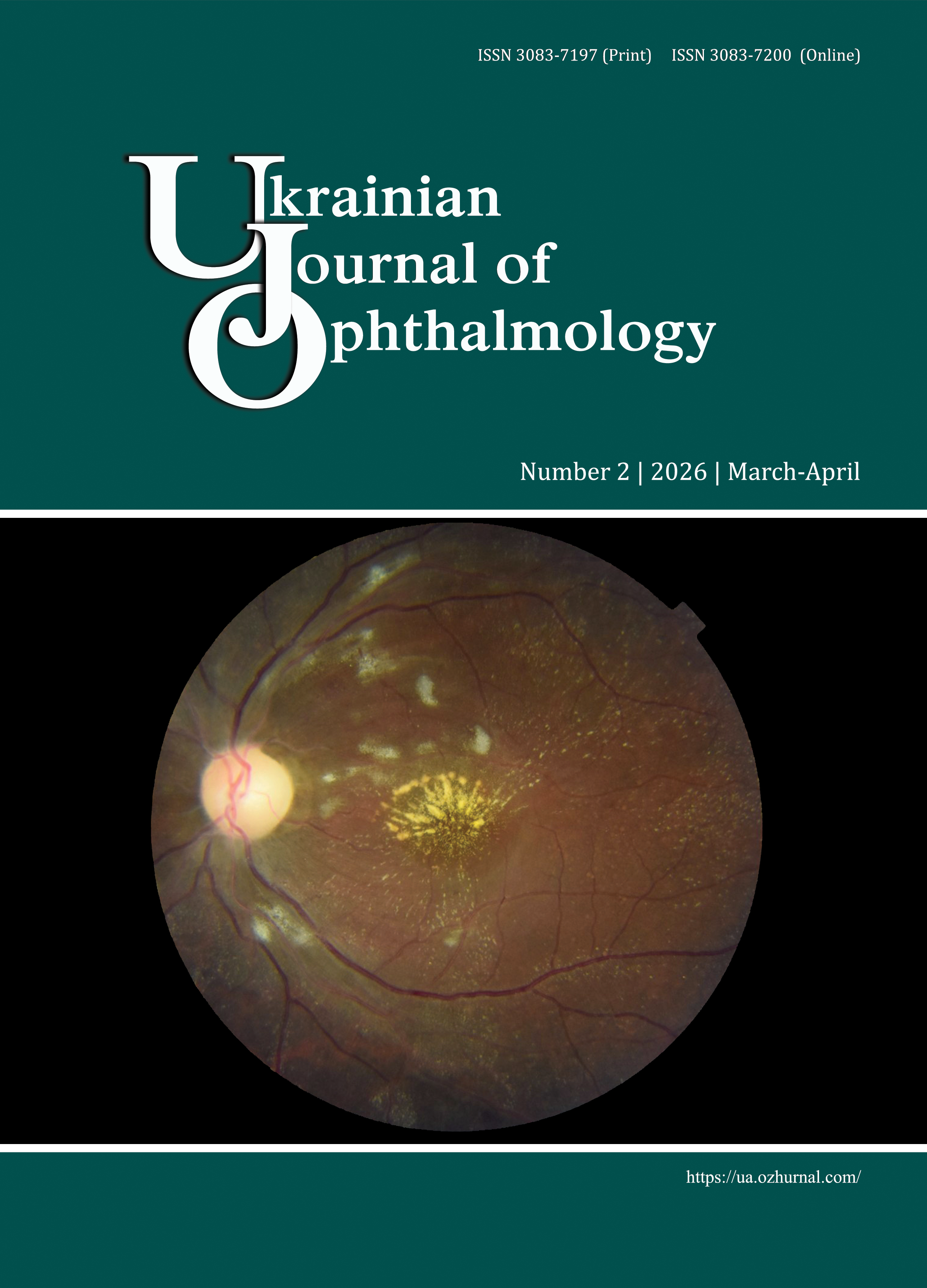
Markers of hypoxia in aqueous humor as factors for assessing the severity of diabetic retinopathy
DOI:
https://doi.org/10.31288/Ukr.j.ophthalmol.20262513Keywords:
diabetic retinopathy, type 2 diabetes mellitus, acute retinal pigment epitheliitis, lactate, hypoxia-inducible factor 1α, biomarkers, aqueous humor, disease progressionAbstract
Purpose. To establish the diagnostic value of lactate versus hypoxia-inducible factor (HIF)-1α concentration in the aqueous humor (AH) for determining the severity of diabetic retinopathy DR.
Material and Methods. Totally, 110 type 2 diabetics with DR (110 eyes) were involved in the study and divided into five groups from no apparent retinopathy (DR0) to proliferative DR (PDR) based on the 2003 international classification. The control group included 25 non-diabetics. Lactate concentrations (mg/mL) were determined in AH samples obtained during cataract surgery, and HIF-1α concentrations (pg/mL) were determined in this cohort of patients in our previous study.
Results. AH lactate level increased with disease progression (p < 0.001), with a median level ranging from 0.32 mg/mL in controls to 6.49 mg/mL in group 5 (PDR). A total accuracy was moderate (~60%) for both markers for the discrimination between all grades of the disease, but a high AH lactate concentration (>8.56 mg/mL) was found to be highly specific (98.9%) for confirming PDR. To assess the risk of DR progression, a total study sample was divided into two categories (mild-to-moderate DR vs severe DR) on the basis of AH HIF-1α concentration, and a threshold of >377 pg/mL provided for a total prediction accuracy of 71.9% and specificity 95.2%.
Conclusion. AH lactate and HIF-1α concentrations reflect a gradient of hypoxic load in DR. Determining AH lactate concentrations (a rule-in marker) is effective for severe conditions, whereas determining AH HIF-1α concentrations should be used for stratifying patients into risk groups to guide planning the intensity of supervision and treatment.
References
GBD 2019 Blindness and Vision Impairment Collaborators; Vision Loss Expert Group of the Global Burden of Disease Study. Causes of blindness and vision impairment in 2020 and trends over 30 years, and prevalence of avoidable blindness in relation to VISION 2020: the Right to Sight: an analysis for the Global Burden of Disease Study. Lancet Glob Health. 2021 Feb;9(2):e144-e160. doi: 10.1016/S2214-109X(20)30489-7. Epub 2020 Dec 1. Erratum in: Lancet Glob Health. 2021 Apr;9(4):e408. https://doi.org/10.1016/S2214-109X(21)00050-4
Moreno A, Lozano M, Salinas P. Diabetic retinopathy. Nutr Hosp. 2013 Mar;28 Suppl 2:53-6. doi: 10.3305/nh.2013.28.sup2.6714.
Magliano DJ, Boyko EJ; IDF Diabetes Atlas 10th edition scientific committee. IDF DIABETES ATLAS [Internet]. 10th ed. Brussels: International Diabetes Federation; 2021.
Tan TE, Wong TY. Diabetic retinopathy: Looking forward to 2030. Front Endocrinol (Lausanne). 2023 Jan 9;13:1077669. https://doi.org/10.3389/fendo.2022.1077669
Wilkinson CP, Ferris FL 3rd, Klein RE, Lee PP, Agardh CD, Davis M et al.; Global Diabetic Retinopathy Project Group. Proposed international clinical diabetic retinopathy and diabetic macular edema disease severity scales. Ophthalmology. 2003 Sep;110(9):1677-82. https://doi.org/10.1016/S0161-6420(03)00475-5
Cao GL, Chen KJ. Evaluation of Social Platform-Based Continuity of Care in Improving Cognitive and Prognostic Effects of Young Patients with Diabetic Retinopathy. Diabetes Metab Syndr Obes. 2023 Jun 27;16:1931-1939. https://doi.org/10.2147/DMSO.S413915
Ong JX, Konopek N, Fukuyama H, Fawzi AA. Deep Capillary Nonperfusion on OCT Angiography Predicts Complications in Eyes with Referable Nonproliferative Diabetic Retinopathy. Ophthalmol Retina. 2023 Jan;7(1):14-23 https://doi.org/10.1016/j.oret.2022.06.018
AttaAllah HR, Mohamed AAM, Ali MA. Macular vessels density in diabetic retinopathy: quantitative assessment using optical coherence tomography angiography. Int Ophthalmol. 2019 Aug;39(8):1845-1859. https://doi.org/10.1007/s10792-018-1013-0
Shinojima A, Lee D, Tsubota K, Negishi K, Kurihara T. Retinal Diseases Regulated by Hypoxia-Basic and Clinical Perspectives: A Comprehensive Review. J Clin Med. 2021 Nov 24;10(23):5496. https://doi.org/10.3390/jcm10235496
Lee D, Tomita Y, Miwa Y, Kunimi H, Nakai A, Shoda C et al. Recent Insights into Roles of Hypoxia-Inducible Factors in Retinal Diseases. Int J Mol Sci. 2024 Sep 21;25(18):10140. https://doi.org/10.3390/ijms251810140
Gui F, You Z, Fu S, Wu H, Zhang Y. Endothelial Dysfunction in Diabetic Retinopathy. Front Endocrinol (Lausanne). 2020 Sep 4;11:591. https://doi.org/10.3389/fendo.2020.00591
Brooks GA, Arevalo JA, Osmond AD, Leija RG, Curl CC, Tovar AP. Lactate in contemporary biology: a phoenix risen. J Physiol. 2022 Mar;600(5):1229-1251. https://doi.org/10.1113/JP280955
Chu KO, Chan TI, Chan KP, Yip YW, Bakthavatsalam M, Wang CC et al. Untargeted metabolomic analysis of aqueous humor in diabetic macular edema. Mol Vis. 2022 Aug 19;28:230-244.
Du X, Yang L, Kong L, Sun Y, Shen K, Cai Y et al. Metabolomics of various samples advancing biomarker discovery and pathogenesis elucidation for diabetic retinopathy. Front Endocrinol (Lausanne). 2022 Oct 27;13:1037164. https://doi.org/10.3389/fendo.2022.1037164
Dolar-Szczasny J, Drab A, Rejdak R. Biochemical Changes in Anterior Chamber of the Eye in Diabetic Patients-A Review. J Clin Med. 2024 Apr 27;13(9):2581. https://doi.org/10.3390/jcm13092581
Chatziralli I, Loewenstein A. Intravitreal Anti-Vascular Endothelial Growth Factor Agents for the Treatment of Diabetic Retinopathy: A Review of the Literature. Pharmaceutics. 2021 Jul 26;13(8):1137. https://doi.org/10.3390/pharmaceutics13081137
Bahr TA, Bakri SJ. Update on the Management of Diabetic Retinopathy: Anti-VEGF Agents for the Prevention of Complications and Progression of Nonproliferative and Proliferative Retinopathy. Life (Basel). 2023 Apr 27;13(5):1098. https://doi.org/10.3390/life13051098
Jakobsen TS, Adsersen RL, Askou AL, Corydon TJ. Functional Roles of Pigment Epithelium-Derived Factor in Retinal Degenerative and Vascular Disorders: A Scoping Review. Invest Ophthalmol Vis Sci. 2024 Dec 2;65(14):41. https://doi.org/10.1167/iovs.65.14.41
Lytvynenko TV. [Hypoxiainducible factor-α (HIF-1α) and progression of diabetic retinopathy]. Medical Science of Ukraine. 2025;21(3):76-84. [in Ukrainian]. https://doi.org/10.32345/2664-4738.3.2025.08
Bouchez CL, Daubon T, Mourier A. NADH-independent enzymatic assay to quantify extracellular and intracellular L-lactate levels. STAR Protoc. 2022 May 16;3(2):101403. https://doi.org/10.1016/j.xpro.2022.101403
Kanda Y. Investigation of the freely available easy-to-use software 'EZR' for medical statistics. Bone Marrow Transplant. 2013 Mar;48(3):452-8. https://doi.org/10.1038/bmt.2012.244
Brownlee J. One-vs-Rest and One-vs-One for Multi-Class Classification [Internet]. Machine Learning Mastery; 2021 [cited 2025 October 5]. Available from: https://machinelearningmastery.com/one-vs-rest-and-one-vs-one-for-multi-class-classification.
Robin X, Turck N, Hainard A, Tiberti N, Lisacek F, Sanchez JC, Müller M. pROC: an open-source package for R and S+ to analyze and compare ROC curves. BMC Bioinformatics. 2011 Mar 17;12:77. https://doi.org/10.1186/1471-2105-12-77
Min J, Zeng T, Roux M, Lazar D, Chen L, Tudzarova S. The Role of HIF1α-PFKFB3 Pathway in Diabetic Retinopathy. J Clin Endocrinol Metab. 2021 Aug 18;106(9):2505-2519.https://doi.org/10.1210/clinem/dgab362
Yu Z, Zhang T, Gong C, Sheng Y, Lu B, Zhou L et al. Erianin inhibits high glucose-induced retinal angiogenesis via blocking ERK1/2-regulated HIF-1α-VEGF/VEGFR2 signaling pathway. Sci Rep. 2016 Sep 28;6:34306. https://doi.org/10.1038/srep34306
Zhang J, Qin Y, Martinez M, Flores-Bellver M, Rodrigues M, Dinabandhu A et al. HIF-1α and HIF-2α redundantly promote retinal neovascularization in patients with ischemic retinal disease. J Clin Invest. 2021 Jun 15;131(12):e139202. https://doi.org/10.1172/JCI139202
Huang H, He J, Johnson D, Wei Y, Liu Y, Wang S et al. Deletion of placental growth factor prevents diabetic retinopathy and is associated with Akt activation and HIF1α-VEGF pathway inhibition. Diabetes. 2015 Jan;64(1):200-12. doi: 10.2337/db14-0016. Epub 2014 Sep 3. Erratum in: Diabetes. 2015 Mar;64(3):1067. https://doi.org/10.2337/db15-er03
Hu P, Liu G, Sun H, Wei W. Expressions of HIF-1α and MiR-210 in aqueous humor of patients with central retinal vein occlusion combined with macular edema. Pak J Med Sci. 2022 May-Jun;38(5):1327-1332. https://doi.org/10.12669/pjms.38.5.5092
Suk-Gyu H, Kang B, Song JS. Increased angiogenic factors in the aqueous and vitreous humors after disinsertion of extraocular muscle and the effects of triamcinolone acetate injection. Sci Rep. Springer Science and Business Media LLC; 2022 Mar 28;12(1):5276.https://doi.org/10.1038/s41598-022-09377-5
Nesper PL, Fawzi AA. Perfusion Deficits in Diabetes Without Retinopathy Localize to the Perivenular Deep Capillaries Near the Fovea on OCT Angiography. Ophthalmol Sci. 2024 Feb 1;4(5):100482. https://doi.org/10.1016/j.xops.2024.100482
Salongcay RP, Aquino LAC, Salva CMG, Peto T, Silva PS. Comparison of Diabetic Retinopathy Lesions Identified Using Ultrawide Field Imaging and Optical Coherence Tomography Angiography. Ophthalmic Res. 2023;66(1):1053-1062. https://doi.org/10.1159/000531723
Adki KM, Kulkarni YA. Potential Biomarkers in Diabetic Retinopathy. Curr Diabetes Rev. 2020;16(9):971-983. https://doi.org/10.2174/1573399816666200217092022
Yeo EJ. Hypoxia and aging. Exp Mol Med. 2019 Jun 20;51(6):1-15. https://doi.org/10.1038/s12276-019-0233-3
Grochowski ET, Pietrowska K, Godlewski A, Gosk W, Buczynska A, Wojnar M, et al. Simultaneous Comparison of Aqueous Humor and Serum Metabolic Profiles of Diabetic and Nondiabetic Patients Undergoing Cataract Surgery-A Targeted and Quantitative Metabolomics Study. Int J Mol Sci. 2023 Aug 11;24(16):12671. https://doi.org/10.3390/ijms241612671
Arai-Okuda M, Murai Y, Maeda H, Kanamori A, Miki T, Naito T, et al. Potentially compromised systemic and local lactate metabolic balance in glaucoma, which could increase retinal glucose and glutamate concentrations. Sci Rep. 2024 Feb 14;14(1):3683. https://doi.org/10.1038/s41598-024-54383-4
Lazzara F, Trotta MC, Platania CBM, D'Amico M, Petrillo F, Galdiero M et al. Stabilization of HIF-1α in Human Retinal Endothelial Cells Modulates Expression of miRNAs and Proangiogenic Growth Factors. Front Pharmacol. 2020 Jul 17;11:1063. https://doi.org/10.3389/fphar.2020.01063
Moshfeghi AA, Khurana RN, Moini H, Sherman S, Reed K, Boucher N et al. Impact of anti-VEGF treatment on development of proliferative diabetic retinopathy in routine clinical practice. BMC Ophthalmol. 2024 May 31;24(1):229. https://doi.org/10.1186/s12886-024-03491-w
Xie X, Lian C, Zhang Z, Feng M, Wang W, Yuan X et al. Aflibercept for long-term treatment of diabetic macular edema and proliferative diabetic retinopathy: a meta-analysis. Front Endocrinol (Lausanne). 2023 May 16;14:1144422. https://doi.org/10.3389/fendo.2023.1144422
Callan A, Heckman J, Tah G, Lopez S, Valdez L, Tsin A. VEGF in Diabetic Retinopathy and Age-Related Macular Degeneration. Int J Mol Sci. 2025 May 22;26(11):4992. https://doi.org/10.3390/ijms26114992
Chong DD, Das N, Singh RP. Diabetic retinopathy: Screening, prevention, and treatment. Cleve Clin J Med. 2024 Aug 1;91(8):503-510.https://doi.org/10.3949/ccjm.91a.24028
Simó R, Franch-Nadal J, Vlacho B, Real J, Amado E, Flores J, et al. Rapid Reduction of HbA1c and Early Worsening of Diabetic Retinopathy: A Real-world Population-Based Study in Subjects With Type 2 Diabetes. Diabetes Care. 2023 Sep 1;46(9):1633-1639. https://doi.org/10.2337/dc22-2521
Lu J, Ma X, Zhang L, Mo Y, Ying L, Lu W et al. Glycemic variability assessed by continuous glucose monitoring and the risk of diabetic retinopathy in latent autoimmune diabetes of the adult and type 2 diabetes. J Diabetes Investig. 2019 May;10(3):753-759. https://doi.org/10.1111/jdi.12957
Zhang B, Zhang B, Zhou Z, Guo Y, Wang D. The value of glycosylated hemoglobin in the diagnosis of diabetic retinopathy: a systematic review and Meta-analysis. BMC Endocr Disord. 2021 Apr 26;21(1):82. https://doi.org/10.1186/s12902-021-00737-2
Yin L, Zhang D, Ren Q, Su X, Sun Z. Prevalence and risk factors of diabetic retinopathy in diabetic patients: A community based cross-sectional study. Medicine (Baltimore). 2020 Feb;99(9):e19236. https://doi.org/10.1097/MD.0000000000019236
Wen X, Ng TK, Zhang G, Chen H, Wu Z, Liu Q et al. Tear lactate improves the evaluation of proliferative diabetic retinopathy in type-2 diabetes patients. Mol Biomed. 2025 Jul 18;6(1):52. https://doi.org/10.1186/s43556-025-00297-0
Zhang J, Zhang J, Zhang C, Zhang J, Gu L, Luo D et al. Diabetic Macular Edema: Current Understanding, Molecular Mechanisms and Therapeutic Implications. Cells. 2022 Oct 25;11(21):3362. https://doi.org/10.3390/cells11213362
Dolar-Szczasny J, Święch A, Flieger J, Tatarczak-Michalewska M, Niedzielski P, Proch Jet al. Levels of Trace Elements in the Aqueous Humor of Cataract Patients Measured by the Inductively Coupled Plasma Optical Emission Spectrometry. Molecules. 2019 Nov 14;24(22):4127. https://doi.org/10.3390/molecules24224127
Downloads
Published
How to Cite
Issue
Section
License
Copyright (c) 2026 Petrenko O.V., Lytvynenko T.V., Babenko M.S., Ziablitsev S.V.

This work is licensed under a Creative Commons Attribution 4.0 International License.
This work is licensed under a Creative Commons Attribution 4.0 International (CC BY 4.0) that allows users to read, download, copy, distribute, print, search, or link to the full texts of the articles, or use them for any other lawful purpose, without asking prior permission from the publisher or the author as long as they cite the source.
COPYRIGHT NOTICE
Authors who publish in this journal agree to the following terms:
- Authors hold copyright immediately after publication of their works and retain publishing rights without any restrictions.
- The copyright commencement date complies the publication date of the issue, where the article is included in.
DEPOSIT POLICY
- Authors are permitted and encouraged to post their work online (e.g., in institutional repositories or on their website) during the editorial process, as it can lead to productive exchanges, as well as earlier and greater citation of published work.
- Authors are able to enter into separate, additional contractual arrangements for the non-exclusive distribution of the journal's published version of the work with an acknowledgement of its initial publication in this journal.
- Post-print (post-refereeing manuscript version) and publisher's PDF-version self-archiving is allowed.
- Archiving the pre-print (pre-refereeing manuscript version) not allowed.


