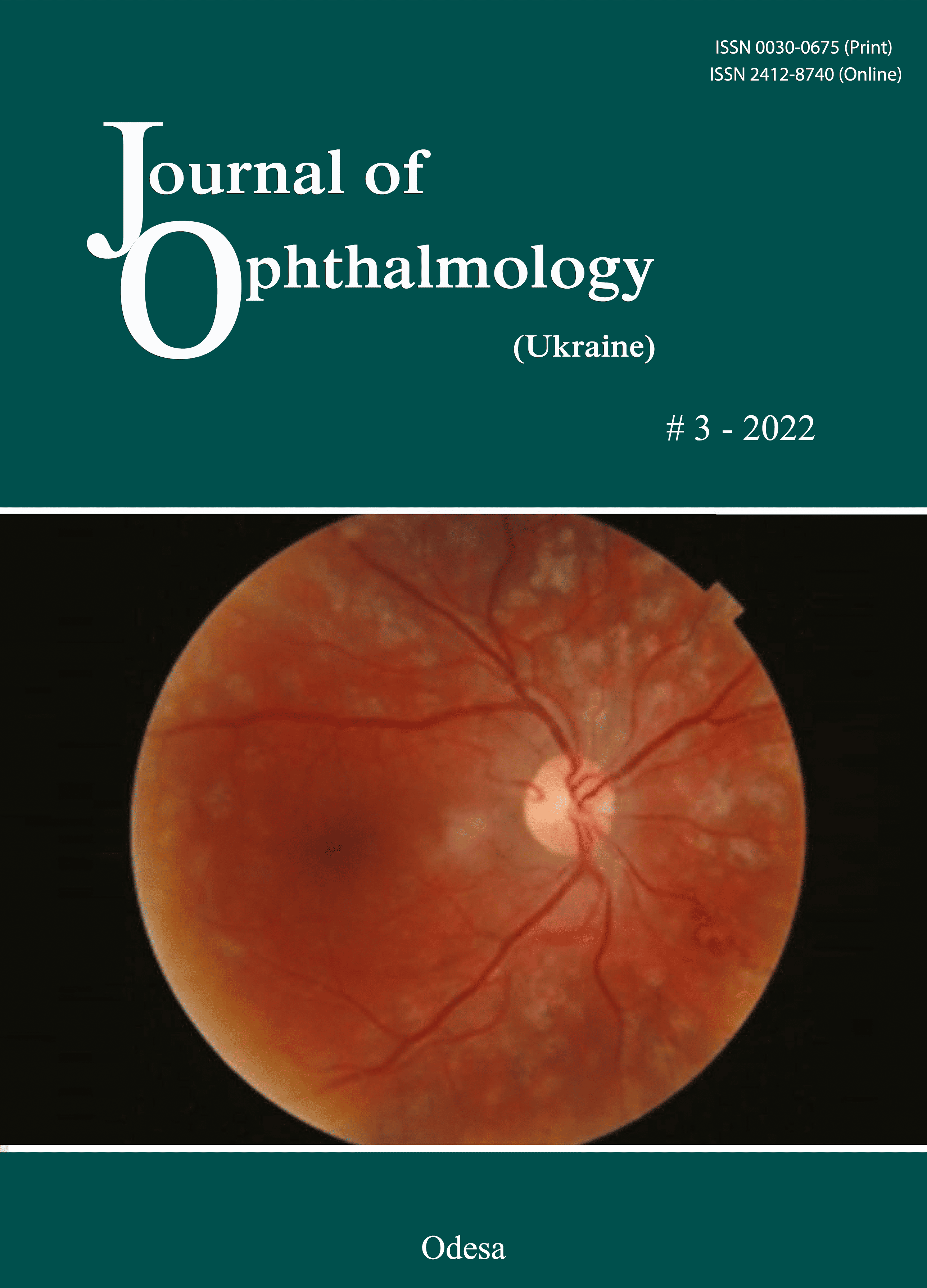
Choroideremia – A clinical insight and differential diagnosis
DOI:
https://doi.org/10.31288/oftalmolzh202235053Ключові слова:
Retinal dystrophy, Choroideremia, Retinitis pigmentosa, Chorioretinal degeneration, Differential diagnosisАнотація
Choroideremia is an X-linked recessive inherited, bilateral progressive chorioretinal dystrophy/degeneration leading to blindness by late adulthood. However, it can be confused occasionally with other conditions, especially retinitis pigmentosa due to their shared clinical manifestations. Since the management and patients' counseling differ between those conditions listed in the differential diagnosis, it is important for clinicians to come to the right diagnosis. This article is trying to make a differential diagnosis between choroideremia and other conditions based on the current knowledge of these disorders.
Посилання
1.Nouraeinejad A. Differential Diagnosis in Optometry and Ophthalmology. Second Edition. Iran: Noruzi Publication; 2017.
2.Katz BJ, Yang Z, Payne M, et al. Fundus appearance of choroideremia using optical coherence tomograpy. Adv Exp Med Biol. 2006;572:57-61. https://doi.org/10.1007/0-387-32442-9_9
3.Zinkernagel MS, MacLaren RE. Recent advances and future prospects in choroideremia. Clin Ophthalmol. 2015;9:2195-2200. https://doi.org/10.2147/OPTH.S65732
4.Freund PR, Sergeev YV, MacDonald IM. Analysis of a large choroideremia dataset does not suggest a preference for inclusion of certain genotypes in future trials of gene therapy. Mol Genet Genomic Med. 2016;4:344-358. https://doi.org/10.1002/mgg3.208
5.Heon E, Alabduljalil T, McGuigan ID, et al. Visual function and central retinal structure in choroideremia. Invest Ophthalmol Vis Sci. 2016;57:377-387. https://doi.org/10.1167/iovs.15-18421
6.Mitsios A, Dubis AM, Moosajee M. Choroideremia: from genetic and clinical phenotyping to gene therapy and future treatments. Ther Adv Ophthalmol. 2018;10:1-18.
https://doi.org/10.1177/2515841418817490
7.Dong S, Tsao N, Hou Q, et al. US Health Resource Utilization and Cost Burden Associated with Choroideremia. Clin Ophthalmol. 2021;15:3459-3465. https://doi.org/10.2147/OPTH.S311844
8.Moosajee M, Ramsden SC, Black GC, et al. Clinical utility gene card for: choroideremia. Eur J Hum Genet. 2014;22(4):e1-e4. https://doi.org/10.1038/ejhg.2013.183
9.Sankila EM, Tolvanen R, van den Hurk JAJM, et al. Aberrant splicing of the CHM gene is a significant cause of choroideremia. Nat Genet. 1992; 1: 109-113. https://doi.org/10.1038/ng0592-109
10.MacDonald IM, Sereda C, McTaggart K, et al. Choroideremia gene testing. Expert Rev Mol Diagn. 2004; 4: 478-484. https://doi.org/10.1586/14737159.4.4.478
11.Coussa RG, Kim J, Traboulsi EI. Choroideremia: effect of age on visual acuity in patients and female carriers. Ophthalmic Genet. 2012; 33: 66-73. https://doi.org/10.3109/13816810.2011.623261
12.Pennesi ME, Birch DG, Duncan JL, et al. Choroideremia: retinal degeneration with an unmet need. Retina. 2019;39:2059-2069. https://doi.org/10.1097/IAE.0000000000002553
13.Bonilha VL, Trzupek KM, Li Y, et al. Choroideremia: analysis of the retina from a female symptomatic carrier. Ophthalmic Genet. 2008; 29: 99-110. https://doi.org/10.1080/13816810802206499
14.Thobani A, Anastasakis A, Fishman GA. Microperimetry and OCT findings in female carriers of choroideremia. Ophthalmic Genet. 2010;31(4):235-9.
https://doi.org/10.3109/13816810.2010.518578
15.Edwards TL, Groppe M, Jolly JK, et al. Correlation of retinal structure and function in choroideremia carriers. Ophthalmology. 2015; 122: 1274-1276. https://doi.org/10.1016/j.ophtha.2014.12.036
16.Khan KN, Islam F, Moore AT, et al. Clinical and genetic features of choroideremia in childhood. Ophthalmology. 2016; 123: 2158-2165. https://doi.org/10.1016/j.ophtha.2016.06.051
17.Zweifel SA, Engelbert M, Laud K, et al. Outer Retinal Tubulation: A Novel Optical Coherence Tomography Finding. Arch Ophthalmol. 2009;127(12):1596-1602. https://doi.org/10.1001/archophthalmol.2009.326
18.Genead MA, Fishman GA. Cystic macular oedema on spectral-domain optical coherence tomography in choroideremia patients without cystic changes on fundus examination. Eye 2011; 25: 84-90. https://doi.org/10.1038/eye.2010.157
19.Shen LL, Ahluwalia A, Sun M, et al. Long-term natural history of visual acuity in eyes with choroideremia: a systematic review and meta-analysis of data from 1004 individual eyes. Br J Ophthalmol. 2021;105:271-8. https://doi.org/10.1136/bjophthalmol-2020-316028
20.Campos-Pavon J, Torres-Pena JL. Choroidal neovascularization secondary to choroideremia. Arch Soc Esp Oftalmol. 2015; 90: 289-291. https://doi.org/10.1016/j.oftal.2014.03.012
21.Yang J, Wang LN, Yu RG, et al. Multimodal imaging of the carriers of choroideremia and X-linked retinitis pigmentosa. Int J Ophthalmol. 2018;11(10):1721-1725.
22.Nanda A, Salvetti A.P, Martinez-Fernandez de la Camara C, MacLaren RE. Misdiagnosis of X-linked retinitis pigmentosa in a choroideremia patient with heavily pigmented fundi. Ophthalmic Genetics. 2018; 39(3):380-383. https://doi.org/10.1080/13816810.2018.1430242
23.Guo H, Li J, Gao F, et al. Whole-exome sequencing reveals a novel CHM gene mutation in a family with choroideremia initially diagnosed as retinitis pigmentosa. BMC Ophthalmol. 2015;15:85. https://doi.org/10.1186/s12886-015-0081-4
24.Lee TKM, McTaggart KE, Sieving PA, et al. Clinical diagnoses that overlap with choroideremia. Can J Ophthalmol. 2003; 38: 364-372. https://doi.org/10.1016/S0008-4182(03)80047-9
25.Hartong DT, Berson EL, Dryja TP. Retinitis pigmentosa. Lancet. 2006; 368: 1795-1809. https://doi.org/10.1016/S0140-6736(06)69740-7
26.Bowne SJ, Humphries MM, Sullivan LS, et al. A dominant Mutation in RPE65 identified by whole-exome sequencing causes retinitis pigmentosa with choroidal involvement. Eur J Hum Genet. 2011; 19: 1074-1081. https://doi.org/10.1038/ejhg.2011.86
27.van den Hurk JA, Schwartz M, van Bokhoven H, et al. Molecular basis of choroideremia (CHM): mutations involving the Rab escort protein-1 (REP-1) gene. Hum Mutat. 1997;9(2):110-7. https://doi.org/10.1002/(SICI)1098-1004(1997)9:2<110::AID-HUMU2>3.0.CO;2-D
28.Genead MA, Fishman GA, Grover S. Hereditary choroidal diseases. In: Retina. 5th ed. Amsterdam: Elsevier, 2012, pp. 891-898. https://doi.org/10.1016/B978-1-4557-0737-9.00043-6
29.Sergouniotis PI, Davidson AE, Lenassi E, et al. Retinal structure, function, and molecular pathologic features in gyrate atrophy. Ophthalmology. 2012; 119: 596-605. https://doi.org/10.1016/j.ophtha.2011.09.017
30.Kabunga P, Lau AK, Phan K, et al. Systematic review of cardiac electrical disease in Kearns-Sayre syndrome and mitochondrial cytopathy. Int J Cardiol. 2015; 181: 303-310. https://doi.org/10.1016/j.ijcard.2014.12.038
31.Halford S, Liew G, MacKay DS, et al. Detailed phenotypic and genotypic characterization of bietti crystalline dystrophy. Ophthalmology. 2014; 121: 1174-1184. https://doi.org/10.1016/j.ophtha.2013.11.042
##submission.downloads##
Опубліковано
Як цитувати
Номер
Розділ
Ліцензія
Авторське право (c) 2025 Ali Nouraeinejad

Ця робота ліцензується відповідно до Creative Commons Attribution 4.0 International License.
Ця робота ліцензується відповідно до ліцензії Creative Commons Attribution 4.0 International (CC BY). Ця ліцензія дозволяє повторно використовувати, поширювати, переробляти, адаптувати та будувати на основі матеріалу на будь-якому носії або в будь-якому форматі за умови обов'язкового посилання на авторів робіт і первинну публікацію у цьому журналі. Ліцензія дозволяє комерційне використання.
ПОЛОЖЕННЯ ПРО АВТОРСЬКІ ПРАВА
Автори, які подають матеріали до цього журналу, погоджуються з наступними положеннями:
- Автори отримують право на авторство своєї роботи одразу після її публікації та назавжди зберігають це право за собою без жодних обмежень.
- Дата початку дії авторського права на статтю відповідає даті публікації випуску, до якого вона включена.
ПОЛІТИКА ДЕПОНУВАННЯ
- Редакція журналу заохочує розміщення авторами рукопису статті в мережі Інтернет (наприклад, у сховищах установ або на особистих веб-сайтах), оскільки це сприяє виникненню продуктивної наукової дискусії та позитивно позначається на оперативності і динаміці цитування.
- Автори мають право укладати самостійні додаткові угоди щодо неексклюзивного розповсюдження статті у тому вигляді, в якому вона була опублікована цим журналом за умови збереження посилання на первинну публікацію у цьому журналі.
- Дозволяється самоархівування постпринтів (версій рукописів, схвалених до друку в процесі рецензування) під час їх редакційного опрацювання або опублікованих видавцем PDF-версій.
- Самоархівування препринтів (версій рукописів до рецензування) не дозволяється.


