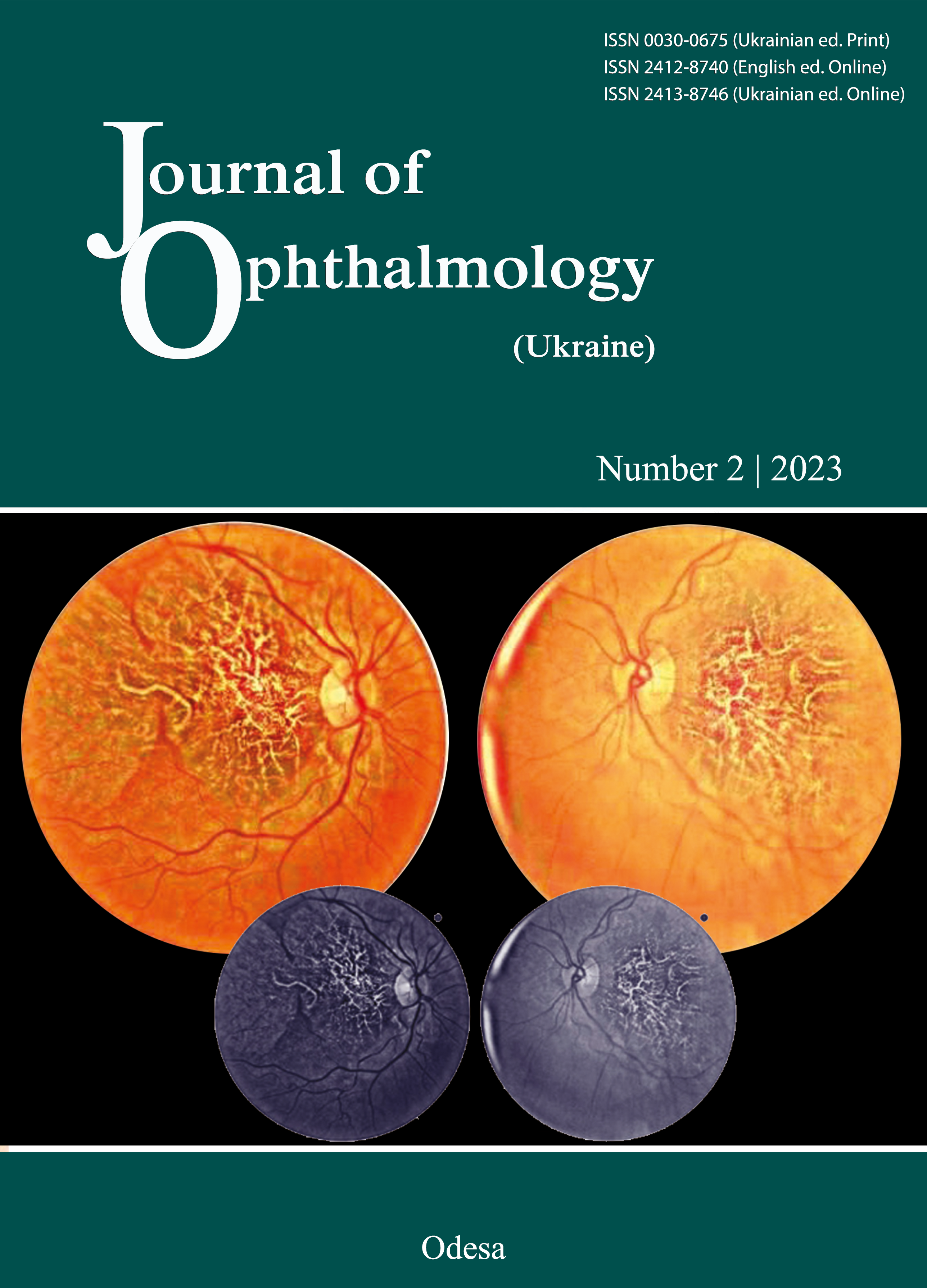
Review on imaging methods in non-infectious posterior uveitis, principles, relevance, and practical clinical applications to disease entities
DOI:
https://doi.org/10.31288/oftalmolzh20233959Keywords:
uveitis, choroiditis, choriocapillaritis, photoreceptoritis, fluorescein angiography (FA), indocyanine green angiography (ICGA), optical coherence angiography (OCT), EDI-OCT, blue light fundus autofluorescence (BAF) OCT-angiographyAbstract
The work-up and diagnosis of posterior uveitis rely heavily on multiple imaging methods that have become available beyond the mere photographic imaging and fluorescein angiography (FA) used to image uveitis in the past. Global assessment and precise follow-up of posterior uveitis were achieved with the development of indocyanine green angiography (ICGA) since the mid-1990ties that, together with FA, made it possible to perform dual FA and ICGA giving information on both the retina and the choroidal compartment. Further non-invasive imaging methods were developed subsequently that contributed to additional valuable information completing the dual FA/ICGA basic appraisal of uveitis, including (1) optical coherence tomography (OCT) giving a quasi-histological morphology of retinal structures of the posterior pole, (2) enhanced-depth imaging OCT (EDI-OCT) allowing to image the choroidal compartment and (3) blue light fundus autofluorescence (BAF) showing the integrity or damage of the retinal pigment epithelium, the photoreceptors and the outer retina. OCT-angiography (OCT-A) became available more recently and presented the advantage to image the retinal and choroidal circulations without needing dye injections, necessary for dual FA/ICGA. This review article will illustrate the principles, relevance and practical applications of these different imaging methods used in uveitis by examining the main categories of non-infectious posterior uveitis entities including (1) retinal inflammatory disorders, inflammatory diseases of the outer retina and of the choriocapillaris (choriocapillaritis) and stromal choroiditis.
References
Novotny HR, Alvis DL. A method of photographing fluorescence in circulating blood in the human retina. Circulation. 1961 Jul;24:82-6. https://doi.org/10.1161/01.CIR.24.1.82
Herbort CP, LeHoang P, Guex-Crosier Y. Schematic interpretation of indocyanine green angiography in posterior uveitis using a standard angiographic protocol. Ophthalmology. 1998 Mar;105(3):432-40. https://doi.org/10.1016/S0161-6420(98)93024-X
Altan-Yaycioglu R, Akova YA, Akca S, Yilmaz G. Inflammation of the posterior uvea: findings on fundus fluorescein and indocyanine green angiography. Ocul Immunol Inflamm. 2006 Jun;14(3):171-9. https://doi.org/10.1080/09273940600660524
Fardeau C, Tran TH, Gharbi B, Cassoux N, Bodaghi B, LeHoang P. Retinal fluorescein and indocyanine green angiography and optical coherence tomography in successive stages of Vogt-Koyanagi-Harada disease. Int Ophthalmol. 2007 Apr-Jun;27(2-3):163-72. https://doi.org/10.1007/s10792-006-9024-7
Herbort CP. Fluorescein and indocyanine green angiography for uveitis. Middle East Afr J Ophthalmol. 2009 Oct;16(4):168-87.
El Ameen A, Herbort CP. Comparison of Retinal and Choroidal Involvement in Sarcoidosis-related Chorioretinitis Using Fluorescein and Indocyanine Green Angiography. J Ophthalmic Vis Res. 2018 Oct-Dec;13(4):426-432. https://doi.org/10.4103/jovr.jovr_201_17
Tugal-Tutkun I, Herbort CP, Khairallah M; Angiography Scoring for Uveitis Working Group (ASUWOG). Scoring of dual fluorescein and ICG inflammatory angiographic signs for the grading of posterior segment inflammation (dual fluorescein and ICG angiographic scoring system for uveitis). Int Ophthalmol. 2010 Oct;30(5):539-52. https://doi.org/10.1007/s10792-008-9263-x
Tanaka R, Kaburaki T, Yoshida A, Takamoto M, Miyaji T, Yamaguchi T. Fluorescein Angiography Scoring System Using Ultra-Wide-Field Fluorescein Angiography Versus Standard Fluorescein Angiography in Patients with Sarcoid Uveitis. Ocul Immunol Inflamm. 2021 Nov 17;29(7-8):1398-1402. https://doi.org/10.1080/09273948.2020.1737141
Sadiq MA, Hassan M, Afridi R, Halim MS, Do DV, Sepah YJ, Nguyen QD; STOP-UVEITIS Investigators. Posterior segment inflammatory outcomes assessed using fluorescein angiography in the STOP-UVEITIS study. Int J Retina Vitreous. 2020 Oct 6;6:47. https://doi.org/10.1186/s40942-020-00245-w
Fujimoto J, Swanson E. The Development, Commercialization, and Impact of Optical Coherence Tomography. Invest Ophthalmol Vis Sci. 2016 Jul 1;57(9):OCT1-OCT13. https://doi.org/10.1167/iovs.16-19963
Antcliff RJ, Stanford MR, Chauhan DS, Graham EM, Spalton DJ, Shilling JS, et al. Comparison between optical coherence tomography and fundus fluorescein angiography for the detection of cystoid macular edema in patients with uveitis. Ophthalmology. 2000 Mar;107(3):593-9. https://doi.org/10.1016/S0161-6420(99)00087-1
Papasavvas I, Mantovani A, Herbort CP Jr. Acute Posterior Multifocal Placoid Pigment Epitheliopathy (APMPPE): A Comprehensive Approach and Case Series: Systemic Corticosteroid Therapy Is Necessary in a Large Proportion of Cases. Medicina (Kaunas). 2022 Aug 8;58(8):1070. https://doi.org/10.3390/medicina58081070
Papasavvas I, Herbort CP Jr. Diagnosis and Treatment of Primary Inflammatory Choriocapillaropathies (PICCPs): A Comprehensive Overview. Medicina (Kaunas). 2022 Jan 21;58(2):165. https://doi.org/10.3390/medicina58020165
Papasavvas I, Neri P, Mantovani A, Herbort CP Jr. Idiopathic multifocal choroiditis (MFC): aggressive and prolonged therapy with multiple immunosuppressive agents is needed to halt the progression of active disease. An offbeat review and a case series. J Ophthalmic Inflamm Infect. 2022 Jan 10;12(1):2. https://doi.org/10.1186/s12348-021-00278-8
Papasavvas I, Mantovani A, Herbort CP Jr. Diagnosis, Mechanisms, and Differentiation of Inflammatory Diseases of the Outer Retina: Photoreceptoritis versus Choriocapillaritis; A Multimodal Imaging Perspective. Diagnostics (Basel). 2022 Sep 9;12(9):2179. https://doi.org/10.3390/diagnostics12092179
Spaide RF, Koizumi H, Pozzoni MC. Enhanced depth imaging spectral-domain optical coherence tomography. Am J Ophthalmol. 2008 Oct;146(4):496-500. https://doi.org/10.1016/j.ajo.2008.05.032
Balci O, Gasc A, Jeannin B, Herbort CP Jr. Enhanced depth imaging is less suited than indocyanine green angiography for close monitoring of primary stromal choroiditis: a pilot report. Int Ophthalmol. 2017 Jun;37(3):737-748. https://doi.org/10.1007/s10792-016-0303-7
Delori F, Keilhauer C, Sparrow JR, Staurenghi G. Origin of fundus Autofluorescence. In: Atlas of fundus autofluorescence imaging, Holz FG, Schmitz-Valckenberg S, Spaide RF, Bird AC, Eds Springer, Heidelberg 2007, pp17-26.
Kramer M, Priel E. Fundus autofluorescence imaging in multifocal choroiditis: beyond the spots. Ocul Immunol Inflamm. 2014 Oct;22(5):349-55. https://doi.org/10.3109/09273948.2013.855797
Mantovani A, Giani A, Herbort CP Jr, Staurenghi G. Interpretation of fundus autofluorescence changes in choriocapillaritis: a multi-modality imaging study. Graefes Arch Clin Exp Ophthalmol. 2016 Aug;254(8):1473-1479. https://doi.org/10.1007/s00417-015-3205-x
Herbort CP Jr, Arapi I, Papasavvas I, Mantovani A, Jeannin B. Acute Zonal Occult Outer Retinopathy (AZOOR) Results from a Clinicopathological Mechanism Different from Choriocapillaritis Diseases: A Multimodal Imaging Analysis. Diagnostics (Basel). 2021 Jun 29;11(7):1184. https://doi.org/10.3390/diagnostics11071184
Spaide RF, Fujimoto JG, Waheed NK, Sadda SR, Staurenghi G. Optical coherence tomography angiography. Prog Retin Eye Res. 2018 May;64:1-55. https://doi.org/10.1016/j.preteyeres.2017.11.003
Rocholz R, Corvi F, Weichsel J, Schmidt S, Staurenghi G. OCT Angiography (OCTA) in Retinal Diagnostics. 2019 Aug 14. In: Bille JF, editor. High Resolution Imaging in Microscopy and Ophthalmology: New Frontiers in Biomedical Optics [Internet]. Cham (CH): Springer; 2019. Chapter 6. https://doi.org/10.1007/978-3-030-16638-0_6
Herbort CP Jr, Papasavvas I, Tugal-Tutkun I. Benefits and Limitations of OCT-A in the Diagnosis and Follow-Up of Posterior Intraocular Inflammation in Current Clinical Practice: A Valuable Tool or a Deceiver? Diagnostics (Basel). 2022 Sep 30;12(10):2384. https://doi.org/10.3390/diagnostics12102384
Pichi F, Hay S. Use of optical coherence tomography angiography in the uveitis clinic. Graefes Arch Clin Exp Ophthalmol. 2022 Jul 16. https://doi.org/10.1007/s00417-022-05763-x
Invernizzi A, Carreño E, Pichi F, Munk MR, Agarwal A, Zierhut M, Pavesio C. Experts Opinion: OCTA vs. FFA/ICG in Uveitis - Which Will Survive? Ocul Immunol Inflamm. 2022 Jul 7:1-8. https://doi.org/10.1080/09273948.2022.2084421
Tian M, Zeng G, Tappeiner C, Zinkernagel MS, Wolf S, Munk MR. Comparison of Indocyanine Green Angiography and Swept-Source Wide-Field Optical Coherence Tomography Angiography in Posterior Uveitis. Front Med (Lausanne). 2022 May 2;9:853315. https://doi.org/10.3389/fmed.2022.853315
Herbort CP Jr, Takeuchi M, Papasavvas I, Tugal-Tutkun I, Hedayatfar A, et al. Optical coherence tomography angiography (OCT-A) in uveitis: a literature review and a reassessment pf its real role. Diagnostics (Basel) 2022: submitted https://doi.org/10.3390/diagnostics13040601
Tang W, Guo J, Liu W, Xu G. Optical Coherence Tomography Angiography of Inflammatory Choroidal Neovascularization Early Response after Anti-VEGF Treatment. Curr Eye Res. 2020 Dec;45(12):1556-1562. https://doi.org/10.1080/02713683.2020.1767790
Perente A, Kotsiliti D, Taliantzis S, Panagiotopoulou EK, Gkika M, Perente I, et al. Serpiginous Choroiditis Complicated with Choroidal Neovascular Membrane Detected using Optical Coherence Tomography Angiography: A Case Series and Literature Review. Turk J Ophthalmol. 2021 Oct 26;51(5):326-333. https://doi.org/10.4274/tjo.galenos.2021.49323
Kongwattananon W, Grasic D, Lin H, Oyeniran E, Sen HN, Kodati S. Role of optical coherence tomography angiography in detecting and monitoring inflammatory choroidal neovascularization. Retina. 2022 Jun 1;42(6):1047-1056. https://doi.org/10.1097/IAE.0000000000003420
Dutheil C, Korobelnik JF, Delyfer MN, Rougier MB. Optical coherence tomography angiography and choroidal neovascularization in multifocal choroiditis: A descriptive study. Eur J Ophthalmol. 2018 Sep;28(5):614-621. https://doi.org/10.1177/1120672118759623
Astroz P, Miere A, Mrejen S, Sekfali R, Souied EH, Jung C, et al. Optical coherence tomography angiography to distinguish choroidal neovascularization from macular inflammatory lesions in multifocal choroiditis. Retina. 2018 Feb;38(2):299-309. https://doi.org/10.1097/IAE.0000000000001617
Gan Y, Zhang X, Su Y, Shen M, Peng Y, Wen F. OCTA versus dye angiography for the diagnosis and evaluation of neovascularisation in punctate inner choroidopathy. Br J Ophthalmol. 2022 Apr;106(4):547-552. https://doi.org/10.1136/bjophthalmol-2020-318191
Abucham-Neto JZ, Torricelli AAM, Lui ACF, Guimarães SN, Nascimento H, Regatieri CV. Comparison between optical coherence tomography angiography and fluorescein angiography findings in retinal vasculitis. Int J Retina Vitreous. 2018 Apr 16;4:15. https://doi.org/10.1186/s40942-018-0117-z
Arias JD, Parra MM, Arango FJ, Hoyos AT, Rangel CM, Sánchez-Ávila RM. Differentiation of Features of Inflammatory Neovascular Membrane Versus Active Posterior Uveitis by SS-OCTA. Ophthalmic Surg Lasers Imaging Retina. 2021 Mar;52(3):129-137. https://doi.org/10.3928/23258160-20210302-03
Smid LM, Vermeer KA, Missotten TOAR, van Laar JAM, van Velthoven MEJ. Parafoveal Microvascular Alterations in Ocular and Non-Ocular Behҫet's Disease Evaluated With Optical Coherence Tomography Angiography. Invest Ophthalmol Vis Sci. 2021 Mar 1;62(3):8. https://doi.org/10.1167/iovs.62.3.8
Mebsout-Pallado C, Orès R, Terrada C, Dansingani KK, Chhablani J, Eller AW, et al. Review of the Current Literature and Our Experience on the Value of OCT-angiography in White Dot Syndromes. Ocul Immunol Inflamm. 2022 Feb 17;30(2):364-378. https://doi.org/10.1080/09273948.2020.1837185
Furino C, Shalchi Z, Grassi MO, Cardoso JN, Keane PA, Niro A, et al. OCT Angiography in Acute Posterior Multifocal Placoid Pigment Epitheliopathy. Ophthalmic Surg Lasers Imaging Retina. 2019 Jul 1;50(7):428-436. https://doi.org/10.3928/23258160-20190703-04
Papasavvas I, Mantovani A, Tugal-Tutkun I, Herbort CP Jr. Multiple evanescent white dot syndrome (MEWDS): update on practical appraisal, diagnosis and clinicopathology; a review and an alternative comprehensive perspective. J Ophthalmic Inflamm Infect. 2021 Dec 18;11(1):45. https://doi.org/10.1186/s12348-021-00279-7
Usui Y, Goto H. Granuloma-like formation in deeper retinal plexus in ocular sarcoidosis. Clin Ophthalmol. 2019 May 27;13:895-896. https://doi.org/10.2147/OPTH.S200519
Herbort CP, Probst K, Cimino L, Tran VT. Differential inflammatory involvement in retina and choroïd in birdshot chorioretinopathy. Klin Monbl Augenheilkd. 2004 May;221(5):351-6. https://doi.org/10.1055/s-2004-812827
Essex RW, Wong J, Jampol LM, Dowler J, Bird AC. Idiopathic multifocal choroiditis: a comment on present and past nomenclature. Retina 2013; 33:1-4. https://doi.org/10.1097/IAE.0b013e3182641860
Skvortsova N, Gasc A, Jeannin B, Herbort CP. Evolution of choroidal thickness over time and effect of early and sustained therapy in birdshot retinochoroiditis. Eye (Lond). 2017 Aug;31(8):1205-1211. https://doi.org/10.1038/eye.2017.54
Knecht PB, Papadia M, Herbort CP Jr. Early and sustained treatment modifies the phenotype of birdshot retinochoroiditis. Int Ophthalmol. 2014 Jun;34(3):563-74. https://doi.org/10.1007/s10792-013-9861-0
Elahi S, Herbort CP Jr. Vogt-Koyanagi-Harada Disease and Birdshot Retinochoroidopathy, Similarities and Differences: A Glimpse into the Clinicopathology of Stromal Choroiditis, a Perspective and a Review. Klin Monbl Augenheilkd. 2019 Apr;236(4):492-510. English. https://doi.org/10.1055/a-0829-6763
Elahi S, Gillmann K, Gasc A, Jeannin B, Herbort CP Jr. Sensitivity of indocyanine green angiography compared to fluorescein angiography and enhanced depth imaging optical coherence tomography during tapering and fine-tuning of therapy in primary stromal choroiditis: A case series. J Curr Ophthalmol. 2019 Jan 17;31(2):180-187. https://doi.org/10.1016/j.joco.2018.12.006
Herbort CP Jr, Abu El Asrar AM, Takeuchi M, Pavésio CE, Couto C, et al. Catching the therapeutic window of opportunity in early initial-onset Vogt-Koyanagi-Harada uveitis can cure the disease. Int Ophthalmol. 2019 Jun;39(6):1419-1425. https://doi.org/10.1007/s10792-018-0949-4
Downloads
Published
How to Cite
Issue
Section
License
Copyright (c) 2023 Ioannis Papasavvas, Carl P. Herbort Jr

This work is licensed under a Creative Commons Attribution 4.0 International License.
This work is licensed under a Creative Commons Attribution 4.0 International (CC BY 4.0) that allows users to read, download, copy, distribute, print, search, or link to the full texts of the articles, or use them for any other lawful purpose, without asking prior permission from the publisher or the author as long as they cite the source.
COPYRIGHT NOTICE
Authors who publish in this journal agree to the following terms:
- Authors hold copyright immediately after publication of their works and retain publishing rights without any restrictions.
- The copyright commencement date complies the publication date of the issue, where the article is included in.
DEPOSIT POLICY
- Authors are permitted and encouraged to post their work online (e.g., in institutional repositories or on their website) during the editorial process, as it can lead to productive exchanges, as well as earlier and greater citation of published work.
- Authors are able to enter into separate, additional contractual arrangements for the non-exclusive distribution of the journal's published version of the work with an acknowledgement of its initial publication in this journal.
- Post-print (post-refereeing manuscript version) and publisher's PDF-version self-archiving is allowed.
- Archiving the pre-print (pre-refereeing manuscript version) not allowed.


