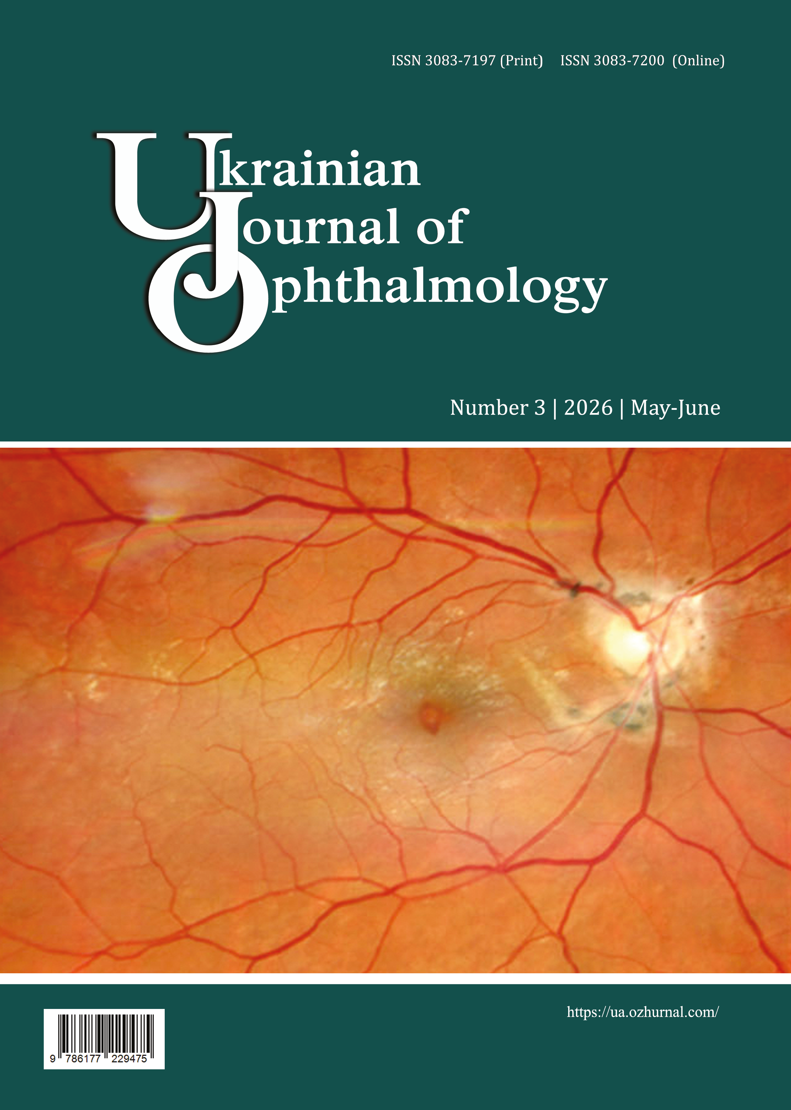
Multimodal characterization of the features of the course of stromal herpetic keratitis according to clinical, functional, laboratory and morphometric indicators
DOI:
https://doi.org/10.31288/Ukr.j.ophthalmol.202631221Keywords:
herpetic keratitis, herpes simplex virus, ocular hemodynamics, tear production, corneal morphometry, corneaAbstract
Purpose: To determine the features of the course of herpetic stromal keratitis (HSK) based on a comprehensive evaluation of clinical, functional, laboratory and morphometric parameters.
Material and Methods: Totally, 60 patients with HSK (mean age, 39.4 ± 9.5 years) were included in the study. Doppler ultrasound (Toshiba Nemio-20) was used to assess hemodynamics in the ophthalmic artery (OA), central retinal artery (CRA) and central retinal vein (CRV). Pentacam AXL (Oculus, Germany) was used for keratometry, pachymetry and corneal densitometry measurements, and optical computed tomography (Optopol REVO NX, Poland), for corneal epithelial thickness measurements. Corneal neovascularization (NVR) was assessed by the area and depth of vascular invasion; SpO2, by pulse oxymetry; and immunoglobulin G (IgG) for herpes simplex virus 1 and 2 (HSV 1/2) and cytomegalovirus (CMV), by enzyme-linked immunosorbent assay. Schirmer II and tear film break-up time tests were used to assess tear production and tear film stability.
Results: In patients with HSK, the incidence of corneal ulceration increased with the area of corneal NVR (p = 0.0006). Peripheral ulceration prevailed in NVR in one quadrant, and central NVR, in ≥2 quadrants (χ² = 4.3; p = 0.04). The incidence of total corneal edema increased with the area of corneal NVR. Corneal densitometry values were by 20.5% higher in the presence of total edema than in the presence of focal edema. The risk of mixed NVR was significantly increased when ≥2 corneal quadrants were affected by NVR (p < 0.001). The incidence of corneal ulceration in superficial, deep and mixed NVR was 25.9%, 16.7% and 100%, respectively. CRA hemodynamics parameters did not depend on the presence of NVR and ulceration, whereas CRV flow rate was 7.2% higher in the presence of NVR (р < 0.05). The presence of NVR was associated with a 23.6% reduced tear film stability (p = 0.004). We found no association of IgG for HSV 1/2 or CMV with characteristics of corneal NVR and ulceration.
Conclusion: Progression of damage to the corneal stroma and development of corneal ulceration were associated with the severity of neovascularization. The incidence of ulceration increased with the number of NVR quadrants (p = 0.0006), and patients with mixed NVR had 4.3 times the odds of ulceration (p < 0.001). NVR depth and keratometry measurements were the most informative for predicting the risk of corneal ulceration. NVR was associated with moderate changes in regional hemodynamics and marked abnormality of tear production with tear film instability.
References
Liesegang TJ, Melton LJ 3rd, Daly PJ, Ilstrup DM. Epidemiology of ocular herpes simplex. Incidence in Rochester, Minn, 1950 through 1982. Arch Ophthalmol. 1989 Aug;107(8):1155-9. https://doi.org/10.1001/archopht.1989.01070020221029
Park PJ, Chang M, Garg N, Zhu J, Chang JH, Shukla D. Corneal lymphangiogenesis in herpetic stromal keratitis. Surv Ophthalmol. 2015 Jan-Feb;60(1):60-71. https://doi.org/10.1016/j.survophthal.2014.06.001
Chodosh J, Ung L. Adoption of Innovation in Herpes Simplex Virus Keratitis. Cornea. 2020 Nov;39 Suppl 1(1):S7-S18.https://doi.org/10.1097/ICO.0000000000002425
Farooq AV, Shukla D. Herpes simplex epithelial and stromal keratitis: an epidemiologic update. Surv Ophthalmol. 2012 Sep;57(5):448-62. https://doi.org/10.1016/j.survophthal.2012.01.005
McCormick I, James C, Welton NJ, Mayaud P, Turner KME, Gottlieb SL, et al. Incidence of herpes simplex virus keratitis and other ocular disease: global review and estimates. Ophthalmic Epidemiol. 2022 Aug;29(4):353-362. https://doi.org/10.1080/09286586.2021.1962919
Young RC, Hodge DO, Liesegang TJ, Baratz KH. Incidence, recurrence, and outcomes of herpes simplex virus eye disease in Olmsted County, Minnesota, 1976-2007: the effect of oral antiviral prophylaxis. Arch Ophthalmol. 2010 Sep;128(9):1178-83. https://doi.org/10.1001/archophthalmol.2010.187
Musa M, Enaholo E, Aluyi-Osa G, Atuanya GN, Spadea L, Salati C, Zeppieri M. Herpes simplex keratitis: A brief clinical overview. World J Virol. 2024 Mar 25;13(1):89934.https://doi.org/10.5501/wjv.v13.i1.89934
Kim GN, Yoo WS, Park MH, Chung JK, Han YS, Chung IY, et al. Clinical Features of Herpes Simplex Keratitis in a Korean Tertiary Referral Center: Efficacy of Oral Antiviral and Ascorbic Acid on Recurrence. Korean J Ophthalmol. 2018 Oct;32(5):353-360. https://doi.org/10.3341/kjo.2017.0131
Lobo AM, Agelidis AM, Shukla D. Pathogenesis of herpes simplex keratitis: The host cell response and ocular surface sequelae to infection and inflammation. Ocul Surf. 2019 Jan;17(1):40-49. https://doi.org/10.1016/j.jtos.2018.10.002
Liesegang TJ. Classification of herpes simplex virus keratitis and anterior uveitis. Cornea. 1999 Mar;18(2):127-43. https://doi.org/10.1097/00003226-199903000-00001
Holland EJ, Schwartz GS. Classification of herpes simplex virus keratitis. Cornea. 1999 Mar;18(2):144-54. https://doi.org/10.1097/00003226-199903000-00002
Nanji A, Redd T, Chamberlain W, Schallhorn JM, Chen S, Ploner S, Maier A, Fujimoto JG, Jia Y, Huang D, Li Y. Application of Corneal Optical Coherence Tomography Angiography for Assessment of Vessel Depth in Corneal Neovascularization. Cornea. 2020 May;39(5):598-604. https://doi.org/10.1097/ICO.0000000000002232
Zheng G, Gastwirth JL. On estimation of the variance in Cochran-Armitage trend tests for genetic association using case-control studies. Stat Med. 2006 Sep 30;25(18):3150-9. https://doi.org/10.1002/sim.2250
Spineli, L. M. Local inconsistency detection using the Kullback-Leibler divergence measure. Systematic Reviews, 13, 261 (2024). https://doi.org/10.1186/s13643-024-02680-4
Wang L, Wang R, Xu C, Zhou H. Pathogenesis of Herpes Stromal Keratitis: Immune Inflammatory Response Mediated by Inflammatory Regulators. Front Immunol. 2020 May 13;11:766. https://doi.org/10.3389/fimmu.2020.00766
Azar DT. Corneal angiogenic privilege: angiogenic and antiangiogenic factors in corneal avascularity, vasculogenesis, and wound healing (an American Ophthalmological Society thesis). Trans Am Ophthalmol Soc. 2006;104:264-302.
Cursiefen C, Küchle M, Naumann GO. Angiogenesis in corneal diseases: histopathologic evaluation of 254 human corneal buttons with neovascularization. Cornea. 1998 Nov;17(6):611-3. https://doi.org/10.1097/00003226-199811000-00008
Abdelfattah NS, Amgad M, Zayed AA, Salem H, Elkhanany AE, Hussein H, Abd El-Baky N. Clinical correlates of common corneal neovascular diseases: a literature review. Int J Ophthalmol. 2015 Feb 18;8(1):182-93. doi: 10.3980/j.issn.2222-3959.2015.01.32.
Sharif Z, Sharif W. Corneal neovascularization: updates on pathophysiology, investigations & management. Rom J Ophthalmol. 2019 Jan-Mar;63(1):15-22.https://doi.org/10.22336/rjo.2019.4
Drzyzga Ł, Śpiewak D, Dorecka M, Wyględowska-Promieńska D. Available Therapeutic Options for Corneal Neovascularization: A Review. Int J Mol Sci. 2024 May 17;25(10):5479. https://doi.org/10.3390/ijms25105479
Ahmad A, Nawaz MI. Molecular mechanism of VEGF and its role in pathological angiogenesis. J Cell Biochem. 2022 Dec;123(12):1938-1965. https://doi.org/10.1002/jcb.30344
Akinsiku S, Shukla D. Molecular Pathways Driving Corneal Neovascularization in Herpes Simplex Keratitis. Pathogens. 2026 Feb 7;15(2):186. https://doi.org/10.3390/pathogens15020186
Giménez F, Suryawanshi A, Rouse BT. Pathogenesis of herpes stromal keratitis--a focus on corneal neovascularization. Prog Retin Eye Res. 2013 Mar;33:1-9. https://doi.org/10.1016/j.preteyeres.2012.07.002
Schmidt VJ, Hilgert JG, Covi JM, Leibig N, Wietbrock JO, et al. Flow increase is decisive to initiate angiogenesis in veins exposed to altered hemodynamics. PLoS One. 2015;10(1):e0117407. https://doi.org/10.1371/journal.pone.0117407
Rao P, Suvas S. Development of Inflammatory Hypoxia and Prevalence of Glycolytic Metabolism in Progressing Herpes Stromal Keratitis Lesions. J Immunol. 2019 Jan 15;202(2):514-526. https://doi.org/10.4049/jimmunol.1800422
Wilhelmus KR, Sugar J, Hyndiuk RA, Stulting RD. Corneal thickness changes during herpes simplex virus disciform keratitis. Cornea. 2004 Mar;23(2):154-7. https://doi.org/10.1097/00003226-200403000-00008
Lu L, Palioura S. Management of Stromal Herpes Simplex Virus Keratitis With Epithelial Ulceration Using Optical Coherence Tomography-Generated Corneal Thickness Maps. Cornea. 2020 Dec;39(12):1566-1570. https://doi.org/10.1097/ICO.0000000000002423
Abtahi MA, Beheshtnejad AH, Latifi G, Akbari-Kamrani M, Ghafarian S, Masoomi A, Sonbolastan SA, Jahanbani-Ardakani H, Atighechian M, Banan L, Nouri H, Abtahi SH. Corneal Epithelial Thickness Mapping: A Major Review. J Ophthalmol. 2024 Jan 2;2024:6674747. https://doi.org/10.1155/2024/6674747
Versura P, Giannaccare G, Pellegrini M, Sebastiani S, Campos EC. Neurotrophic keratitis: current challenges and future prospects. Eye Brain. 2018 Jun 28;10:37-45.https://doi.org/10.2147/EB.S117261
Messmer EM. The pathophysiology, diagnosis, and treatment of dry eye disease. Dtsch Arztebl Int. 2015 Jan 30;112(5):71-81; quiz 82. https://doi.org/10.3238/arztebl.2015.0071
Humayun S, Noor M, Shahid M, Naqvi SAH, Ishaq M, Humayun Q. Diagnosis of Dry Eye Syndrome Using Ocular Surface Disease Index, Tear Film Break-up Time, and Schirmer Test. J Coll Physicians Surg Pak. 2024 Mar;34(3):308-312. https://doi.org/10.29271/jcpsp.2024.03.308
Ma X, Lu Y. Bilateral tear film alterations in patients with unilateral quiescent herpes simplex keratitis. Acta Ophthalmol. 2017 Sep;95(6):629-633. https://doi.org/10.1111/aos.13329
M'Garrech M, Rousseau A, Kaswin G, Sauer A, Barreau E, Bourcier T, Labetoulle M. Impairment of lacrimal secretion in the unaffected fellow eye of patients with recurrent unilateral herpetic keratitis. Ophthalmology. 2013 Oct;120(10):1959-67. https://doi.org/10.1016/j.ophtha.2013.02.037
Shoji J, Sakimoto T, Inada N, Kamei Y, Matsubara M, Takamura E, Sawa M. A diagnostic method for herpes simplex keratitis by simultaneous measurement of viral DNA and virus-specific secretory IgA in tears: an evaluation. Jpn J Ophthalmol. 2016 Jul;60(4):294-301. https://doi.org/10.1007/s10384-016-0448-y
Chen S, Peng Q, Wang H, Xie J, Cao H, Xu X. Seroprevalence and Clinical Insights of Ocular Herpesvirus Infections: A Cross-Sectional Study Evaluating ELISA as a Diagnostic Tool. Infect Drug Resist. 2025 Jun 19;18:3063-3070. https://doi.org/10.2147/IDR.S527047
Wang J, Cherfan DG, Goshe JM. Utility of HSV Serology for Chronic Corneal Pathology. Eye Contact Lens. 2020 May;46(3):190-193. https://doi.org/10.1097/ICL.0000000000000635
Downloads
Published
How to Cite
Issue
Section
License
Copyright (c) 2026 Maksymova I. R., Khramenko N. I.

This work is licensed under a Creative Commons Attribution 4.0 International License.
This work is licensed under a Creative Commons Attribution 4.0 International (CC BY 4.0) that allows users to read, download, copy, distribute, print, search, or link to the full texts of the articles, or use them for any other lawful purpose, without asking prior permission from the publisher or the author as long as they cite the source.
COPYRIGHT NOTICE
Authors who publish in this journal agree to the following terms:
- Authors hold copyright immediately after publication of their works and retain publishing rights without any restrictions.
- The copyright commencement date complies the publication date of the issue, where the article is included in.
DEPOSIT POLICY
- Authors are permitted and encouraged to post their work online (e.g., in institutional repositories or on their website) during the editorial process, as it can lead to productive exchanges, as well as earlier and greater citation of published work.
- Authors are able to enter into separate, additional contractual arrangements for the non-exclusive distribution of the journal's published version of the work with an acknowledgement of its initial publication in this journal.
- Post-print (post-refereeing manuscript version) and publisher's PDF-version self-archiving is allowed.
- Archiving the pre-print (pre-refereeing manuscript version) not allowed.


