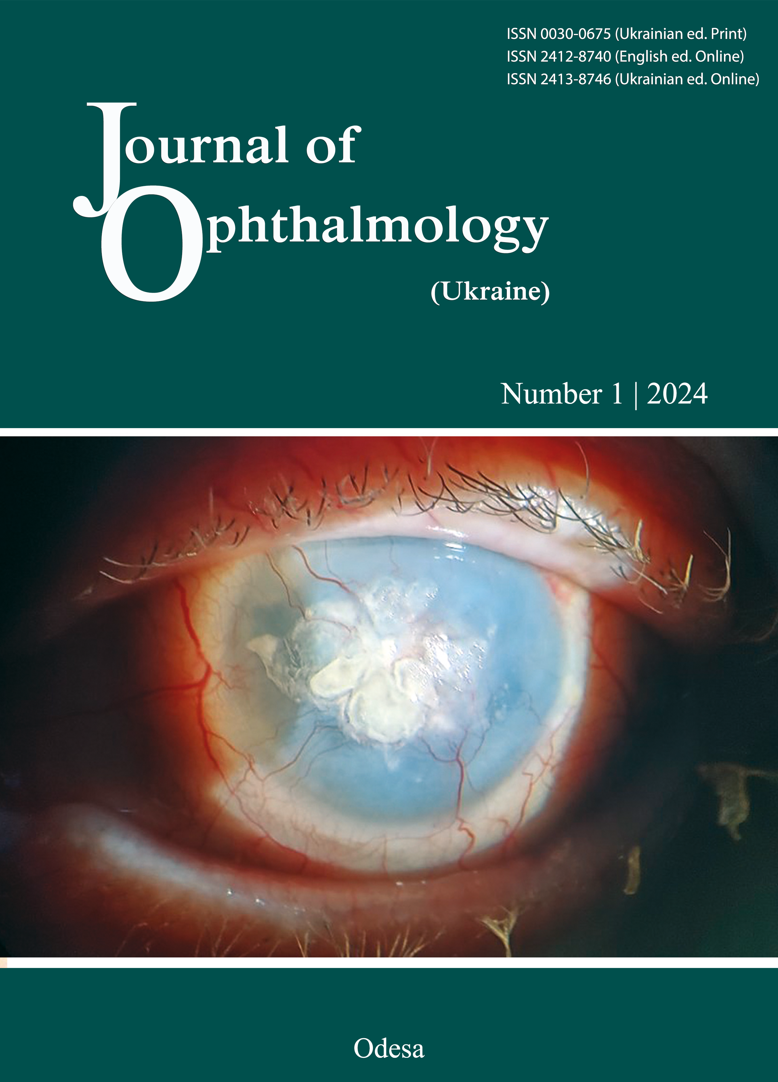
Tear lactoferrin and ceruloplasmin levels in patients with traumatic and recurrent corneal erosions
DOI:
https://doi.org/10.31288/oftalmolzh20241814Keywords:
corneal trauma, lactoferrin, ceruloplasmin, recurrent corneal erosionAbstract
Background: Studies on the mechanisms of corneal wound healing are still important. Apart from the integrity of the corneal epithelium, tear fluid is important for maintaining homeostasis of the ocular surface; it is composed of a variety of proteins, lipids and metabolites. Studies on changes in concentrations of biochemical tear components are important for the diagnosis and treatment of corneal injuries.
Purpose: To assess changes in tear lactoferrin (Lf) and ceruloplasmin (Cp) levels over the course of comprehensive treatment for patients with traumatic corneal erosions (TCE) and recurrent corneal erosions (RCE).
Material and Methods: The study sample included 62 patients (19 to 65 years of age; mean age plus or minus standard deviation, 43.5 ± 2.4 years). Group 1 included 44 patients with TCE, and group 2, 18 patients with recurrent RCE. Each patient group was divided into two subgroups on the basis of the treatment method. Subgroup 1 was administered eye broad-spectrum antibiotic (AB) eye drops and dexpantenol over a course of treatment. Subgroup 2 received AB eye drops and dexpantenol plus adjunct lactoferrin (Lf)-containing eye drops. An eye examination included visual acuity, biomicroscopy and fluorescein test. Monospecific antibodies were used to determine tear Lf and Cp levels. Tears from healthy volunteers were used as controls.
Results: At baseline, the tear Lf level in patients with TCR was lower than in controls (3.94 ± 0.45 arbitrary units (a.u.) versus 10.3 ± 0.4 a.u., respectively; p < 0.05), resulting in reduced ocular surface protection. In subgroup 1 of the TCE group, after treatment with an AB plus dexpantenol only, the tear Lf level increased to 6.38 ± 0.55 a.u. (p < 0.05), and the mean period of treatment was 7.6 ± 0.43 days (p ≥ 0.1). In subgroup 2 of the TCE group, after treatment with an AB plus dexpantenol plus Lf-containing eye drops, the tear Lf level was 12.23 ± 0.6 a.u. (p < 0.05) and the mean period of treatment was 6.0 ± 0.23 days. The presence of Cp in the tear fluid prior to treatment for TCE or RCE indicated activation of acute inflammation; at baseline, the tear Cp level in patients with TCE was 2.37 ± 0.25 a.u. compared to controls (p < 0.05), and in those with RCE, 1.78 ± 0.2 a.u. On completion of treatment with Lf-containing eye drops, the tear Lf level increased and the tear Cp level decreased to the levels in controls, and there was a negative correlation between the tear Lf level and the tear Cp level (r = -0.491, p < 0.001).
Conclusion: The results confirmed the feasibility of utlizing Lf-containing eye drops as an adjunct in the treatment of TCE and RCE. This approach contributed to the restoration of ocular surface homeostasis, thus promoting corneal epithelialization and enabling a reduction in treatment duration.
References
Barrientez B, Nicholas SE, Whelchel A, Sharif R, Hjortdal J, Karamichos D. Corneal injury: Clinical and molecular aspects. Exp Eye Res. 2019 Sep;186:107709. https://doi.org/10.1016/j.exer.2019.107709
Willmann D, Fu L, Melanson SW. Corneal Injury. 2023 Jul 17. In: Stat Pearls [Internet]. Treasure Island (FL): Stat Pearls Publishing; 2023. PMID: 29083785.
Nuzzi A, PozzoGiuffrida F, Luccarelli S, Nucci P. Corneal Epithelial Regeneration: Old and New Perspectives. Int J Mol Sci. 2022 Oct 28;23(21):13114. https://doi.org/10.3390/ijms232113114
Wilson SE, Torricelli AAM, Marino GK. Corneal epithelial basement membrane: Structure, function, and regeneration. Exp Eye Res. 2020 May;194:108002. https://doi.org/10.1016/j.exer.2020.108002
Miller DD, Hasan SA, Simmons NL, Stewart MW. Recurrent corneal erosion: a comprehensive review. Clin Ophthalmol. 2019 Feb 11;13:325-335. https://doi.org/10.2147/OPTH.S157430
Jan RL, Tai MC, Ho CH, Chu CC, Wang JJ, Tseng SH, et al. Risk of recurrent corneal erosion in patients with diabetes mellitus in Taiwan: a population-based cohort study. BMJ Open. 2020;10:e035933. https://doi.org/10.1136/bmjopen-2019-035933
Paley GL, Wagoner MD, Afshari NA, Pineda R, Huang AJW, Kenyon KR. Corneal Wound Healing, Recurrent Corneal Erosions, and Persistent Epithelial Defects. In: Albert DM, Miller JW, Azar, DT, Young LH. (eds). Albert and Jakobiec's Principles and Practice of Ophthalmology. Springer, 2022. Cham. https://doi.org/10.1007/978-3-030-42634-7_212
Zhan X, Li J, Guo Y, Golubnitschaja O. Mass spectrometry analysis of human tear fluid biomarkers specific for ocular and systemic diseases in the context of 3P medicine. EPMA J. 2021 Dec 3;12(4):449-75. https://doi.org/10.1007/s13167-021-00265-y
Liu Z, Wang M, Zhang C, Zhou S, Ji G. Molecular Functions of Ceruloplasmin in Metabolic Disease Pathology. Diabetes Metab Syndr Obes. 2022 Mar 3;15:695-711. https://doi.org/10.2147/DMSO.S346648
Wang B, Timilsena YP, Blanch E, Adhikari B. Lactoferrin: Structure, function, denaturation and digestion. Crit Rev Food Sci Nutr. 2019;59(4):580-596. https://doi.org/10.1080/10408398.2017.1381583
Drozhzhyna GI, Riazanova LIu, Khramenko NI, Velychko LM. Lactoferrin concentration in tears of patients with chronic conjunctivitis and effect of Lacto eyedrops in the multicomponent treatment for this disorder. J of Ophthalnology (Ukraine). 2023;1:39-46. https://doi.org/10.31288/oftalmolzh202313945
Vagge A, Senni C, Bernabei F, Pellegrini M, Scorcia V, Traverso CE, Giannaccare G. Therapeutic Effects of Lactoferrin in Ocular Diseases: From Dry Eye Disease to Infections. Int J MolSci. 2020 Sep 12;21(18):6668. https://doi.org/10.3390/ijms21186668
Orzheshkovskyi VV, Trishchynska MA. Ceruloplasmin: its role in the physiological and pathological processes. Neurophysiology. 2019; 51:141-149. https://doi.org/10.1007/s11062-019-09805-9
Bonaccorsidi Patti MC, Cutone A, Polticelli F, Rosa L, Lepanto MS, Valenti P, Musci G. The ferroportin-ceruloplasmin system and the mammalian iron homeostasis machine: regulatory pathways and the role of lactoferrin. Biometals. 2018 Jun;31(3):399-414. https://doi.org/10.1007/s10534-018-0087-5
Tykhomyrov A, Yusova O, Kapustianenko L, Bilous V, Drobotko T, Gavryliak I, et al. Production of anti-lactoferrin antibodies and their application in the analysis of the tear fluid. Biotech Acta. 2022; 15(5):31-40. https://doi.org/10.15407/biotech15.05.031
Zeitler AF, Gerrer KH, Haas R, Jiménez-Soto LF. Optimized semi-quantitative blot analysis in infection assays using the Stain-Free technology. J Microbiol Methods. 2016 Jul;126:38-41. https://doi.org/10.1016/j.mimet.2016.04.016
Wilson SE. Corneal wound healing. Exp Eye Res. 2020 Aug;197:108089. https://doi.org/10.1016/j.exer.2020.108089
Thompson MW. Regulation of zinc-dependent enzymes by metal carrier proteins. Biometals. 2022 Apr;35(2):187-213. https://doi.org/10.1007/s10534-022-00373-w
Kell DB, Heyden EL, Pretorius E. The Biology of Lactoferrin, an Iron-Binding Protein That Can Help Defend Against Viruses and Bacteria. Front Immunol. 2020 May 28;11:1221. https://doi.org/10.3389/fimmu.2020.01221
Drozhzhyna GI, Velyksar TA. [Lactoferrin: an invisible eye protector]. Oftalmologiia. Ukrainskyi zhurnal. 2021;1(12):73-85. Ukrainian. https://doi.org/10.30702/Ophthalmology31032021-12.1.73-84/048.8
Ohradanova-Repic A, Praženicová R, Gebetsberger L, Moskalets T, Skrabana R, Cehlar O, et al. Time to Kill and Time to Heal: The Multifaceted Role of Lactoferrin and Lactoferricin Host Defense. Pharmaceutics. 2023 Mar 24;15(4):1056. https://doi.org/10.3390/pharmaceutics15041056
Regueiro U, López-López M, Varela-Fernández R, Sobrino T, Diez-Feijoo E, Lema I. Immunomodulatory Effect of Human Lactoferrin on Toll-like Receptors 2 Expression as Therapeutic Approach for Keratoconus. Int J MolSci. 2022 Oct15;23(20):12350. https://doi.org/10.3390/ijms232012350
Higuchi A, Inoue H, Kaneko, Y. et al. Selenium-binding lactoferrin is taken into corneal epithelial cells by a receptor and prevents corneal damage in dry eye model animals. Sci Rep. 2016; 6: 36903. https://doi.org/10.1038/srep36903
Burcel M, Constantin M, Ionita G, Gabriela, Covilitir V. Levels of lactoferrin, lysozyme and albumin in the tear film of keratoconus patients and their correlations with important parameters of the disease. Revista Română de Medicină de Laborator. 2020; 28:2. https://doi.org/10.2478/rrlm-2020-0018
Downloads
Published
How to Cite
Issue
Section
License
Copyright (c) 2024 Gavrylyak I. V., Zhaboiedov D. G., Greben N. K., Tykhomyrov A. O.

This work is licensed under a Creative Commons Attribution 4.0 International License.
This work is licensed under a Creative Commons Attribution 4.0 International (CC BY 4.0) that allows users to read, download, copy, distribute, print, search, or link to the full texts of the articles, or use them for any other lawful purpose, without asking prior permission from the publisher or the author as long as they cite the source.
COPYRIGHT NOTICE
Authors who publish in this journal agree to the following terms:
- Authors hold copyright immediately after publication of their works and retain publishing rights without any restrictions.
- The copyright commencement date complies the publication date of the issue, where the article is included in.
DEPOSIT POLICY
- Authors are permitted and encouraged to post their work online (e.g., in institutional repositories or on their website) during the editorial process, as it can lead to productive exchanges, as well as earlier and greater citation of published work.
- Authors are able to enter into separate, additional contractual arrangements for the non-exclusive distribution of the journal's published version of the work with an acknowledgement of its initial publication in this journal.
- Post-print (post-refereeing manuscript version) and publisher's PDF-version self-archiving is allowed.
- Archiving the pre-print (pre-refereeing manuscript version) not allowed.


