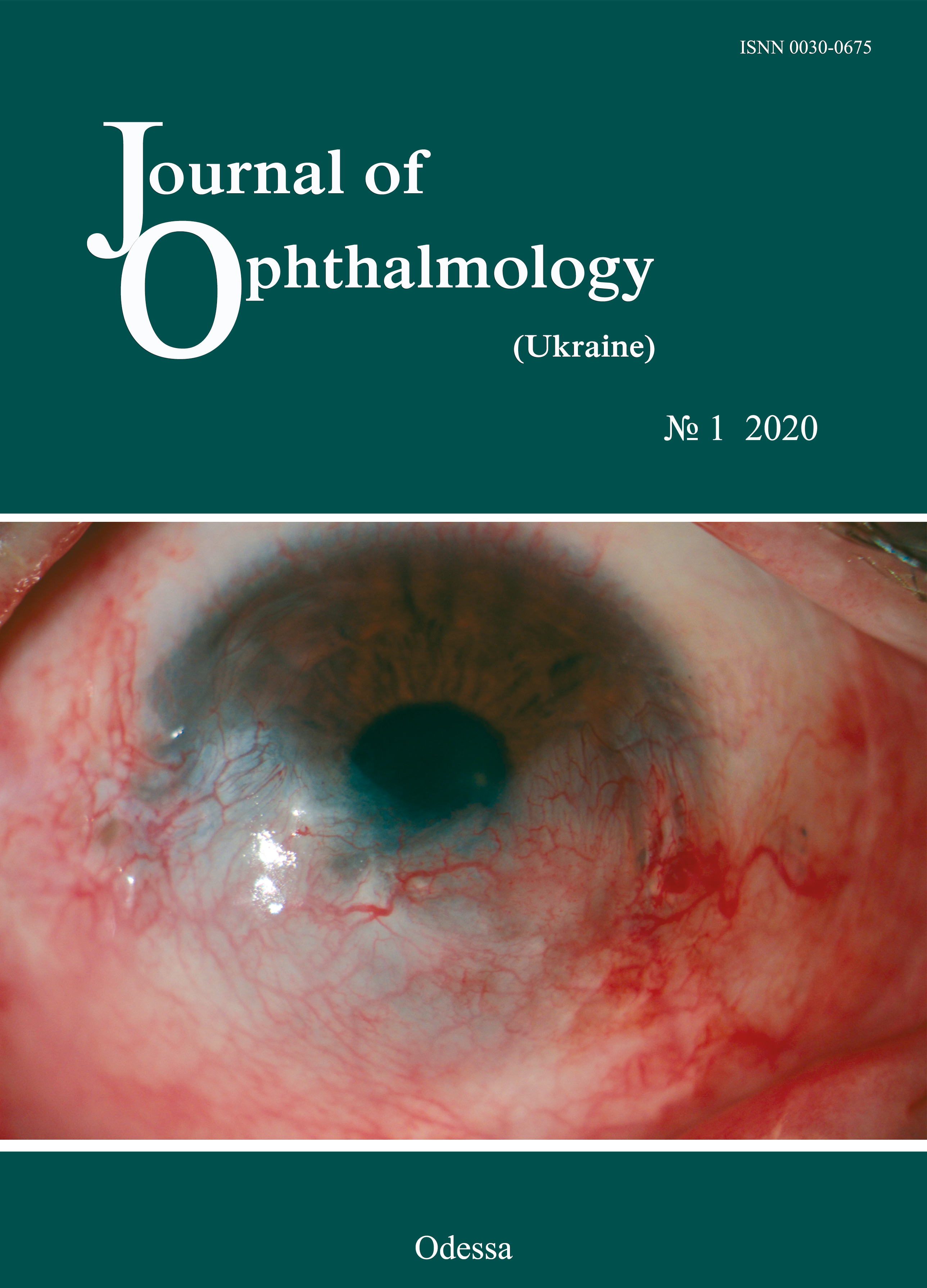
Post-Streptococcal Uveitis
DOI:
https://doi.org/10.31288/oftalmolzh202014953Keywords:
uveitis, streptococcus, diagnosis, treatmentAbstract
Post-streptococcal uveitis refers to the most severe inflammatory conditions of the eye. It often affects young people leading to incapacity for work and physical disability. The clinical features, diagnosis, and treatment of uveitis after a streptococcal infection are described in the paper.
References
1.Arbenyeva NS, Chekhova TA, Bratko GV, Chernakh VV. [Comparative analysis of diseases incidence in patients with uveitis]. [The Annual All-Russian Scientific Practical Conference of Young Scientists «Current Problems of Ophthalmology». Abstract book]. M.: Oftalmologiia; 2012: 28-29. In Russian.
2.Drozdova EA. [The classification and epidemiology of uveitis]. RMZh. Klinicheskaia Oftalmologiia; 2016;3:155-9. In Russian.
3.Panova IE, Drozdova EA. [Uveitis: Guidance for practitioners]. M.: Meditsinskoie informatsionnoie agenstvo; 2014. 144p. In Russian.
4.Burkholder BM, Moradi A, Thorne JE, Dunn JP. (2015) The Dexamethasone intravitreal implant for noninfectious uveitis: practice patterns among uveitis specialists. Ocular Immunology and Inflammation. 2015;23(6):444-53. https://doi.org/10.3109/09273948.2015.1070180
5.Chan CC, Inrig T, Molloy CB et al. Prevalence of inflammatory back pain in a cohort of patients with anterior uveitis. Am. J. Ophthalmol. 2012; 153(6):1025-30. https://doi.org/10.1016/j.ajo.2011.11.016
6.Daguano CR, Bochnia CR, Gehlen M. Anterior uveitis in the absence of scleritis in a patient with rheumatoid arthritis: case report. Arq. Bras. Oftalmol. 2011;74(2):132-133.https://doi.org/10.1590/S0004-27492011000200014
7.Darke C, Coates E. One-tube HLA-B27/B2708 typing by flow cytometry using two "Anti-HLA-B27" monoclonal antibody reagents. Cytometry B Clin Cytom. 2010; Jan; 78(1):21-30. https://doi.org/10.1002/cyto.b.20490
8.Dayani PN. Posterior uveitis: an overview. Advanced ocular care. 2011;1: 32-4.
9.Din NM, Taylor SR, Isa H et al. Evaluation of retinal nerve fiber layer thickness in eyes with hypertensive uveitis. JAMA Ophthalmology. 2014;132(7):859-65. https://doi.org/10.1001/jamaophthalmol.2014.404
10.El Maghraoui A. Extra-articular manifestations of ankylosing spondylitis: prevalence, characteristics and therapeutic implications. Eur. J. Intern. Med. 2011; 22(6):554-60. https://doi.org/10.1016/j.ejim.2011.06.006
11.Fardeau C, Champion E, Massamba N, LeHoang P. Uveitic macular edema. Journal Fran?ais d'Ophtalmologie. 2015;38:74-81.https://doi.org/10.1016/j.jfo.2014.09.001
12.Fonollosa A, Adan A. Uveitis: a multidisciplinary approach. Arch. Soc. Esp. Oftalmol. 2011;86(12):393-4.https://doi.org/10.4321/S0365-66912011001200001
13.Lee SY, Chung WT, Jung WJ et al. Retrospective study on the effects of immunosuppressive therapy in uveitis associated with rheumatic diseases in Korea. Rheumatol. Int. 2011;24(12):77-83. https://doi.org/10.1007/s00296-011-2294-z
14.Moore DB, Jaffe GJ, Asrani S. Retinal nerve fiber layer thickness measurements: uveitis, a major confounding factor. Ophthalmology. 2015;122(3):511-7. https://doi.org/10.1016/j.ophtha.2014.09.008
15.Nobre-Cardoso J, Champion E, Darugar A et al. Treatment of noninfectious uveitic macular edema with the intravitreal Dexamethasone implant. Ocul. Immunol. Inflamm. 2016. https://doi.org/10.3109/09273948.2015.1132738
16.Morovi?-Vergles J, Culo MI. Extra-articular manifestations of seronegative spondyloarthritides. Reumatizam. 2011;58(2):54-6.
17.N?lle B, Both M, Heller M et al. Typical questions from the rheumatologist to the ophthalmologist and cooperating radiologist. Z. Rheumatol. 2008;67(5):360-71. https://doi.org/10.1007/s00393-008-0336-z
18.Rosenbaum JT, Rosenzweig HL. Spondyloarthritis: the eyes have it: uveitis in patients with spondyloarthritis. Nat. Rev. Rheumatol. 2012;8(5):249-50.https://doi.org/10.1038/nrrheum.2012.43
19.Seo BY, Won DI.Flow cytometric human leukocyte antigen-B27 typing with stored samples for batch testing. Ann Lab Med. 2013 May;33(3):174-83. https://doi.org/10.3343/alm.2013.33.3.174
20.Zurutuza A, Andonegui J, Ber?stegui L et al. Bilateral posterior sclerites. An. Sist. Sanit. Navar. 2011;34(2):313-5.https://doi.org/10.4321/S1137-66272011000200019
Downloads
Published
How to Cite
Issue
Section
License
Copyright (c) 2025 Н. В. Коновалова, В. В. Савко, Н. И. Храменко, О. В. Гузун, С. Б. Слободяник, Ю. А. Журавок, В. В. Савко (мл)

This work is licensed under a Creative Commons Attribution 4.0 International License.
This work is licensed under a Creative Commons Attribution 4.0 International (CC BY 4.0) that allows users to read, download, copy, distribute, print, search, or link to the full texts of the articles, or use them for any other lawful purpose, without asking prior permission from the publisher or the author as long as they cite the source.
COPYRIGHT NOTICE
Authors who publish in this journal agree to the following terms:
- Authors hold copyright immediately after publication of their works and retain publishing rights without any restrictions.
- The copyright commencement date complies the publication date of the issue, where the article is included in.
DEPOSIT POLICY
- Authors are permitted and encouraged to post their work online (e.g., in institutional repositories or on their website) during the editorial process, as it can lead to productive exchanges, as well as earlier and greater citation of published work.
- Authors are able to enter into separate, additional contractual arrangements for the non-exclusive distribution of the journal's published version of the work with an acknowledgement of its initial publication in this journal.
- Post-print (post-refereeing manuscript version) and publisher's PDF-version self-archiving is allowed.
- Archiving the pre-print (pre-refereeing manuscript version) not allowed.


