Value of morphological OCT changes in the posterior lens capsule in optimization of surgical treatment for posterior capsular cataracts
DOI:
https://doi.org/10.31288/oftalmolzh2022639Keywords:
posterior capsular cataract, optical coherence tomographyAbstract
Background: It is reasonable and important to optimize the diagnostic and surgical measures for faster visual rehabilitation of patients with posterior capsular cataract (PCC).
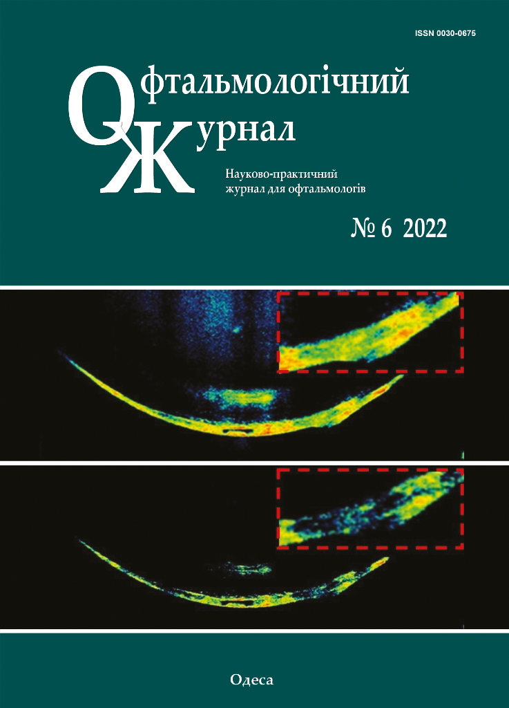
Purpose: To investigate morphological changes (as assessed by optical coherence tomography (OCT)) in the posterior lens capsule (PLC) in PCC, and to assess their value in the optimization of phacoemulsification for these cataracts.
Material and Methods: Of the 1780 eyes (1200 patients) with cataract examined, 512 eyes (28.8%) were diagnosed with PCC. Patients had OCT of the anterior segment (AS-OCT) and phacoemulsification. The morphological changes in the PLC as assessed by OCT, the course of surgical treatment and perioperative complications were reviewed.
Results: Three types of morphological changes in the PLC were identified. Type 1 changes were characterized by a clear PLC margin and found in 312 of the 512 eyes (61%). A posterior capsule-sparing phacoemulsification (i.e., with low power, vacuum and irrigation settings) was used only in 28% of eyes. In type 2 changes, the PLC margin was not uniform in reflectivity and thickness, but appeared clear at the retrolenticular space. Type 2 changes were found in 185 of the 512 eyes (36%). We refrained from hydrodissection in 95.1% of cases, used a posterior capsule-sparing phacoemulsification in 97% of cases and posterior capsule polishing in all cases of PLC with type 2 changes. Type 3 changes were characterized by PLC bulging into the retrolenticular space, and were found in 15 eyes (3%). A capsule rupture was observed in 53% of eyes with type 3 changes, although we used a posterior capsule-sparing phacoemulsification with viscodilation in these cases.
Conclusion: AS-OCT allows detailed assessment of morphological changes in the PLC and identification of three types of these changes. It is reasonable to take in account these facts when selecting a surgical treatment strategy.
References
1.de Silva SR, Evans JR, Kirthi V, Ziaei M, Leyland M. Multifocal versus monofocal intraocular lenses after cataract extraction [Internet]. Vol. 2016, Cochrane Database of Systematic Reviews. 2016 Dec 12 [cited 2022 Aug 10];12(12):CD003169. https://doi.org/10.1002/14651858.CD003169.pub4
2.Yang S, Jiang H, Nie K, Feng L, Fan W. Effect of capsular tension ring implantation on capsular stability after phacoemulsification in patients with weak zonules: a randomized controlled trial. CTR implantation in cataract patients with weak zonules. BMC Ophthalmol [Internet]. 2021 Jan 7 [cited 2022 Aug 10];21(1):1-11. https://doi.org/10.1186/s12886-020-01772-8
3.Yang Y, Chen H, An J, Fan W. Long-term effects of phacoemulsification and intraocular lens implantation in a patient with pathologic myopia and extremely long axial length: A case report. Medicine (Baltimore) [Internet]. 2020 Sep 11 [cited 2022 Aug 10];99(37):e22081. https://doi.org/10.1097/MD.0000000000022081
4.Fontana L, Coassin M, Iovieno A, Moramarco A, Cimino L. Cataract surgery in patients with pseudoexfoliation syndrome: current updates. Clin Ophthalmol [Internet]. 2017 Jul 31 [cited 2022 Aug 10];11:1377-83. https://doi.org/10.2147/OPTH.S142870
5.Panchapakesan J, Mitchell P, Tumuluri K, Rochtchina E, Foran S, Cumming RG. Five year incidence of cataract surgery: the Blue Mountains Eye Study. Br J Ophthalmol [Internet]. 2003 Feb 1 [cited 2022 May 15];87(2):168-72.
6.Chan E, Omar F, Mahroo AR, Bchir MB, Spalton DJ, Frcp F, et al. Complications of cataract surgery. Clin Exp Optom [Internet]. 2010 Nov 1 [cited 2022 Mar 19];93(6):379-89. https://doi.org/10.1111/j.1444-0938.2010.00516.x
7.Feng H, Aadelman RA. Cataract formation following vitreoretinal procedures. Clin Ophthalmol [Internet]. 2014 Sep 23 [cited 2022 Mar 19];8:1957-65. https://doi.org/10.2147/OPTH.S68661
8.Richardson RB, Ainsbury EA, Prescott CR, Lovicu FJ. Etiology of posterior subcapsular cataracts based on a review of risk factors including aging, diabetes, and ionizing radiation. Int J Radiat Biol [Internet]. 2020 Nov 1 [cited 2022 Mar 19];96(11):1339-61. https://doi.org/10.1080/09553002.2020.1812759
9.Rim THT, Kim MH, Kim WC, Kim TI, Kim EK. Cataract subtype risk factors identified from the Korea National Health and Nutrition Examination survey 2008-2010. BMC Ophthalmol [Internet]. 2014 Jan 10 [cited 2022 May 9];14(1):1-15. https://doi.org/10.1186/1471-2415-14-4
10.Klein BEK, Klein R, Moss SE. Incident cataract surgery: The Beaver Adam Eye Study. Ophthalmology. 1997;104(4):573-80. https://doi.org/10.1016/S0161-6420(97)30267-X
11.Osher RH, Yu BCY, Koch DD. Posterior polar cataracts: a predisposition to intraoperative posterior capsular rupture. J Cataract Refract Surg [Internet]. 1990 [cited 2022 Aug 10];16(2):157-62. https://doi.org/10.1016/S0886-3350(13)80724-9
12.Vasavada A, Singh R. Phacoemulsification in eyes with posterior polar cataract. J Cataract Refract Surg [Internet]. 1999 Feb[cited 2022 Aug 10];25(2):238-45. https://doi.org/10.1016/S0886-3350(99)80133-3
13.Hayashi K, Hayashi H, Nakao F, Hayashi F. Outcomes of surgery for posterior polar cataract. J Cataract Refract Surg [Internet]. 2003 Jan 1 [cited 2022 Aug 10];29(1):45-9. https://doi.org/10.1016/S0886-3350(02)01692-9
14.Kumar S, Ram J, Sukhija J, Severia S. Phacoemulsification in posterior polar cataract: does size of lens opacity affect surgical outcome? Clin Experiment Ophthalmol [Internet]. 2010 Dec 1 [cited 2022 Aug 10];38(9):857-61. https://doi.org/10.1111/j.1442-9071.2010.02354.x
15.Guo Y, Lu C, Wu B, Gao J, Li J, Yuan X, et al. Application of 25MHz B-Scan Ultrasonography to Determine the Integrity of the Posterior Capsule in Posterior Polar Cataract. J Ophthalmol [Internet]. 2018 [cited 2022 Aug 10];23:1-5. https://doi.org/10.1155/2018/9635289
16.Chan TCY, Li EYM, Yau JCY. Application of anterior segment optical coherence tomography to identify eyes with posterior polar cataract at high risk for posterior capsule rupture. J Cataract Refract Surg. 2014 Dec 1;40(12):2076-81. https://doi.org/10.1016/j.jcrs.2014.03.033
17.Kymionis GD, Diakonis VF, Liakopoulos DA, Tsoulnaras KI, Klados NE, Pallikaris IG. Anterior segment optical coherence tomography for demonstrating posterior capsular rent in posterior polar cataract. Clin Ophthalmol [Internet]. 2014 Jan 10 [cited 2022 Mar 19];8:215-7. https://doi.org/10.2147/OPTH.S55763
18.Dhami A, Dhami N, Dhami S. Role of ASOCT in Preoperative Assessment of Posterior Polar Cataract. Acta Sci Ophthalmol. 2020;3:2582-3191. https://doi.org/10.31080/ASOP.2020.03.0183
19.Siatiri H, Moghimi S. Posterior polar cataract: minimizing risk of posterior capsule rupture. Eye [Internet]. 2005 Oct 28 [cited 2022 Aug 10];20(7):814-6. https://doi.org/10.1038/sj.eye.6702023
20.Fine IH, Packer M, Hoffman RS. Management of posterior polar cataract. J Cataract Refract Surg. 2003 Jan 1;29(1):16-9. https://doi.org/10.1016/S0886-3350(02)01616-4
21.Vasavada AR, Raj SM, Vasavada V, Shrivastav S. Surgical approaches to posterior polar cataract: a review. Eye [Internet]. 2012 Mar 23 [cited 2022 Apr 19];26(6):761-70. https://doi.org/10.1038/eye.2012.33
22.Vasavada AR, Raj SM. Inside-out delineation. J Cataract Refract Surg [Internet]. 2004 Jun [cited 2022 Aug 10];30(6):1167-9. https://doi.org/10.1016/j.jcrs.2003.10.034
23.Allen D, Wooda C. Minimizing risk to the capsule during surgery for posterior polar cataract. J Cataract Refract Surg [Internet]. 2002 [cited 2022 Aug 10];28(5):742-4. https://doi.org/10.1016/S0886-3350(02)01244-0
24.Taskapili M, Gulkilik G, Kocabora MS, Ozsutcu M. Phacoemulsification with viscodissection in posterior polar cataract: minimizing risk of posterior capsule tear. Ann Ophthalmol (Skokie) [Internet]. 2007 Jun [cited 2022 Aug 10];39(2):145-9. https://doi.org/10.1007/s12009-007-0004-y
25.Salahuddin A. Inverse horse-shoe technique for the phacoemulsification of posterior polar cataract. Can J Ophthalmol. 2010;45(2):154-6. https://doi.org/10.3129/i09-231
Downloads
Published
How to Cite
Issue
Section
License
Copyright (c) 2025 Lutsenko N. S., Isakova O. A., Rudycheva O. A., Kyrylova T. S., Iatsun G. V.

This work is licensed under a Creative Commons Attribution 4.0 International License.
This work is licensed under a Creative Commons Attribution 4.0 International (CC BY 4.0) that allows users to read, download, copy, distribute, print, search, or link to the full texts of the articles, or use them for any other lawful purpose, without asking prior permission from the publisher or the author as long as they cite the source.
COPYRIGHT NOTICE
Authors who publish in this journal agree to the following terms:
- Authors hold copyright immediately after publication of their works and retain publishing rights without any restrictions.
- The copyright commencement date complies the publication date of the issue, where the article is included in.
DEPOSIT POLICY
- Authors are permitted and encouraged to post their work online (e.g., in institutional repositories or on their website) during the editorial process, as it can lead to productive exchanges, as well as earlier and greater citation of published work.
- Authors are able to enter into separate, additional contractual arrangements for the non-exclusive distribution of the journal's published version of the work with an acknowledgement of its initial publication in this journal.
- Post-print (post-refereeing manuscript version) and publisher's PDF-version self-archiving is allowed.
- Archiving the pre-print (pre-refereeing manuscript version) not allowed.


