Visualization of intraocular foreign bodies in the projection of the ciliary body by transpalpebral near-infrared transillumination
DOI:
https://doi.org/10.31288/oftalmolzh201942327Keywords:
penetrating globe injury, intraocular foreign body, infrared radiation, transilluminationAbstract
Background: Despite advances in intraocular foreign body (IOFB) diagnosis, detecting IOFB is a challenge for the ocular traumatologist.
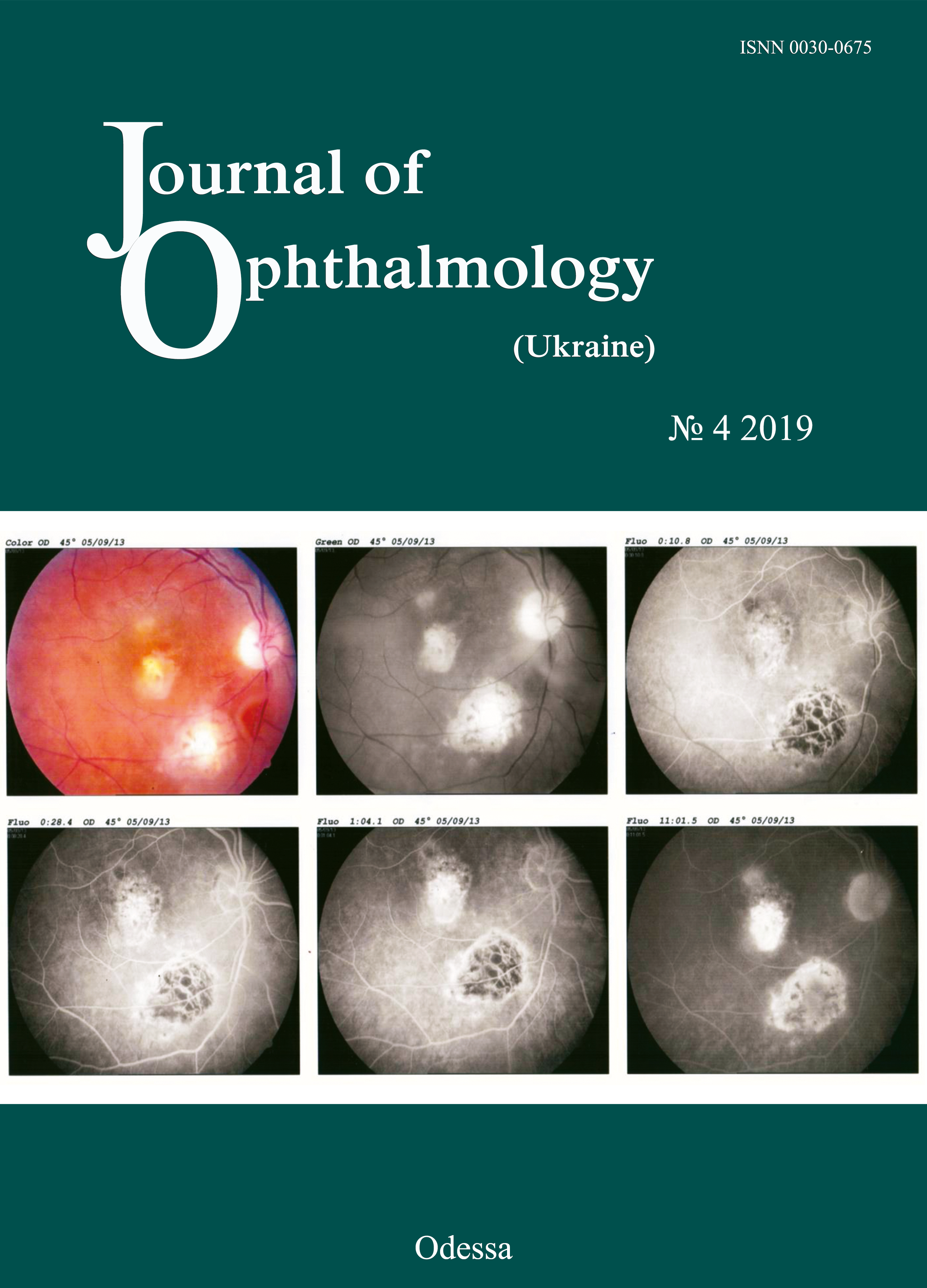
Purpose: To assess the potential of near-infrared transpalpebral transillumination (NIR TPT) for visualization of IOFB in the projection of the ciliary body to the sclera.
Materials and Methods: Ten patients (10 eyes) with penetrating globe injuries associated with IOFB were under observation. All patients underwent an X-ray examination with frontal and lateral projections (both with and without using the Komberg-Baltin prosthesis), ultrasound scanning of the anterior eye and posterior eye; ultrasound distant perimetry; metal detector examination; and NIR TPT.
Results: In all patients, NIR TPT visualized scleral shadows of the ciliary body pars plicata and pars plana as well as the IOFB shadow. The IOFB was localized in relation to ciliary body structures in all cases.
Conclusion: NIR TPT enables non-invasive visualization of IOFBs in the anterior segment, as well as localization of these IOFBs in relation to ciliary body structures in patients with penetrating globe injury.
References
1.Erikitola O, Shahid S, Waqar S, Hewick S. Ocular trauma: classification, management and prognosis. Brit J Hosp Med. 2013;74:108-11. https://doi.org/10.12968/hmed.2013.74.Sup7.C108
2.Loporchio D, Mukkamala L, Gorukanti K, Zarbin M, Langer P, Bhagat N. Intraocular foreign bodies. Surv Ophthalmol. 2016; 61(5):582-96.https://doi.org/10.1016/j.survophthal.2016.03.005
3.Lima-G?mez V, Blanco-Hern?ndez D, Rojas-Dosal J. Ocular trauma score at the initial evaluation of ocular trauma.CIR CIR. 2010 May-Jun;78(3):209-13.
4.Arora R, Sanga L, Kumar M, Taneja M. Intralenticular foreign bodies: report ofeight cases and review of management. Indian J Ophthalmol. 2000;48:119-22.
5.Zhang Y, Zhang M, Jiang C, Qiu HY. Intraocular foreign bodies in Сhina: clinical characteristics, prognostic factors, and visual outcomes in 1,421 eyes. Am J Ophthalmol. 2011;152(1):66-73.https://doi.org/10.1016/j.ajo.2011.01.014
6.Raina U, Kumar V, Kumar V, Sud R, Goel N, Ghosh B. Metallic intraocular foreignbody retained for four years - an unusual presentation. Cont Lens Anterior Eye. 2010;33:202-4.https://doi.org/10.1016/j.clae.2010.01.005
7.Gundorova RA, Stepanov AV, Kurbanova NF. [Current ocular traumatology]. Moscow: Meditsina; 2007. Russian.
8.Pandey A.N. Ocular Foreign Bodies: A Review. J Clin Exp Ophthalmol. 2017; 8: 645.https://doi.org/10.4172/2155-9570.1000645
9.Gundarova R, Chentsova E, Leparskaya N, Lugovkina K, Pavlova V, Shaldin P. [Ultrasound biomicroscopy and laser Doppler fluormetry study of the ciliary body in traumatic retinal detachment due to ocular contusion]. Rus Ophthalmol J. 2012;3:14-8. Russian.
10.Kaushik S, Ichhpujani P, Ramasubramanian A, Pandav SS. Occult intraocular foreign body: ultrasound biomicroscopy holds the key. Int Ophthalmol. 2008;28:71-3.https://doi.org/10.1007/s10792-007-9110-5
11.Koch FHJ, Deuchler S, Singh P, Hessling M. Diaphanoskopie am Auge. Ophthalmologe. 2017;114(9):857-64.https://doi.org/10.1007/s00347-017-0470-6
12.Zadorozhnyy O, Alibet Yassine, Kryvoruchko A, Levytska G, Pasyechnikova N. Dimensions of ciliary body structures in various axial lengths in patients with rhegmatogenous retinal detachment. Journal of Ophthalmology (Ukraine). 2017;6:32-6.https://doi.org/10.31288/oftalmolzh201763236
13.Zadorozhnyy O, Korol A, Nevska A, Kustryn T, Pasyechnikova N. Ciliary body imaging with transpalpebral near-infrared transillumination - a pilot study. Klinika oczna. 2016;3:184-6.
14.Wylegala E, Dobrowolski D, Nowinska A, Tarnawska D. Anterior segment optical coherence tomography in eye injuries. Graef Arch Clin. Exp.2009;247:451-5.https://doi.org/10.1007/s00417-008-0937-x
15.Pasyechnikova N, Naumenko V, Korol A, Zadorozhnyy O. Digital imaging of the fundus with long-wave illumination. Klinika oczna. 2009;111(1-3):18-20.
Downloads
Published
How to Cite
Issue
Section
License
Copyright (c) 2025 М. Б. Коган, О. С. Задорожный, О. С. Петрецкая, Т. А. Красновид, А. Р. Король, Н.В. Пасечникова

This work is licensed under a Creative Commons Attribution 4.0 International License.
This work is licensed under a Creative Commons Attribution 4.0 International (CC BY 4.0) that allows users to read, download, copy, distribute, print, search, or link to the full texts of the articles, or use them for any other lawful purpose, without asking prior permission from the publisher or the author as long as they cite the source.
COPYRIGHT NOTICE
Authors who publish in this journal agree to the following terms:
- Authors hold copyright immediately after publication of their works and retain publishing rights without any restrictions.
- The copyright commencement date complies the publication date of the issue, where the article is included in.
DEPOSIT POLICY
- Authors are permitted and encouraged to post their work online (e.g., in institutional repositories or on their website) during the editorial process, as it can lead to productive exchanges, as well as earlier and greater citation of published work.
- Authors are able to enter into separate, additional contractual arrangements for the non-exclusive distribution of the journal's published version of the work with an acknowledgement of its initial publication in this journal.
- Post-print (post-refereeing manuscript version) and publisher's PDF-version self-archiving is allowed.
- Archiving the pre-print (pre-refereeing manuscript version) not allowed.


