Clinical manifestation of congenital aniridia in Ukrainian children and adolescents
DOI:
https://doi.org/10.31288/oftalmolzh202553643Keywords:
congenital aniridia, aniridia-associated keratopathy, cararact, nystagmusAbstract
Purpose: To investigate clinical manifestations of congenital aniridia (CA) in Ukrainian children and adolescents.
Material and Methods: Twenty four children and adolescents (47 eyes; group 1 of children under 11 years and group 2 of adolescents aged 11 to 18 years) and 5 adult parents (age, 38 to 48 years; group 3) of patients with CA were included in the study. Of these, 72.4% had familial aniridia and 27.6%, sporadic aniridia. The eye examination included gonioscopy, keratometry, ultrasound biometry, ultrasound examination of the anterior and posterior segments, phosphene assessment, and optical computed tomography.
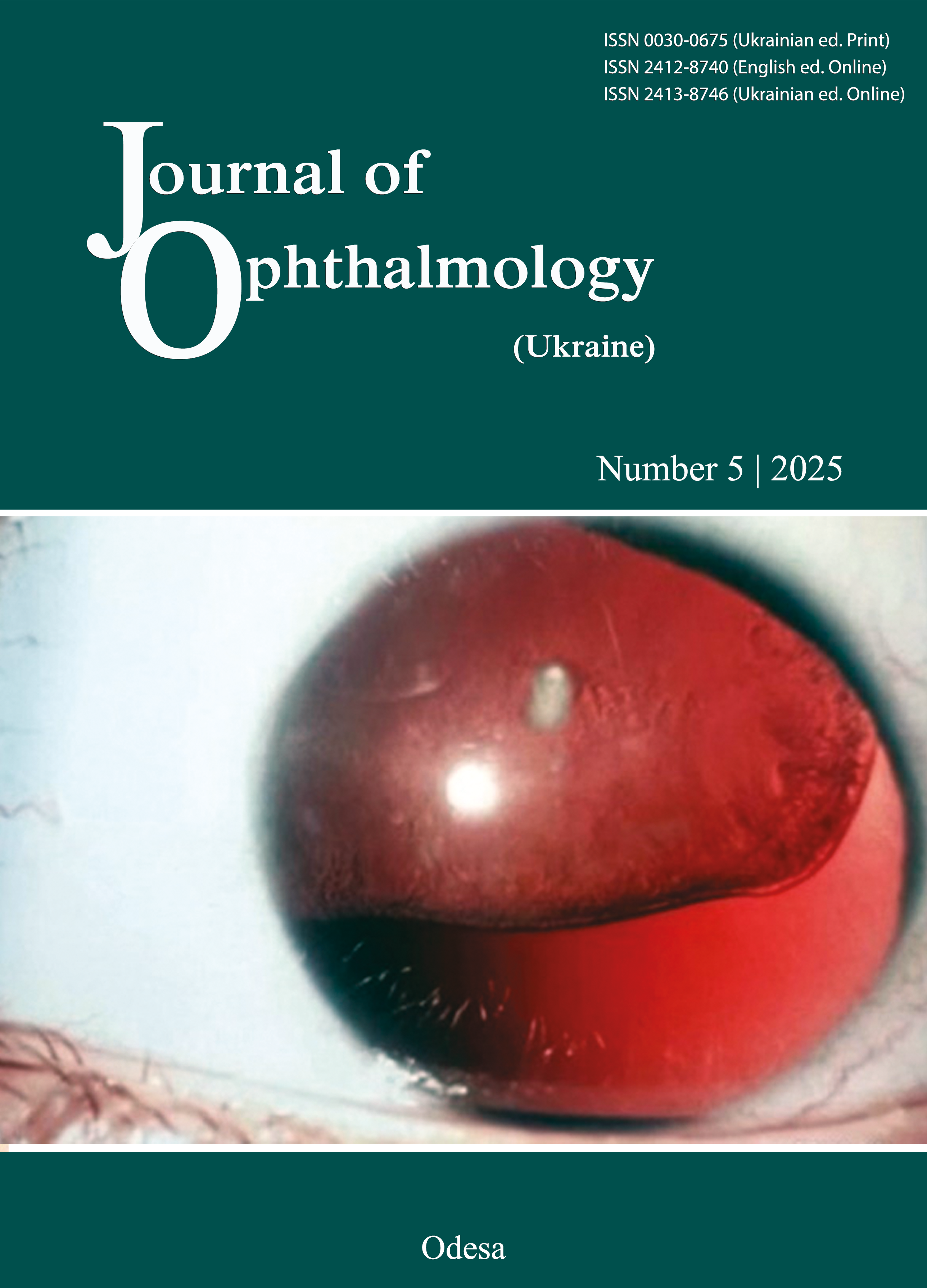
Results: Most patients (75.0% in group 1, 86.7% in group 2, and 100% in group 3) had biomicroscopic evidence of complete aniridia. Aniridia-associated keratopathy (AAK) was found in all groups, with differences in clinical manifestations and severity of AAK between groups. Rates of stages A, B, C and D AAK were 0%, 12.5%, 0%, and 0%, respectively, for group 1, 0%, 40%, 20%, and 13.3%, respectively, for group 2, and 0%, 0%, 70%, and 30%, respectively, for group 3. Congenital cataract was found in 81.3% of eyes in group 1, and progressive cataract, in 86.7% of eyes in group 2. The rate of congenital glaucoma with corneal edema and buphthalmos was 12.5% in group 1. The rate of secondary glaucoma in groups 2 and 3 was 60%. Congenital foveal hypoplasia was observed in 93.75% and 93.3% of the eyes in children and adolescents, respectively. In groups 1, 2 and 3, visual acuity was more commonly ≤ 0.1, and 0.1-0.25 in 37.5%, 26.7%, and 20.0% of eyes, respectively.
Conclusion: CA is a rare bilateral panocular genetic disorder, which in 93.3-93.8% of our cases was associated with foveal hypoplasia, causing low vision. Clinical manifestations of CA (AAK, glaucoma, cataract, foveal hypoplasia, nystagmus, etc.) were found to increase in severity with age. Parents of aniridic children exhibited the worst state of the eye. Clinical manifestations of CA were more severe, with higher rates of complications, in eyes with familial CA, compared to eyes with sporadic CA.
References
Bobrova NF, Vit VV. [Atlas of congenital and hereditary diseases of the eye]. Odesa: Palmira; 2006. Russian.
Bobrova NF, Skrypnychenko ZM. [Toxic, congenital and secondary cataracts]. Odesa: Feniks; 2017. Russian.
Landsend ECS, Pedersen HR, Utheim OA, et al. Characteristics and Utility of Fundus Autofluorescence in Congenital Aniridia Using Scanning Laser Ophthalmoscopy. Invest Ophthalmol Vis Sci. 2019; 60 (13): 4120-4128. https://doi.org/10.1167/iovs.19-26994
Moosajee M, Hingorani M, Moore AT. PAX6-Related Aniridia. In: Adam MP, Ardinger HH, Pagon RA, Wallace SE, Bean LJH, Stephens K, Amemiya A, editors. GeneReviews((R)). University of Washington: Seattle, DC; 2018.
Hingorani M, Hanson I, van Heyningen V. Aniridia. Eur J Hum Genet. 2012; 20:1011-7. https://doi.org/10.1038/ejhg.2012.100
Landsend ECS, Lagali N, Utheim TP. Surv Ophthalmol. 2022 Mar-Apr;67(2):629-630. https://doi.org/10.1016/j.survophthal.2021.11.001
Kasmann-Kellner B, Seitz B. Aniridia syndrome: clinical findings, problematic courses and suggestions for optimization of care ("aniridia guide"). Ophthalmologe. 2014;111(12):1145-56.
Kit V, Cunha DL, Hagag AM, Moosajee M. Longitudinal genotype-phenotype analysis in 86 patients with PAX6-related aniridia. JCI Insight. 2021 Jul 22;6(14):e148406.https://doi.org/10.1172/jci.insight.148406
The UN Convention on the Rights of the Child as amended by General Assembly resolution 50/155 of 21 December 1995.
Mackman G, Brightbill FS, Optiz JM. Corneal changes in aniridia. Am J Ophthalmol. 1979;87(4):497-502.https://doi.org/10.1016/0002-9394(79)90238-1
Gronskov K, Olsen J, Sand A et al. Population-based risk estimates of Wilms tumor in sporadic aniridia. A comprehensive mutation screening procedure of PAX6 identifies 80% of mutations in aniridia. Hum Genet 2001;109:11-8. https://doi.org/10.1007/s004390100529
Eden U, Beijar C, Riise R et al. Aniridia among children and teenagers in Sweden and Norway. Acta Ophthalmol. 2008; 86: 730-4. https://doi.org/10.1111/j.1755-3768.2008.01310.x
Lee NY, Lee YE, Mok J, et al. Three cases with unusual ophthalmic phenotypes of congenital aniridia. Can J Ophthalmol. 2013; 48(4):340-342. https://doi.org/10.1016/j.jcjo.2013.02.009
Schanilec P, Biernacki R. Aniridia: a comparative overview. Am Orthopt J. 2014; 64:98-104. https://doi.org/10.3368/aoj.64.1.98
Netland PA, Scott ML, Boyle 4th JW, et al. Ocular and systemic findings in a survey of aniridia subjects. J AAPOS. 2011;15(6):562-566. https://doi.org/10.1016/j.jaapos.2011.07.009
Hingorani M, Williamson KA, Moore AT, et al. Detailed ophthalmologic evaluation of 43 individuals with PAX6 mutations. Invest Ophthalmol Vis Sci. 2009;50(6):2581-2590. https://doi.org/10.1167/iovs.08-2827
Lopez-Garcia J, Garcia-Lozano I, Rivas L, Martinez-Garchitorena J. Congenital aniridia keratopathy treatment. Arch Soc Esp Oftalmol. 2006;81:435. https://doi.org/10.4321/S0365-66912006000800004
Shiple D, Finklea B, Lauderdale J, Netland P. Keratopathy, cataract, and dry eye in a survey of aniridia subjects. Clin Ophthalmol. 2015; 9: 291-5. https://doi.org/10.2147/OPTH.S74648
Park SH, Park YG, Lee MY, Kim MS. Clinical features of Korean patients with congenital aniridia. Korean J Ophthalmol. 2010;24(5):291-296. https://doi.org/10.3341/kjo.2010.24.5.291
Eden U, Lagali N, Dellby A, et al. Cataract development in Norwegian patients with congenital aniridia. Acta Ophthalmol. 2014;92(2):165-e167. https://doi.org/10.1111/aos.12225
Singh B, Mohamed A, Chaurasia S, et al. Clinical manifestations of congenital aniridia. J Pediatr Ophthalmol Strabismus. 2014; 51(1):59-62. https://doi.org/10.3928/01913913-20131223-01
Baulmann DС, Oplmann A, Flugel-Koch C, et al. PAX6 heterozygous eyes show defects in chamber angle differentiation that are associated with a wide spectrum of other anterior eye segment abnormalities. Mech Dev. 2002;118:3-17. https://doi.org/10.1016/S0925-4773(02)00260-5
Thomas MG, Kumar A, Mohammad S et al. Structural grading of foveal hypoplasia using spectral-domain optical coherence tomography a predictor of visual acuity. Ophthalmology. 2011;118(8):1653-1660.https://doi.org/10.1016/j.ophtha.2011.01.028
Idrissi SBA, Moutei H, Bennis A, Chraibi F, Abdellaoui M, Andaloussi IB. Shedding light on congenital aniridia: an in-depth study of two cases. JFO Open Ophthalmology. 2024; 8(6):100146.https://doi.org/10.1016/j.jfop.2024.100146
Hall J, Corton M et al. Comprehensive Analysis of Congenital Aniridia and Differential Diagnoses: Genetic Insights and Clinical Manifestations. Ophthalmol Ther. 2025 May;14(5):835-856.https://doi.org/10.1007/s40123-025-01122-1
Downloads
Published
How to Cite
Issue
Section
License
Copyright (c) 2025 Bobrova N. F., Romanova T. V., Demnovetska G. M., Sorochynska T. A., Vdovichenko K. S., Dovhan O. D.

This work is licensed under a Creative Commons Attribution 4.0 International License.
This work is licensed under a Creative Commons Attribution 4.0 International (CC BY 4.0) that allows users to read, download, copy, distribute, print, search, or link to the full texts of the articles, or use them for any other lawful purpose, without asking prior permission from the publisher or the author as long as they cite the source.
COPYRIGHT NOTICE
Authors who publish in this journal agree to the following terms:
- Authors hold copyright immediately after publication of their works and retain publishing rights without any restrictions.
- The copyright commencement date complies the publication date of the issue, where the article is included in.
DEPOSIT POLICY
- Authors are permitted and encouraged to post their work online (e.g., in institutional repositories or on their website) during the editorial process, as it can lead to productive exchanges, as well as earlier and greater citation of published work.
- Authors are able to enter into separate, additional contractual arrangements for the non-exclusive distribution of the journal's published version of the work with an acknowledgement of its initial publication in this journal.
- Post-print (post-refereeing manuscript version) and publisher's PDF-version self-archiving is allowed.
- Archiving the pre-print (pre-refereeing manuscript version) not allowed.


