Improving the technique for controlled cryogenic destruction of conjunctival tumors located in the projection of the ciliary body onto the sclera: a preliminary report
DOI:
https://doi.org/10.31288/oftalmolzh201856065Keywords:
epibulbar tumors, cryogenic destruction, infrared thermography, modelAbstract
Background: In case of controlled cryogenic destruction of an epibulbar tumor, making direct temperature measurements in the tumor and surrounding tissues during the treatment process is difficult and dangerous. Infrared thermography (IRT) is, however, capable of imaging and recording only superficial-tissue temperature changes. Mathematical modeling can be used to assess the temperature distribution in the underlying structures based on their thermal physical characteristics.
Purpose: To develop a model for temperature distribution in the ocular tunics in cryogenic destruction of conjunctival tumors located within a zone involving the ciliary body, in order to determine the tunic freezing parameters enabling reduced risk of complications while meeting the principles of ablastics.
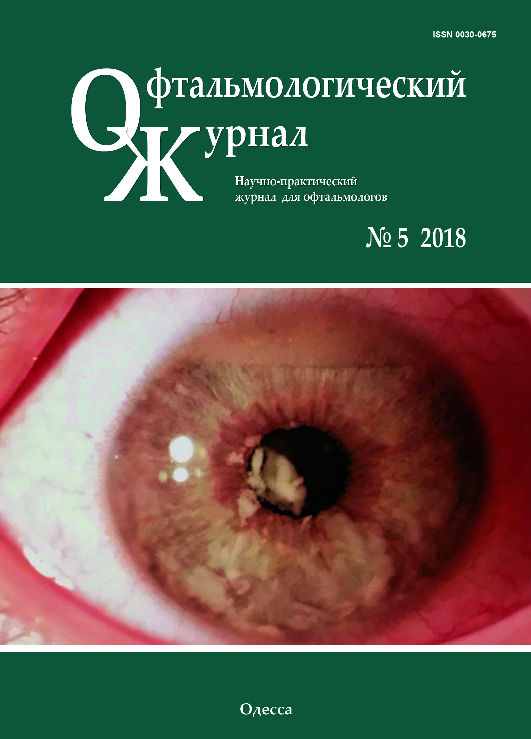
Materials and Methods: Twenty-five patients (25 eyes) underwent real-time IRT during controlled cryogenic destruction of benign and malignant epibulbar lesions located in the projection of the ciliary body onto the sclera. A model for temperature distribution in the ocular tunics in cryogenic destruction of conjunctival tumors located within a ciliary body zone was implemented using Microsoft Quick BASIC Version 4.5.
Results: IRT-based analysis of temperature distribution found that, in cryogenic destruction of epibulbar tumors located in the projection of the ciliary body onto the sclera, initially, the sclera surrounding the centre of exposure gradually cooled down, while the cornea started cooling down rapidly only in 30 to 60 seconds, depending on tumor size and cryogenic unit parameters. The model (a) was developed while taking into account differences in heat conduction and heat capacity between the sclera, ciliary body and cornea, and (b) was also in agreement with the above finding.
Conclusion: The method for IRT-based temperature field monitoring in cryogenic destruction of epibulbar tumors, and the thermal physical model developed will allow determining the individual cryogenic exposure parameters that would prevent excessive freezing of the surrounding structures (in particular, the ciliary body and corneal endothelium). IRT allows for the assessment of post-freezing increases in ocular tissue temperature to the baseline temperature, thus contributing to reduced complication rate, should a repeat cycle of cryogenic destruction of the tumor be required. Further studies should match individual cryogenic exposure durations computed by the model to the data relating to long-term clinical outcomes.
References
Divine RD, Anderson RL. Nitrous oxide cryotherapy for intraepithelial epithelioma of the conjunctiva. Arch Ophthalmol. 1983;101(5):782-6. https://doi.org/10.1001/archopht.1983.01040010782018
Fraunfelder FT, Wingfield D. Management of intraepithelial conjunctival tumors and squamous cell carcinomas. Am J Ophthalmol. 1983 Mar;95(3):359-63.https://doi.org/10.1016/S0002-9394(14)78306-0
Peksayar G, Soyturk MK, Demiryont M. Long-term results of cryotherapy on malignant epithelial tumors of the conjunctiva. Am J Ophthalmol. 1989 Apr 15;107(4):337-40.https://doi.org/10.1016/0002-9394(89)90655-7
Buiko AS, Safronenkova IA, Elagina VA. [Methodological guidelines for cryogenic and radiocryogenic surgical treatment for malignant epithelial eyelid tumors]. Odessa: Astroprint; 2015. Russian
Basti S, Macsai MS. Ocular surface squamous neoplasia: a review. Cornea. 2003;22:687-704.https://doi.org/10.1097/00003226-200310000-00015
Kenawy N, Lake SL, Coupland SE, et al. Conjunctival melanoma and melanocytic intraepithelial neoplasia. Eye (Lond). 2013;27(2):142-52.https://doi.org/10.1038/eye.2012.254
Peksayar G, Altan-Yaycioglu R, Onal S. Excision and cryosurgery in the treatment of conjunctival malignant epithelial tumours. Eye (Lond). 2003;17(2): 228-32.https://doi.org/10.1038/sj.eye.6700331
Buschmann W. Kryochirurgie von Tumoren in der Augenregion. Stuttgart, New York: Thieme; 1999. pp.56-104.
Buĭko AS, Karpovskii EYa, Safronenkova IA et al. [Epithelial tumors of the eyelids: cryosurgery or scalpel?]. Oftalmol Zh. 1991;(6):338-44. Russian
Kaczmarek M, Nowakowski A, Suchowirski M, et al. Active dynamic thermography in cardiosurgery. Quant Infr Therm J. 2007;4(1):107-23 https://doi.org/10.3166/qirt.4.107-123
Kawasaki S, Mizoue S, Yamaguchi M, Shiraishi A, et al. Evaluation of filtering bleb function by thermography. Br J Ophthalmol. 2009 Oct;93(10):1331-6. https://doi.org/10.1136/bjo.2008.152066
Tan JH, Ng EYK, Rajendra Acharya U. Infrared thermography on ocular surface temperature: a review. Infrared Phys Technol. 2009;52:97-108. https://doi.org/10.1016/j.infrared.2009.05.002
Anatychuk LI, Pasechnikova NV, Kobylianskyi RR, et al. Computer simulation of thermal processes in human eye. Journal of Thermoelectricity. 2017;(5):41-58. Russian
Ooi EH, Ng EYK. Ocular temperature distribution: a mathematical perspective. J Mech Med Biol. 2009;9(2):199-227. https://doi.org/10.1142/S0219519409002936
Buĭko AS, Elagina VA, Landa Iu. [Potentials for increasing the effectiveness of cryogenic treatment of eyelid tumors using a device based on an adjustable balloon throttle microcryogenic system]. Oftalmol Zh. 1987;(5):272-6. Russian
Zadorozhnyy OS, Guzun OV, Brarishko AIu, et al. Infrared thermography of external ocular surface in patients with absolute glaucoma in transscleral cyclophotocoagulation: a pilot study. J Ophthalmol (Ukraine). 2018; (2):23-8. https://doi.org/10.31288/oftalmolzh/2018/2/2328
Mapstone R. Determinants of ocular temperature. Br J Ophthalmol. 1968;52:729-41. https://doi.org/10.1136/bjo.52.10.729
Vit VV. [The structure of the human visual system]. Odessa: Astroprint; 2003. Russian
Scott JA. A finite element model of heat transport in the human eye. Phys Med Biol. 1988; 33: 227-41. https://doi.org/10.1088/0031-9155/33/2/003
Karlslow HS, Jaeger JC. [Conduction of Heat in Solids]. Moscow: Nauka; 1964. Russian
Savin SN. [Modeling the epoxy cure processes in spherical layers]. Visnyk ONU. Khimiia. 2013;18(4):38-45. Russian https://doi.org/10.18524/2304-0947.2013.4(48).37846
Segerlind LJ. [Applied Finite Element Analysis]. Moscow: Mir; 1979. Russian
Altoiz BA, Savin NV, Shatagina EA. [Effect of heat release in a microinterlayer of a liquid on the measurement of its viscosity]. Zhurnal Tekhnicheskoi Fiziki. 2014; 59 (5):21-7. Russian https://doi.org/10.1134/S1063784214050028
Lee GA, Hirst LW. Ocular surface squamous neoplasia. Surv Ophthalmol. 1995 May-Jun;39(6):429-50. https://doi.org/10.1016/S0039-6257(05)80054-2
Downloads
Published
How to Cite
Issue
Section
License
Copyright (c) 2026 О. С. Задорожный, Н. В. Савин, А. С. Буйко

This work is licensed under a Creative Commons Attribution 4.0 International License.
This work is licensed under a Creative Commons Attribution 4.0 International (CC BY 4.0) that allows users to read, download, copy, distribute, print, search, or link to the full texts of the articles, or use them for any other lawful purpose, without asking prior permission from the publisher or the author as long as they cite the source.
COPYRIGHT NOTICE
Authors who publish in this journal agree to the following terms:
- Authors hold copyright immediately after publication of their works and retain publishing rights without any restrictions.
- The copyright commencement date complies the publication date of the issue, where the article is included in.
DEPOSIT POLICY
- Authors are permitted and encouraged to post their work online (e.g., in institutional repositories or on their website) during the editorial process, as it can lead to productive exchanges, as well as earlier and greater citation of published work.
- Authors are able to enter into separate, additional contractual arrangements for the non-exclusive distribution of the journal's published version of the work with an acknowledgement of its initial publication in this journal.
- Post-print (post-refereeing manuscript version) and publisher's PDF-version self-archiving is allowed.
- Archiving the pre-print (pre-refereeing manuscript version) not allowed.


