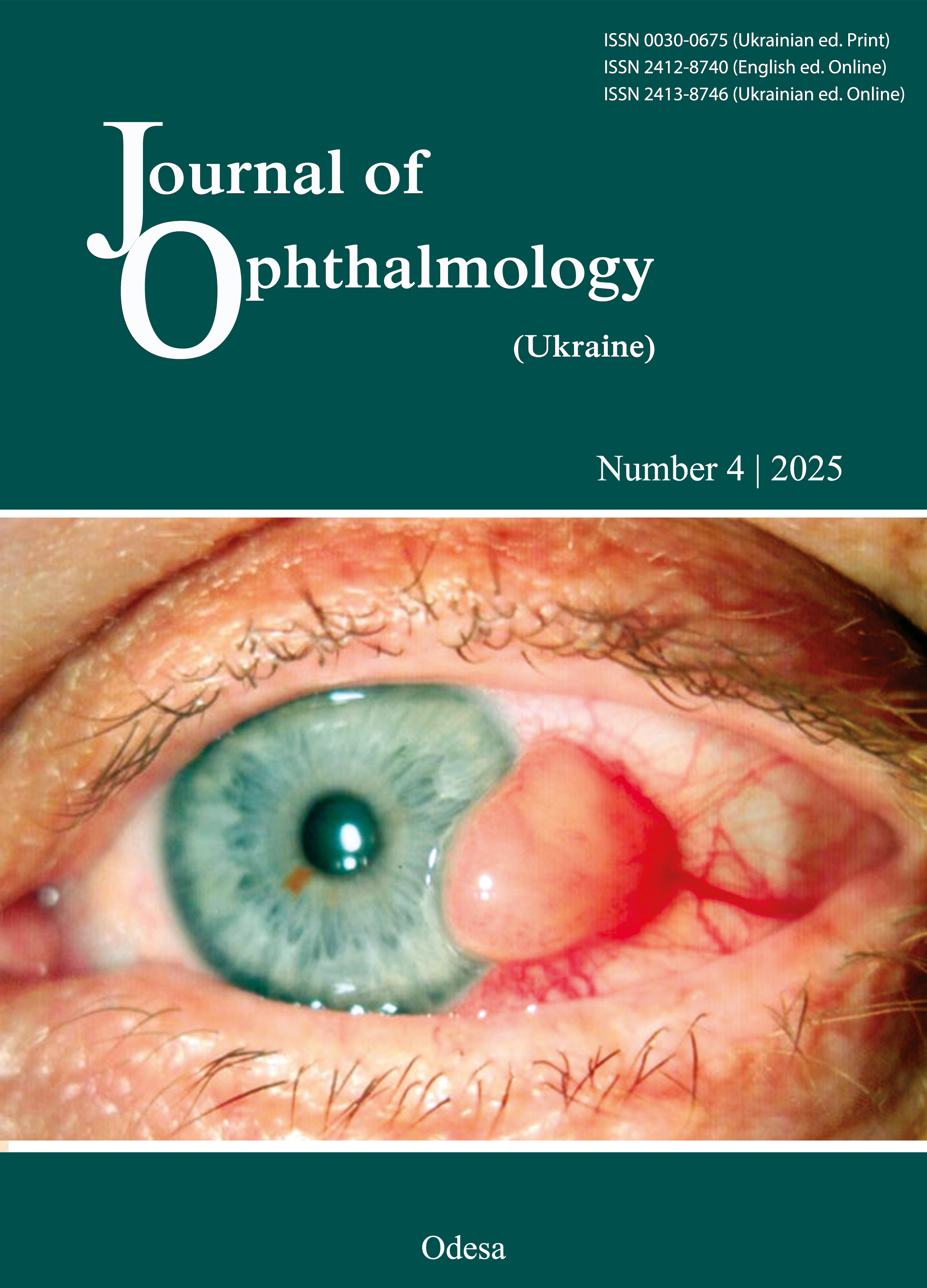
Morphometric analysis of retinal structural components in СВА/С57 mice in experimental diabetes mellitus
DOI:
https://doi.org/10.31288/oftalmolzh20254954Keywords:
diabetic retinopathy, diabetes mellitus, retina, morphometry, experiment, neurodegenerationAbstract
Purpose. To perform morphometric analysis of retinal structural components in СВА/С57 mice in experimental diabetes mellitus (DM) in an attempt to obtain measurable characteristics of possible retinal neurodegenerative changes required for comparison with those in the diabetic retina of other species.
Material and Methods. We retrospectively reviewed hematoxylin-and-eosin (H&E) stained specimens of eyes from eight СВА/С57 mice with diabetes duration of 6 months or less and healthy control animals. Total retinal thickness and thicknesses of the following retinal layers were measured: the photoreceptor layer (PRL), outer nuclear layer (ONL), outer plexiform layer (OPL), inner nuclear layer (INL), inner plexiform layer (IPL), ganglion cell layer (GCL), and nerve fiber layer (NFL). The numbers of neural-cell rows in both nuclear layers were calculated visually.
Results. The mean total thickness of the retina was 197.2 ± 6.32 µm for healthy controls and 227.8 ± 6.2 µm for diabetic animals. In controls and diabetic animals, the mean thicknesses for particular retinal layers were as follows: PRL, 54.4 ± 2.49 µm and 54.9 ± 1.69 µm, respectively; ONL, 47.1 ± 1.73 µm and 49.8 ± 1.69 µm, respectively; OPL, 15.7 ± 1.08 µm and 16.5 ± 0.75 µm, respectively; INL, 24.6 ± 1.094 µm and 32.1 ± 1.46 µm, respectively; IPL, 41.6 ± 1.79 µm and 52.9 ± 1.73 µm, respectively; and GCL plus NFL, 13.8 ± 0.92 µm and 21.6 ± 1.05 µm, respectively. In addition, the numbers of neural-cell rows in the ONL were 9.1 ± 0.32, and 10.0 ± 0.38, respectively, and in the INL, 3.6 ± 0.13 µm, and 3.8 ± 0.16, respectively. In diabetic mice, thicknesses of all inner retinal layers (INL, IPL, and GCL plus NFL) were increased due to edema (р ˂ 0.01 for all cases), resulting in a 30.6-µm increase in the total thickness of the retina. There was no microscopic evidence of neurodegeneration.
Conclusion: Microscopic images of the retina and measurements of thickness of individual retinal layers and numbers of neural-cell rows in retinal nuclear layers for mice with diabetes duration of 6 months or less indicated that a mouse model of streptozotocin-induced diabetes cannot be considered well suited for H&E staining assisted histological assessment of neurodegenerative changes.
References
Khan MAB, Hashim MJ, King JK, Govender RD, Mustafa H, Al Kaabi J. Epidemiology of Type 2 Diabetes - Global Burden of Disease and Forecasted Trends. J Epidemiol Glob Health. 2020 Mar;10(1):107-111. https://doi.org/10.2991/jegh.k.191028.001
Malachkova NV, Kyryliuk ML, Komarovskaia IV. [Blood pressure features in patients with diabetic retinopathy, type 2 diabetes mellitus and obesity]. Arkhiv oftalmologii Ukrainy. 2017; 5(1):32-37. Russian.
Xu Y, Jiang Z, Huang J, et al. The association between toll-like receptor 4 polymorphisms and diabetic retinopathy in Chinese patients with type 2 diabetes. Br J Ophthalmol. 2015;99(9):1301-1305. https://doi.org/10.1136/bjophthalmol-2015-306677
Giyasova AO, Yangieva NR. Comparing the effectiveness of brolucizumab therapy alone versus that combined with subthreshold micropulse laser exposure in the treatment of diabetic macular edema. Oftalmol Zh. 2023;2:16-20. https://doi.org/10.31288/oftalmolzh202321620
Alifanov IS, Sakovych VM. Prognostic risk factors for diabetic retinopathy in patients with type 2 diabetes mellitus. J Ophthalmol (Ukraine). 2022;6:19-23. https://doi.org/10.31288/oftalmolzh202261923
Guzman-Vilca WC, Carrillo-Larco RM. Number of people with type 2 diabetes mellitus in 2035 and 2050: A modelling study in 188 countries. Current Diabetes Reviews. 2025; 21(1), E120124225603. https://doi.org/10.2174/0115733998274323231230131843
Huang X, Wu Y, Ni Y, Xu H, He Y. Global, regional, and national burden of type 2 diabetes mellitus caused by high BMI from 1990 to 2021, and forecasts to 2045: analysis from the global burden of disease study 2021. Frontiers in Public Health. 2025; 13: 1515797. https://doi.org/10.3389/fpubh.2025.1515797
Korobov KV. Progression of the initial stages of non-proliferative diabetic retinopathy and glycation markers in type 2 diabetes mellitus. Arkhiv of-talmologii Ukrainy. 2022; 10(1):17-24. Ukrainian. https://doi.org/10.22141/2309-8147.10.1.2022.287
Chugaev DI. [Features of the development of diabetic macular edema and different stages of diabetic retinopathy in type 2 diabetes]. Arkhiv of-talmologii Ukrainy. 2022;10(3):42-48. Ukrainian.
Nevska AO, Pogosian OA, Goncharuk KO, et al. Detecting diabetic retinopathy using an artificial intelligence-based software platform: a pilot study. J Ophthalmol (Ukraine). 2024;1: 27-31. https://doi.org/10.31288/oftalmolzh202412731
Rykov SO, Galytska IeP, Zhmuryk DV, Dufynets VA. Association of TLR4 rs1927911 polymorphism with diabetic retinopathy and diabetic macular edema in type 2 diabetic patients. https://doi.org/10.31288/oftalmolzh202412026
Walker J, Rykov SA, Suk SA, et al. [Diabetic retinopathy: complexity made simple: a monograph]. Kyiv: Biznes-Logika; 2013. Russian.
Cho H, Alwassia AA, Regiatieri CV, et al. Retinal neovascularization secondary to proliferative diabetic retinopathy characterized by spectral domain optical coherence tomography. Retina. 2013;33(3):542-547. https://doi.org/10.1097/IAE.0b013e3182753b6f
Dorokhova OE, Samoilenko LI, Maltsev EV, Zborovska OV. Morphometric analysis of retinal structural components in Wistar rats in experimental diabetes mellitus. J Ophthalmol (Ukraine). 2025;1:41-46. https://doi.org/10.31288/oftalmolzh202514146
Maltsev EV, Zborovska OV, Dorokhova OE. [Fundamental aspects of the development and treatment of diabetic retinopathy]. Odesa: Astroprint; 2018. Russian.
Glantz SA. Primer of Biostatistics. 4th ed. New York: McGraw-Hill; 1997. 455 p.
Alder VA, Su EN, Yu DY, et al. Overview of studies on metabolic and vascular regulatory changes in early diabetic retinopathy. Aust N Z J Ophthalmol. 1998;26(2):141-148. https://doi.org/10.1111/j.1442-9071.1998.tb01530.x
Kern TS, Engerman RL. Comparison of retinal lesions in alloxan-diabetic rats and galactose-fed rats. Curr Eye Res. 1994;13(12):863-867. https://doi.org/10.3109/02713689409015087
Quiroz J, Yazdanyar A. Animal models of diabetic retinopathy. Ann Transl Med. 2021;9(15):1272. https://doi.org/10.21037/atm-20-6737
Barber AJ. A new view of diabetic retinopathy: a neurodegenerative disease of the eye. Prog Neuropsychopharmacol Biol Psychiatry. 2003;27(2):283-290. https://doi.org/10.1016/S0278-5846(03)00023-X
Lieth E, Gardner TW, Barber AJ, et al. Retinal neurodegeneration: early pathology in diabetes. Clin Exp Ophthalmol. 2000;28(1):3-8.https://doi.org/10.1046/j.1442-9071.2000.00222.x
Downloads
Published
How to Cite
Issue
Section
License
Copyright (c) 2025 Dorokhova O. E., Samoilenko L. I., Maltsev E. V., Zborovska O.V.

This work is licensed under a Creative Commons Attribution 4.0 International License.
This work is licensed under a Creative Commons Attribution 4.0 International (CC BY 4.0) that allows users to read, download, copy, distribute, print, search, or link to the full texts of the articles, or use them for any other lawful purpose, without asking prior permission from the publisher or the author as long as they cite the source.
COPYRIGHT NOTICE
Authors who publish in this journal agree to the following terms:
- Authors hold copyright immediately after publication of their works and retain publishing rights without any restrictions.
- The copyright commencement date complies the publication date of the issue, where the article is included in.
DEPOSIT POLICY
- Authors are permitted and encouraged to post their work online (e.g., in institutional repositories or on their website) during the editorial process, as it can lead to productive exchanges, as well as earlier and greater citation of published work.
- Authors are able to enter into separate, additional contractual arrangements for the non-exclusive distribution of the journal's published version of the work with an acknowledgement of its initial publication in this journal.
- Post-print (post-refereeing manuscript version) and publisher's PDF-version self-archiving is allowed.
- Archiving the pre-print (pre-refereeing manuscript version) not allowed.


