Removal of Persisting Pupillary Membrane with Refractive Lens Exchange
DOI:
https://doi.org/10.31288/oftalmolzh/2018/2/6066Keywords:
persistant pupilеae membrane, refractive lens exchange, childrenAbstract
Background. Clinical characteristics, histological structure, and surgical technique of persisting pupillary membrane (PPM) have been presented in the modern literature in insufficient and, in some cases, contradictory reports.
Purpose. To describe clinical characteristics and histological structure of PPM, as well as PPM surgery for restoring the transparency of the optic axis and eliminating anisometropia through refractive lens exchange.
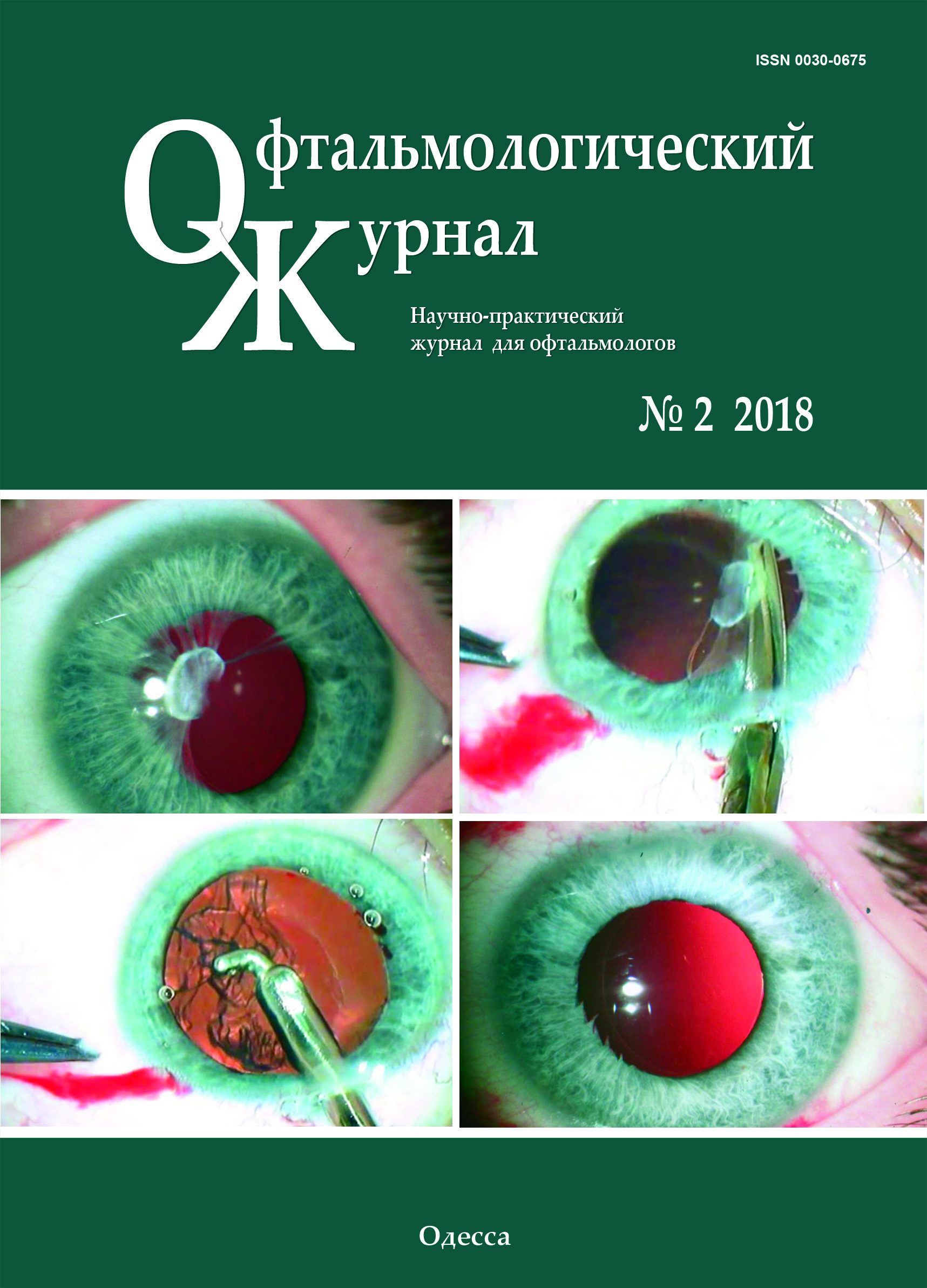
Material and Methods. Complete ophthalmic examination including ultrasound biometry and scanning followed by surgical removal of PPM and refractive exchange of the clear lens was performed in an 11-year-old patient with high myopia, anisometropia and myopia
Results. The PPM case reported is a combination of multiple strands, aligning with the iris in color, involved in the stroma and the inner half of the iris and attached to the anterior lens. The strands, thus, obscured 2/3 of the iris. By sequential surgical dissection of the PPM strands, the membrane was detached from the anterior capsule and removed from the eye. Refractive lens exchange was performed using a standard technique. Histological studies showed a dual character of the removed PPM: the presence of both thick fibrous connective tissue and loose fibrous tissue, resembling the stroma of the iris with the presence of collapsed capillaries.
Conclusions. 1. The rare clinical case described is an example of combined type 1 and type 2 PPM according to Duke-Elder’s classification (1964) or a combination of accessory iris membrane and persistant pupilеae membrane. With the point of Duke-Elder’s classification, this membrane can be considered a special variant of type 2 PPM whereas there is no connection with the lens capsule. 2. Attachement of PPM to the anterior lens capsule, which seemed quite tight, was false since PPM, after being dissected, spontaneously detached from the lens with the latter preserved intact and transparent, which contributed to recover the transparency of the optic axis. 3. One-stage refractive replacement of the lens made it possible to eliminate high myopia and high anisometropia, which made it possible to increase visual acuity of the eye and to create maximal favorable conditions for amblyopia treatment that followed.
References
Bobrova NF, Vit VV. [Atlas of congenital and hereditary diseases of the eye]. Odessa:Palmira;2006. 140p. Russian
Bobrova NF, Dembovetskaia AN, Romanova TV, Sorochinskaia TA. [Preserving the lens transparent is the main task of pupillary membrane surgery]. [Collection of scientific papers "Modern technologies of cataract and refractive surgery - 2013] M.;2013: 47. Russian.
Bobrova N F, Dumbrova NE, Dembovetskaya AN, Romanova TV, Molchanuk NI. [Microinvasive interventions with preservation of the transparent lens in obscure pupillary membranes]. Oftalmol Zh.2014;1:12-17. Russian.
Bobrova NF, Skripnichenko ZM. [Cataract: toxic, congenital, secondary]. Odessa:Feniks; 2017. 320p. Russian.
Vit VV. [The structure of the human visual system]. Odessa: Astroprint; 2003. 664 p. Russian.
Voino-Yasenetskii VV. [Growth and variability of eye tissues in case of eye diseases and injuries]. Kyiv:Vyshcha shkola; 1979. 224p. Russian.
Bhatti S, Kapoor H. Bilateral accessory iris membrane. Indian Journal of Ophtalmology.1998;46:110-1.
Brueckner A. Ueber Persistenz von Resten der tunica vasculosa lentis. Arch. Fur Augenheilk. 1907;56: 5-149.
Duke-Elder S. System of Ophthalmology. 1st ed. Vol.3. Henry Kimpton, London; 1964. 587-775.
Gavris M, Horge J, Avram E et al. Persistant pupillary membrane or accessory iris membrane. Romaian Journal of Ophthalmol. 2015;59:184-7.
Gupta R, Kumar S, Sonika S. Laser and surgical management of hyperplastic persistent pupillary membrane. Ophthalmic Surgery. 2003;34:136-9. https://doi.org/10.3928/1542-8877-20030301-12
Han K, Kim C, Chung J et al. Successful argon laser photocoagulation of diffuse epithelial in growth following concomitant persistant pupillary membrane removal and phacoemulsification. J Cataract Refract Surg. 2012;38:906-11.https://doi.org/10.1016/j.jcrs.2012.02.005
Kapoor K, Baratz K, Barkmeier A. Laser photocoagulation of an avulsed persistent pupillary membrane vessel causing recurrent hyphema. Clin Experiment Ophtalmol. 2013;41:513-515.https://doi.org/10.1111/ceo.12049
Kolin T, Murphee L. Hyperplastic persistent pupillary membrane. Am Journal of Ophthalmol. 1997;123(6):839-41.https://doi.org/10.1016/S0002-9394(14)71137-7
Kraus C, Lueder G. Clinical characteristics and surgical approach to visually significant persistent pupillary membranes. JAAPOS. 2014;18:596-9.https://doi.org/10.1016/j.jaapos.2014.09.005
Kumar H, Sakhuja N, Sadchev M. Hyperplastic pupillary membrane and laser therapy. Ophthalmic Surgery. 1994;25:189-90.https://doi.org/10.3928/1542-8877-19940301-14
Levy W. Congenital iris lesion. British Journal of Ophtalmol. 1957;41:120-23.https://doi.org/10.1136/bjo.41.2.120
Lim K, Yu Y. Surgical management for persistent pupillary membrane with vitreous scissors. Korean journal of ophthalmol. 1996;10(2):124-6.https://doi.org/10.3341/kjo.1996.10.2.124
Pandey S, Ram J, JainA et al. Surgical management of complete hyperplastic persistent pupillary membrane. J Pediatr Ophtalmol Strabismus. 1999;36:221-3.https://doi.org/10.3928/0191-3913-19990701-14
Reynolds J, Hiles D, Johnson B, Biglan A. Hyperplastic persistent pupillary membrane - surgical management. J Pediatr Ophtalmol Strabismus. 1983;20:149-52.https://doi.org/10.3928/0191-3913-19830701-06
Suh D. 2 cases of accessory iris membrane. Journal of the Korean Ophtalmol Society. 1979;20 (1):107-111.
Vega L, Sabates R. Neodymium: YAG laser treatment of persistent pupillary membrane. Ophthalmic Surgery. 1987;18(6):452-4.https://doi.org/10.3928/1542-8877-19870601-14
Downloads
Published
How to Cite
Issue
Section
License
Copyright (c) 2025 Н. Ф. Боброва, А. В. Артемов, Т. В. Романова, Д. В. Смаглий

This work is licensed under a Creative Commons Attribution 4.0 International License.
This work is licensed under a Creative Commons Attribution 4.0 International (CC BY 4.0) that allows users to read, download, copy, distribute, print, search, or link to the full texts of the articles, or use them for any other lawful purpose, without asking prior permission from the publisher or the author as long as they cite the source.
COPYRIGHT NOTICE
Authors who publish in this journal agree to the following terms:
- Authors hold copyright immediately after publication of their works and retain publishing rights without any restrictions.
- The copyright commencement date complies the publication date of the issue, where the article is included in.
DEPOSIT POLICY
- Authors are permitted and encouraged to post their work online (e.g., in institutional repositories or on their website) during the editorial process, as it can lead to productive exchanges, as well as earlier and greater citation of published work.
- Authors are able to enter into separate, additional contractual arrangements for the non-exclusive distribution of the journal's published version of the work with an acknowledgement of its initial publication in this journal.
- Post-print (post-refereeing manuscript version) and publisher's PDF-version self-archiving is allowed.
- Archiving the pre-print (pre-refereeing manuscript version) not allowed.


