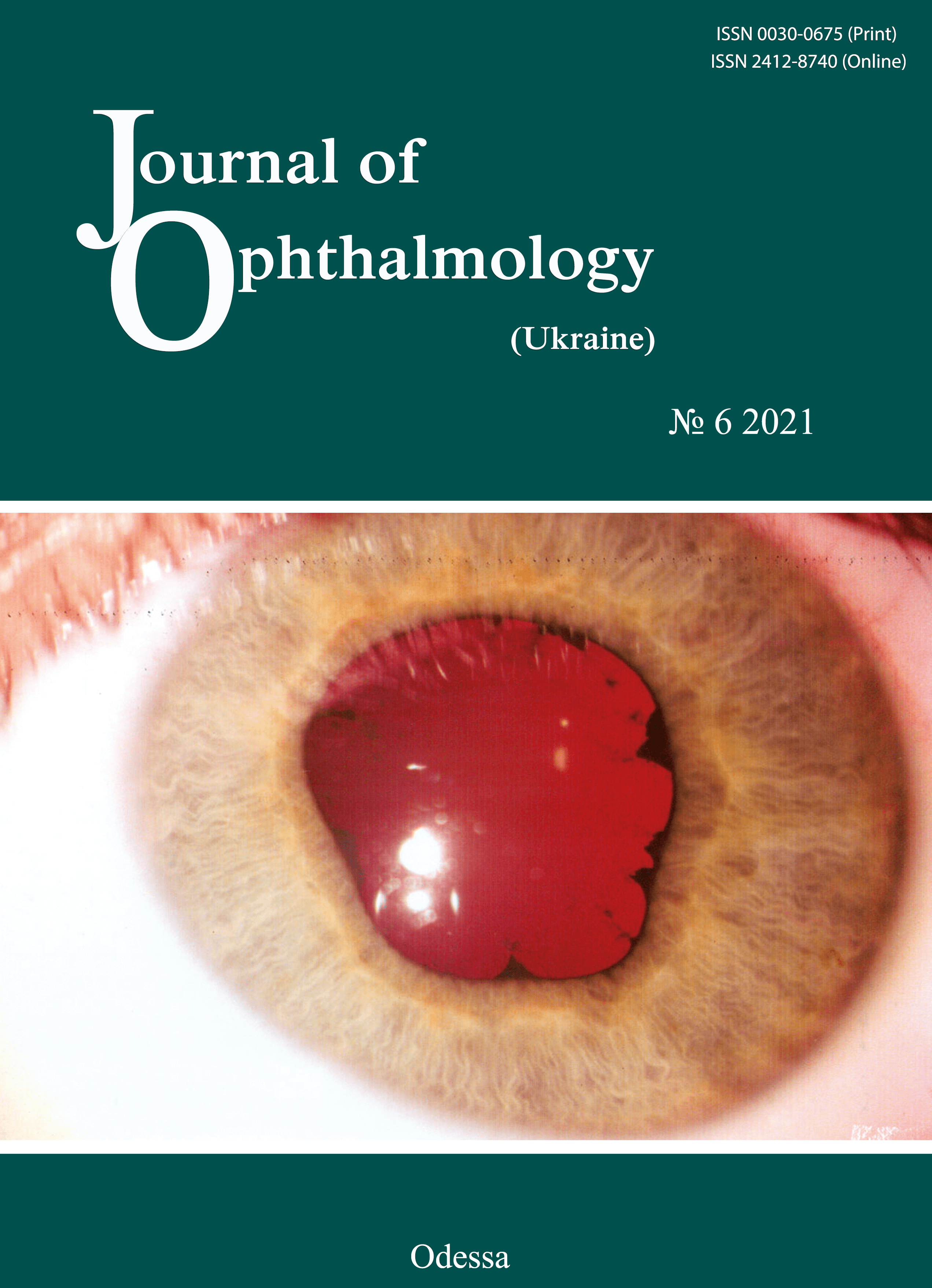
A case of OCT evidence of retinal breaks in a myopic patient woman at 38 weeks of gestation
DOI:
https://doi.org/10.31288/oftalmolzh202166972Keywords:
peripheral retinal degeneration, rhegmatogenous retinal detachment, spectral domain ocular coherence tomography, myopia, pregnancy, retinal laser photocoagulationAbstract
Background: An eye examination of a pregnant woman is a mandatory phase of her preparation for delivery, with a special attention given to ophthalmic symptoms and relevant recommendations for delivery management.
Purpose: To examine spectral domain ocular coherence tomography (SD-OCT) changes in retinal periphery and to provide grounds for the tactics of preparing a pregnant woman for delivery by performing preventive peripheral laser photocoagulation (PPLP).
Material and Methods: A 35-year-old myopic woman at 38 weeks of gestation underwent a routine eye examination (visual acuity assessment, refractometry, tonometry, perimetry, biomicroscopy, and ophthalmoscopy), ultrasound biometry and SD-OCT (Heidelberg Engineering).
Results: The patient was diagnosed with mild myopia along with peripheral lattice degeneration and peripheral cystic degeneration of the retina in both eyes and local retinal detachment with atrophic retinal breaks in the right eye. The peripheral vitreoretinal lattice degeneration was arrested and the local retinal detachment was limited by laser-induced chorioretinal adhesions.
Conclusion: Spectral domain ocular coherence tomography provides an objective picture of the state of both the macular and the periphery of the retina, and, along with biomicroscopy and ophthalmoscopy, provides grounds for the tactics of care for pregnant women for preventing retinal detachment. The outcome of timely preventive laser treatment allowed us to conclude that the patient had no ocular contraindications to vaginal delivery.
References
1.[Order of the Minister of Health of Ukraine No. 977 of December 27, 2002 "An Obstetrics Protocol for Cesarean Delivery"]. Ukrainian.
2.Pasyechnikova NV. [Laser treatment for pathology of the fundus]. Kyiv: Naukova Dumka; 2007. Ukrainian.
3.Popova NV, Fabrikantov OL, Goidin AP. [Frequencies of various clinical forms of peripheral vitreoretinal lattice degeneration for myopia of different severities]. Vestnik TGU. 2017;22(6):1484-6. Russian.
4.Astakhov IS, Lukovskaia NG. [Retinoschisis. 1. Diagnosis, classification, examination methods]. Vestn Oftalmol. Jan-Feb 2004;120(1):26-9. Russian.
5.Neroev VV, Zakharova GY, Kondratieva YP. [Laser photocoagulation for retinoschisis]. In: [Proceedings of the 6th Russian National Ophthalmological Forum: Collection of Papers]. Moscow; October 1-3, 2013. p.59-62. Russian.
6.Shaimova VA, Pozdeeva OG, Shaimov TB, et cal. [Optical hoherence tomography in the diagnosis of peripheral vitreoretinal degeneration]. Oftalmologiia. 2013;10(4):32-9. Russian.
7.Dragoumis I, Richards A, Alexander P, et al. Retinal detachment in severe myopia. Lancet. Lancet. 2017 Jul 8;390(10090):124.https://doi.org/10.1016/S0140-6736(17)31614-8
8.Kottow M. Peripheral retinal degenerations and breaks. Albrecht Von Graefes Arch Klin Exp Ophthalmol. 1980;214(1):53-60.https://doi.org/10.1007/BF00414537
9.Kolenko OV, Sorokin EL, Fil AA. [Ophthalmological criteria for choice of optimal mode of delivery in pregnant women with myopia]. Akusherstvo, ginekologiia i reproduktsiia. 2019;13(2):155-163. Russian.https://doi.org/10.17749/2313-7347.2019.13.2.156-163
Downloads
Published
How to Cite
Issue
Section
License
Copyright (c) 2025 Е. В. Иваницкая, О. Ю. Терлецкая, Г. В. Левицкая

This work is licensed under a Creative Commons Attribution 4.0 International License.
This work is licensed under a Creative Commons Attribution 4.0 International (CC BY 4.0) that allows users to read, download, copy, distribute, print, search, or link to the full texts of the articles, or use them for any other lawful purpose, without asking prior permission from the publisher or the author as long as they cite the source.
COPYRIGHT NOTICE
Authors who publish in this journal agree to the following terms:
- Authors hold copyright immediately after publication of their works and retain publishing rights without any restrictions.
- The copyright commencement date complies the publication date of the issue, where the article is included in.
DEPOSIT POLICY
- Authors are permitted and encouraged to post their work online (e.g., in institutional repositories or on their website) during the editorial process, as it can lead to productive exchanges, as well as earlier and greater citation of published work.
- Authors are able to enter into separate, additional contractual arrangements for the non-exclusive distribution of the journal's published version of the work with an acknowledgement of its initial publication in this journal.
- Post-print (post-refereeing manuscript version) and publisher's PDF-version self-archiving is allowed.
- Archiving the pre-print (pre-refereeing manuscript version) not allowed.


