Corneal macular dystrophy. Case Presentation.
DOI:
https://doi.org/10.31288/oftalmolzh202265961Keywords:
сorneal macular dystrophy, keratoplasty, CHST6, keratin sulfateAbstract
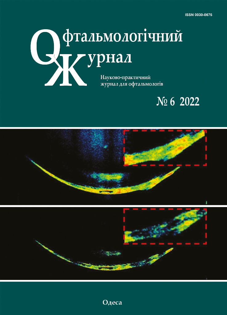
Among the stromal corneal dystrophies corneal macular dystrophy is one of the most frequent. It is an autosomal recessive disorder linked to chromosome 16, in which a mutation occurs in the CHST6 gene, causing an alteration in keratan sulfate metabolism. This alteration produces extracellular deposits of glycosaminoglycans between the stromal lamellae of the cornea, as well as in the cytoplasm of the endothelial cells. Clinically, the presence of centrally predominant white-greyish focal stromal corneal opacities is observed in early stages. Symptoms begin between the second and third decade of life and consist of progressive decrease in visual acuity and photophobia. In this work, we present the clinical case of a 56-year-old male patient who came to the clinic due to progressive decrease in visual acuity and photophobia. On physical examination, multiple intrastromal macules, whitish in color, were found by biomicroscopy in both eyes that were accentuated in greater quantity in the central 5 mm of the cornea. According to the findings obtained in the examination, the diagnosis of corneal macular dystrophy is established.
References
1.Bowling B., Capítulo 6: Córnea. Kanski. Oftalmología clínica. Un enfoque sistemático. Sydney, Australia, ELSEVIER, 2016. 168 p.
2.Weiss JS., Møller HU., Aldave AJ., et al. IC3D Classification of Corneal Dystrophies-Edition 2. Cornea. 2015; 34: 117-159p. https://doi.org/10.1097/ICO.0000000000000307
3.Singh S., Das S., Kannabiran C., et al. Macular Corneal Dystrophy: An Updated Review. Current Eye Research. 2020; 1-6. https://doi.org/10.1080/02713683.2020.1849727
4.Gulias-Cañizo R, et al. Distrofia macular corneal: Características clínicas, histopatológicas y ultraestructurales. ARCH SOC ESP OFTALMOL. 2006; 81: 315-320. https://doi.org/10.4321/S0365-66912006000600004
5.Shields M., Craig JE., Souzeau E., Gupta A. Bilateral phototherapeutic keratectomy for corneal macular dystrophy in an adolescent: Case report and review of the literature. Ophthalmic Genetics. 2020; 41:4, 368-372. https://doi.org/10.1080/13816810.2020.1776335
6.Alemán Suárez IO., Suárez Ojeda V., Armengol Oramas Y., Hernández N. Queratoplastia penetrante con fines ópticos. Presentación de cuatro casos. Rev. Med. Electrón. 2020; 42:3, 1889-1899.
Downloads
Published
How to Cite
Issue
Section
License
Copyright (c) 2025 Leopoldo Garduño-Vieyra, Bruno Flores Escobar, Isabel De la Fuente Batta

This work is licensed under a Creative Commons Attribution 4.0 International License.
This work is licensed under a Creative Commons Attribution 4.0 International (CC BY 4.0) that allows users to read, download, copy, distribute, print, search, or link to the full texts of the articles, or use them for any other lawful purpose, without asking prior permission from the publisher or the author as long as they cite the source.
COPYRIGHT NOTICE
Authors who publish in this journal agree to the following terms:
- Authors hold copyright immediately after publication of their works and retain publishing rights without any restrictions.
- The copyright commencement date complies the publication date of the issue, where the article is included in.
DEPOSIT POLICY
- Authors are permitted and encouraged to post their work online (e.g., in institutional repositories or on their website) during the editorial process, as it can lead to productive exchanges, as well as earlier and greater citation of published work.
- Authors are able to enter into separate, additional contractual arrangements for the non-exclusive distribution of the journal's published version of the work with an acknowledgement of its initial publication in this journal.
- Post-print (post-refereeing manuscript version) and publisher's PDF-version self-archiving is allowed.
- Archiving the pre-print (pre-refereeing manuscript version) not allowed.


