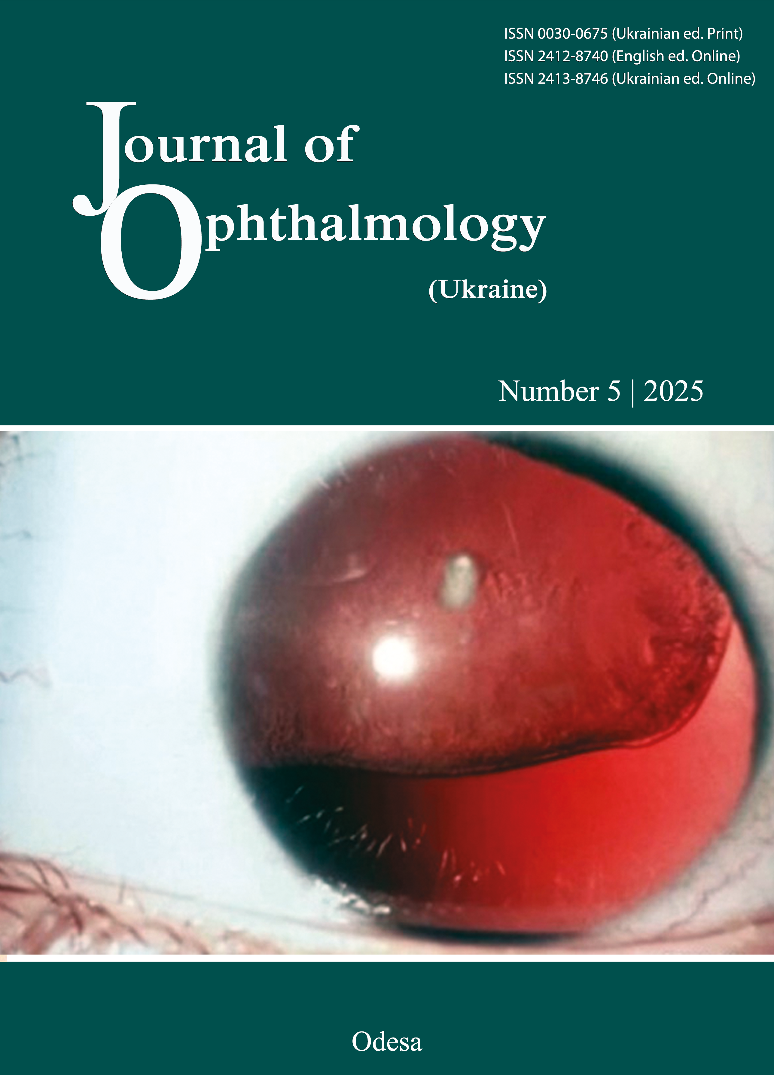
Features of retinal bioelectrical activity in the fellow eye of patients with rhegmatogenous retinal detachment associated with choroidal detachment
DOI:
https://doi.org/10.31288/oftalmolzh202554452Keywords:
retinal detachment, choroidal detachment, electroretinography, myopia, retinal degenerationAbstract
Purpose: To determine, based on full-field electroretinogram (ffERG) data, the features of bioelectrical activity in the fellow eye of moderate and high myopes with rhegmatogenous retinal detachment (RRD) associated with choroidal detachment (CD) (RRD+CD).
Material and Methods: Fifty-two fellow eyes were examined three months after surgery for RRD in the first eye (32 eyes with RRD only and 20 eyes with RRD+CD). A group of 14 normal individuals (28 eyes) was used as a control group. Patients underwent ophthalmological examination and International Society for Clinical Electrophysiology of Vision (ISCEV) ffERG testing.
Results: The severity of myopia and the presence of CD in the affected eye were found to be the factors that influenced the dark-adapted (DA) 0.01 ERG b-wave amplitude (F = 3.83, р = 0.01 and F = 5.0, р = 0.03, respectively) and DA 3.0 ERG b-wave amplitude (F = 4.65, р = 0.012 and F = 9.18, р = 0.005, respectively). In the fellow eye of RRD+CD patients, light-adapted (LA) 3.0 ERG a-wave and b-wave peak times were by 14% longer (р = 0.005), and by 12.3% longer (р < 0.05), respectively, compared to the controls. The presence of CD in the affected eye was found to be a factor that influenced the LA 3.0 ERG a-wave peak time (F = 10.2, р = 0.003): the peak time in the fellow eye for high myopes with RRD+CD was by 19% longer than for those with RRD only. The presence of myopia in the affected eye was found to be a factor that influenced the LA 3.0 ERG b-wave amplitude (F = 3.02, р = 0.042): compared to the controls, the b-wave amplitude in the fellow eye was reduced by 32.5% (р = 0.01) for high myopes with RRD only, and by 40% (р = 0.005) for those with RRD+CD.
Conclusion: We found substantial difference between groups of myopes with RRD+CD and groups of myopes with RRD only in terms of electrical activity of the peripheral retina (the rod photoreceptor layer and layers of inner retinal bipolar cells) in the fellow eye. Based on the findings of our study of the fellow eye in patients with a severity of myopia in the fellow eye being similar to that in the first eye, it may be hypothesized that CD develops in RRD patients with more severe abnormalities in ERG.
References
Sharma T, Challa JK, Ravishankar KV, Murugesan R. Scleral buckling for retinal detachment. Predictors for anatomic failure. Retina. 1994;14(4):338-43. https://doi.org/10.1097/00006982-199414040-00008
Burton TC. Preoperative factors influencing anatomic success rates following retinal detachment surgery. Transact Sec Ophthalmol Am Acad Ophthalmol Otolaryngol. 1977;83(3 Pt 1):OP499-505.
De Smedt S, Sullivan P. Massive choroidal detachment masking overlying primary rhegmatogenous retinal detach¬ment: a case series. Bull Soc Belge Ophtalmol. 2001; 282: 51-55.
Girard P, Mimoun G, Karpouzas I, Montefiore G. Clinical risk factors for proliferative vitreoretinopathy after retinal detachment surgery. Retina. 1994; 14: 417-424.https://doi.org/10.1097/00006982-199414050-00005
Li Z, Li Y, Huang X, Cai XY, Chen X, Li S, Huang Y, Lu L. Quantitative analysis of rhegmatogenous retinal detachment associated with choroidal detachment in Chinese using UBM. Retina. 2012;32(10):2020-5.https://doi.org/10.1097/IAE.0b013e3182561f7c
Kang JH, Park KA, Shin WJ, Kang SW. Macular hole as a risk factor of choroidal detachment in rhegmatogenous retinal detachment. Korean J Ophthalmol. 2008;22(2):100-3.https://doi.org/10.3341/kjo.2008.22.2.100
Wei Y, Wang N, Chen F, et al. Vitrectomy combined with periocular/intravitreal injection of steroids for rhegmatogenous retinal detachment associated with choroidal detachment. Retina. 2014; 34(1): 136-14.https://doi.org/10.1097/IAE.0b013e3182923463
Alibet Y, Levystka G, Umanets N, Pasyechnikova N, Henrich PB. Ciliary body thiсkness changes after preoperative anti-inflammatory treatment in rhegmatogenous retinal detachment complicated by choroidal detachment. Graefe's Arch. Clin. Exp. Ophthalmol. 2017. 255(8): 1503-1508.https://doi.org/10.1007/s00417-017-3673-2
Seelenfreund MH, Kraushar MF, Schepens CL, Freilich DB. Choroidal detachment associated with primary retinal detachment. Arch Ophthalmol. 1974;91(4):254-8.https://doi.org/10.1001/archopht.1974.03900060264003
Rahman N, Harris GS. Choroidal detachment associated with retinal detachment as a presenting finding. Can J Ophthalmol. 1992;27(5):245-8.
Yu Y, An M, Mo B et al. Risk factors for choroidal detachment following rhegmatogenous retinal detachment in a chinese population. BMC Ophthalmol. 2016; 16: 140. https://doi.org/10.1186/s12886-016-0319-9
Wong YL, Ding Y, Sabanayagam C et al. Longi¬tudinal changes in disc and retinal lesions among highly myopic adolescents in Singapore Over a 10-Year period. Eye Contact Lens. 2018; 44:286-291.https://doi.org/10.1097/ICL.0000000000000466
Zahra S, Murphy MJ, Crewther ShG, Riddell N. Flash Electroretinography as a Measure of Retinal Function in Myopia and Hyperopia: A Systematic Review. Vision (Basel). 2023 Feb 27;7(1):15. https://doi.org/10.3390/vision7010015
Gupta SK, Chakraborty R, VerkicharlaPK. Electroretinogram responses in myopia: a review. Doc Ophthalmol. 2021 Nov 17;145(2):77-95.https://doi.org/10.1007/s10633-021-09857-5
Sun J, Zhou J, Zhao P, et al. High prevalence of myopia and high myopia in 5060 Chinese university students in Shanghai. Invest. Ophthalmol. Vis. Sci. 2012; 53: 7504-7509.https://doi.org/10.1167/iovs.11-8343
Purves D, Augustine GJ, Fitzpatrick D et al. Vision: the eye. In: Purves D, Augustine GJ, Fitzpatrick D et al (eds) Neuroscience, 3rd edn. Sinauer Associates Inc, Sunderland, Massachusetts (USA), 2004. pp 229-257.
Quinn N, Csincsik L, Flynn E et al. The clinical relevance of visualising the peripheral retina. Prog Retin Eye Res. 2019; 68:83-109.https://doi.org/10.1016/j.preteyeres.2018.10.001
Robson AG, Nilsson J, Li S et al. ISCEV guide to visual electrodiagnostic procedures. Doc Ophthalmol 2018; 136:1-26.https://doi.org/10.1007/s10633-017-9621-y
McCulloch DL, Marmor MF, Brigell MG et al. ISCEV Standard for full-field clinical electroretinography (2015 update). Doc Ophthalmol. 2015. 130:1-12.https://doi.org/10.1007/s10633-014-9473-7
Frishman LJ. Origins of the electroretinogram. In: Heckenlively JR, Arden GB (eds). Principles and practice of clinical electrophysiology of vision, 2nd edn. The MIT Press, Cambridge, MA, 2006. pp 139-183.https://doi.org/10.7551/mitpress/5557.003.0019
Perlman I. The Electroretinogram: ERG. In: Kolb H, Fernandez E, Nelson R (eds) Webvision: the organi¬zation of the retina and visual system. University of Utah Health Sciences Center, Salt Lake City (UT), 2020. pp 1371-1412.
Robson AG, Frishman LJ, Grigg J, Hamilton R, Jeffrey BG, Kondo M, Li S, McCulloch DL. ISCEV Standard for full-field clinical electroretinography (2022 update). Doc Ophthalmol. 2022 Jun;144(3):165-177. https://doi.org/10.1007/s10633-022-09872-0
Wu De Zheng, Liu Yan. Atlas of testing and clinical application for Roland Electrophysiological Instrument. BeijingScience and TechnologyPress, 2006. р.5-19.
Flitcroft DI, Adams GG, Robson AG et al. Retinal dysfunction and refractive errors: electrophysiological study of children. Br J Ophthalmol. 2005. 89: 484-488].https://doi.org/10.1136/bjo.2004.045328
Marmor MF, Fulton AB, Holder GE, Miyake Y, Brigell M, Bach M. ISCEV Standard for full-field clinical electroretinography (2008 update). Doc Ophthalmol (2009) 118:69-77. https://doi.org/10.1007/s10633-008-9155-4
Westall CA, Dhaliwal HS, Panton CM et al. Values of electroretinogram responses according to axial length. Doc Ophthalmol. 2001; 102:115-130. https://doi.org/10.1023/A:1017535207481
Koh V, Tan C, Nah G et al. Correlation of structural and electrophysiological changes in the retina of young high myopes. Ophthalmic Physiol Opt. 2014; 34:658-666.https://doi.org/10.1111/opo.12159
Kader MA. Electrophysiological study of myopia. Saudi J Ophthalmol. 2012; 26:91-99.https://doi.org/10.1016/j.sjopt.2011.08.002
Perlman I, Meyer E, Haim T et al. Retinal function in high refractive error assessed electroretinographically. Br J Ophthalmol. 1984; 68:79-84.https://doi.org/10.1136/bjo.68.2.79
Wang P, Xiao X, Huang L et al. Cone-rod dysfunction is a sign of early-onset high myopia. Optom Vis Sci. 2013; 90:1327-1330.https://doi.org/10.1097/OPX.0000000000000072
Ishikawa M, Miyake Y, Shiroyama N. Focal mac¬ular electroretinogram in high myopia. Nippon Ganka Gakkai Zasshi. 1990; 94:1040-1047.
Blach RK, Jay B, Kolb H. Electrical activity of the eye in high myopia. Brit J Ophthalmol. 1966; 50:629-641.https://doi.org/10.1136/bjo.50.11.629
Khramenko NI, Umanets MM, Rozanova ZA, Levytska GV. Ocular hemodynamics in patients with rhegmatogenous retinal detachment complicated by choroidal detachment. J of Ophthalmol (Ukraine). 2021; 5: 28-34.https://doi.org/10.31288/oftalmolzh202152834
Downloads
Published
How to Cite
Issue
Section
License
Copyright (c) 2025 Levytska G.V., Khramenko N.I.

This work is licensed under a Creative Commons Attribution 4.0 International License.
This work is licensed under a Creative Commons Attribution 4.0 International (CC BY 4.0) that allows users to read, download, copy, distribute, print, search, or link to the full texts of the articles, or use them for any other lawful purpose, without asking prior permission from the publisher or the author as long as they cite the source.
COPYRIGHT NOTICE
Authors who publish in this journal agree to the following terms:
- Authors hold copyright immediately after publication of their works and retain publishing rights without any restrictions.
- The copyright commencement date complies the publication date of the issue, where the article is included in.
DEPOSIT POLICY
- Authors are permitted and encouraged to post their work online (e.g., in institutional repositories or on their website) during the editorial process, as it can lead to productive exchanges, as well as earlier and greater citation of published work.
- Authors are able to enter into separate, additional contractual arrangements for the non-exclusive distribution of the journal's published version of the work with an acknowledgement of its initial publication in this journal.
- Post-print (post-refereeing manuscript version) and publisher's PDF-version self-archiving is allowed.
- Archiving the pre-print (pre-refereeing manuscript version) not allowed.


