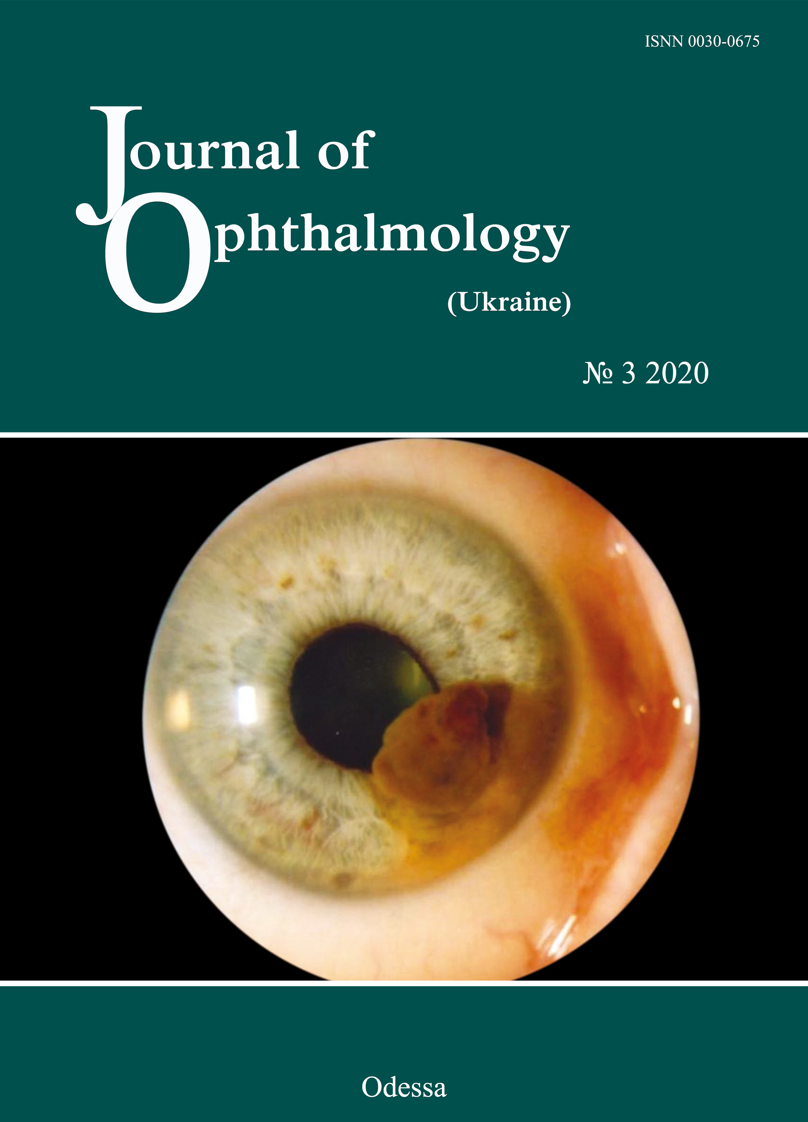
A method of marking the limbus during X-ray localization of IOFB in the presence of apparent chemosis or scleral subconjunctival hemorrhage
DOI:
https://doi.org/10.31288/oftalmolzh202036164Keywords:
X-ray localization of intraocular foreign bodies, ring-shaped limbus markerAbstract
The method proposed for marking the limbus during X-ray localization of IOFB in the presence of apparent chemosis or scleral subconjunctival hemorrhage is based on the use of a ring-shaped limbus marker made from 0.5-0.7-mm stainless steel wire. Rings with external diameters of 10, 11 and 12 mm are used for examining eyes of different sizes. A ring of the appropriate size is placed on the cornea against the limbus to perform X-ray examination. It is well fit in the specified position due to hypertrophy of the conjunctiva surrounding the cornea, and corresponds to the conjunctival attachment to the limbus. Orbit X-ray is performed, quality of X-ray films is assessed and IOFB position in the eye is calculated using the technique of Comberg-Baltin or Abalikhin-Pivovarov. In 12 patients, IOFB was successfully removed based on the data obtained from the use of this method. The accuracy of IOFB X-ray localization by means of the ring-shaped limbus marker was found to be the same as that by means of the Baltin’s prosthesis.
References
1.Gundorova RA, Stepanov AV, Kurbanova NF. [Contemporaty ocular traumatology]. Moscow: Meditsina; 2007. Russian.
2.Gundorova RA, Neroev VV, Kashnikov VV, editors. [Ocular injuries]. Moscow: GEOTAR-Media; 2009. p. 90-91. Russian.
3.Baltin MM. [X-ray diagnostics and therapy in ophthalmology]. Moscow: Medgiz; 1951. Russian.
4.Vainstein ES. [Basic X-ray diagnostics in ophthalmology]. Moscow: Meditsina; 1967. Russian.
5.Poliak BL. [Military field ophthalmology: A tutorial for ophthalmologists]. Leningrad: Medgiz; 1957. Russian.
6.Poliak BL. [Injuries to the eye]. Moscow: Meditsina; 1972. Russian.
7.Danilichev VF, editor. [Contemporaty ophthalmology]. St Petersburg: Piter; 2000. Russian.
8.Belkin M. А retention modification for the limbal ring method of foreign body localization. Am J Ophthalmol. 1979 Jul;88(1):124-5.https://doi.org/10.1016/0002-9394(79)90769-4
9.Ross JA. Use of a fine metal ring in the localisation of intra-ocular foreign bodies. Br J Ophthlmol. 1945 Oct;29(10):545.https://doi.org/10.1136/bjo.29.10.545
10.Stallard HB. War Surgery of the Eye. Br Med J. 1942 Nov 28;2(4273):629-31.https://doi.org/10.1136/bmj.2.4273.629
11.Gardona H, Trokel SL. Soft vacuum lens for foreign body localization. Am J Ophthalmol. 1972 Aug; 74(2):296-8.https://doi.org/10.1016/0002-9394(72)90548-X
Downloads
Published
How to Cite
Issue
Section
License
Copyright (c) 2025 Н. В. Малачкова, І. А. Габрук, І. І. Габрук, О. О. Андрушкова

This work is licensed under a Creative Commons Attribution 4.0 International License.
This work is licensed under a Creative Commons Attribution 4.0 International (CC BY 4.0) that allows users to read, download, copy, distribute, print, search, or link to the full texts of the articles, or use them for any other lawful purpose, without asking prior permission from the publisher or the author as long as they cite the source.
COPYRIGHT NOTICE
Authors who publish in this journal agree to the following terms:
- Authors hold copyright immediately after publication of their works and retain publishing rights without any restrictions.
- The copyright commencement date complies the publication date of the issue, where the article is included in.
DEPOSIT POLICY
- Authors are permitted and encouraged to post their work online (e.g., in institutional repositories or on their website) during the editorial process, as it can lead to productive exchanges, as well as earlier and greater citation of published work.
- Authors are able to enter into separate, additional contractual arrangements for the non-exclusive distribution of the journal's published version of the work with an acknowledgement of its initial publication in this journal.
- Post-print (post-refereeing manuscript version) and publisher's PDF-version self-archiving is allowed.
- Archiving the pre-print (pre-refereeing manuscript version) not allowed.


