Visualization of anterior eye structures by near-infrared transillumination in patients with contusions of the globe
DOI:
https://doi.org/10.31288/oftalmolzh202024549Keywords:
contusion of the globe, ciliary body, near-infrared transilluminationAbstract
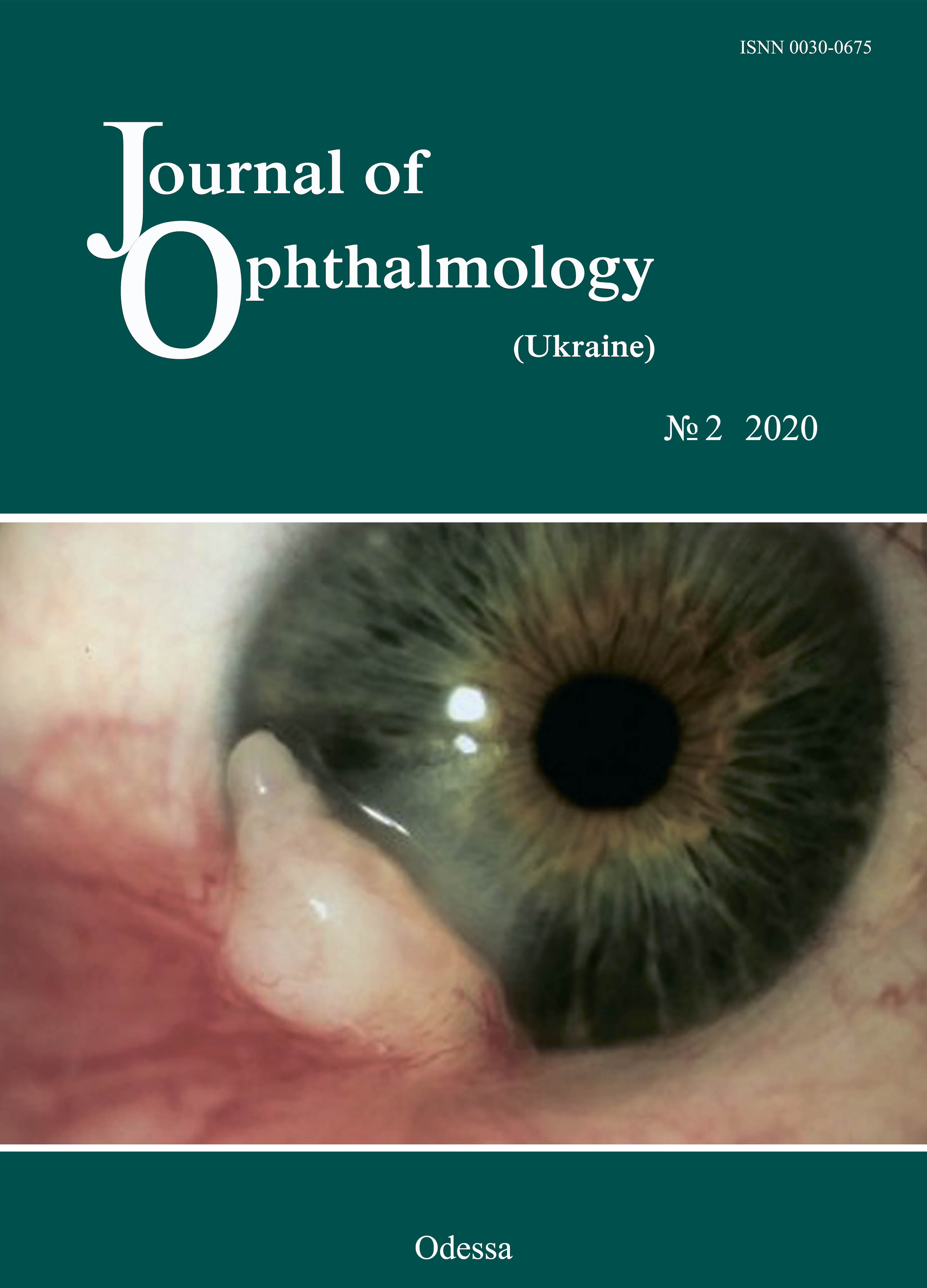
Background: Contusion of the globe is an injury resulting from a blunt object. Despite advances in imaging techniques, the detection of the pathology in contusion of the eye is still a challenge for the ophthalmologist.
Purpose: To assess visualization of anterior eye structures by near-infrared transpalpebral transillumination (NIR TPT) in patients with contusions of the globe.
Material and Methods: Twenty-five patients with a moderate or severe uniocular contusion who presented 2 to 15 days after the traumatic event were under observation. The fellow eye was intact in all cases. Patients underwent visual acuity assessment; biomicroscopy; ophthalmoscopy; intraocular pressure (IOP) measurements; ultrasound scanning of the anterior eye and posterior eye; ultrasound distant perimetry; X-ray examination; color photography and NIR TPT.
Results: NIR TPT enabled identifying various types of damage to the iris, lens and sclera, including subconjunctival scleral ruptures. Pars plicata width was larger in affected eyes than in intact fellow eyes.
Conclusion: NIR TPT enables non-invasive visualization of various types of damage to the anterior eye like iridodialysis in the iris root area, tears of the pupillary margin, lenticular opacity, subluxated lens and subconjunctival scleral ruptures in patients with contusion of the globe. In addition, for these study patients, pars plicata width was larger in affected eyes than in intact fellow eyes.
References
1.Erikitola OO, Shahid SM, Waqar S, Hewick SA. Ocular trauma: classification, management and prognosis. Brit J Hosp Med. 2013;74:108-11. https://doi.org/10.12968/hmed.2013.74.Sup7.C108
2.Yucel O, Demir S, Niyaz L. Clinical characteristics and prognostic factors of scleral rupture due to blunt ocular trauma. Eye. 2016;30:1606-13.https://doi.org/10.1038/eye.2016.194
3.Maiya AS, Dharmesh AM, Jayaram RJ. Clinical profile of ocular blunt trauma in a rural hospital. Clin Ophthalmol Res. 2018;6:3-7.https://doi.org/10.4103/2320-3897.223566
4.Navon SE. Management of the ruptured globe. Int Ophthalmol Clin. 1995;35:71-91.https://doi.org/10.1097/00004397-199503510-00009
5.Zadorozhnyy O, Alibet Yassine, Kryvoruchko A, Levytska G, Pasyechnikova N. Dimensions of ciliary body structures in various axial lengths in patients with rhegmatogenous retinal detachment. J Ophthalmol (Ukraine). 2017;6:32-6.https://doi.org/10.31288/oftalmolzh201763236
6.Kogan MB, Zadorozhnyy OS, Petretska OS, Krasnovid TA, Korol AR, Pasyechnikova NV. Visualization of intraocular foreign bodies in the projection of the ciliary body by transpalpebral near-infrared transillumination. J Ophthalmol (Ukraine). 2019;4:23-27.https://doi.org/10.31288/oftalmolzh201942327
7.Pasyechnikova N, Naumenko V, Korol A, Zadorozhnyy O. Digital imaging of the fundus with long-wave illumination. Klinika oczna. 2009;111(1-3):18-20.
8.Krohn J, Ulltang E, Kjersem B. Near-infrared transillumination photography of intraocular tumours. Br J Ophthalmol. 2013;97:1244-6.https://doi.org/10.1136/bjophthalmol-2013-303090
9.Krohn J, Seland JH, Monge OR. Transillumination for accurate placement of radioactive plaques in brachytherapy of choroidal melanoma. Am J Ophthalmol. 2001;132:418-9.https://doi.org/10.1016/S0002-9394(01)00971-0
10.Thomson ES. The Sachs lamp for transillumination of the eye. Trans Am Ophthalmol Soc. 1905;10:456-60.
11.Saari M, Nieminen H. Fluorescein angiography and infra-red transilluminationstereo technique for studying the ciliary body and iris.In: Proceedings of the 5th Congress of the European Society of Ophthalmology; April 5-9, 1976; Hamburg, Germany.
12.Saari M, Vuorre I, Nieminen H. Infrared transillumination stereophotography of normal iris. Can J Ophthalmol. 1977;12:308-11.
13.Zadorozhnyy O, Korol A, Nevska A, Kustryn T, Pasyechnikova N. Ciliary body imaging with transpalpebral near-infrared transillumination - a pilot study. Klin oczna. 2016;3:184-6.
14.Cherry PM. Indirect traumatic rupture of the globe. Arch Ophthalmol. 1978;96:252-6.https://doi.org/10.1001/archopht.1978.03910050120003
15.Weissman JL, Beatty RL, Hirsch WL, Curtin HD. Enlarged anterior chamber: CT finding of a ruptured globe. AJNR Am J Neuroradiol. 1995;16(4):936-8.
16.Wei WB, Xu L, Jonas JB, et al. Subfoveal choroidal thickness: the Beijing Eye Study. Ophthalmology. 2013;120:175-80.https://doi.org/10.1016/j.ophtha.2012.07.048
17.Hairston RJ, Maguire AM, Vitale S. Morphometric analysis of pars plana development in humans. Retina. 1997;17(2):135-8.https://doi.org/10.1097/00006982-199703000-00009
18.Oliveira C, Tello C, Liebmann JM. Ciliary body thickness increases with increasing axial myopia. Am J Ophthalmol. 2005;140(2):324-5.https://doi.org/10.1016/j.ajo.2005.01.047
Downloads
Published
How to Cite
Issue
Section
License
Copyright (c) 2025 М. Б. Коган

This work is licensed under a Creative Commons Attribution 4.0 International License.
This work is licensed under a Creative Commons Attribution 4.0 International (CC BY 4.0) that allows users to read, download, copy, distribute, print, search, or link to the full texts of the articles, or use them for any other lawful purpose, without asking prior permission from the publisher or the author as long as they cite the source.
COPYRIGHT NOTICE
Authors who publish in this journal agree to the following terms:
- Authors hold copyright immediately after publication of their works and retain publishing rights without any restrictions.
- The copyright commencement date complies the publication date of the issue, where the article is included in.
DEPOSIT POLICY
- Authors are permitted and encouraged to post their work online (e.g., in institutional repositories or on their website) during the editorial process, as it can lead to productive exchanges, as well as earlier and greater citation of published work.
- Authors are able to enter into separate, additional contractual arrangements for the non-exclusive distribution of the journal's published version of the work with an acknowledgement of its initial publication in this journal.
- Post-print (post-refereeing manuscript version) and publisher's PDF-version self-archiving is allowed.
- Archiving the pre-print (pre-refereeing manuscript version) not allowed.


