Persistent dry eye syndrome after and late functional outcomes of excimer laser correction for myopia
DOI:
https://doi.org/10.31288/oftalmolzh202311926Keywords:
excimer laser correction, myopia, persistent dry eye syndrome, myopic regressionAbstract
Background: Laser In Situ Keratomileusis (LASIK) is the laser vision correction procedure of choice for refractive surgeons and accounts for 80% to 85% of the procedures. Dry eye syndrome (DES) is the most common complication of LASIK, with 20% of patients having this complication at 6 months after intervention. Chronic DES after LASIK can cause epithelial hyperplasia, which may be associated with myopic regression.
Purpose: To assess the impact of DES after ELC for myopia on the late functional outcomes.
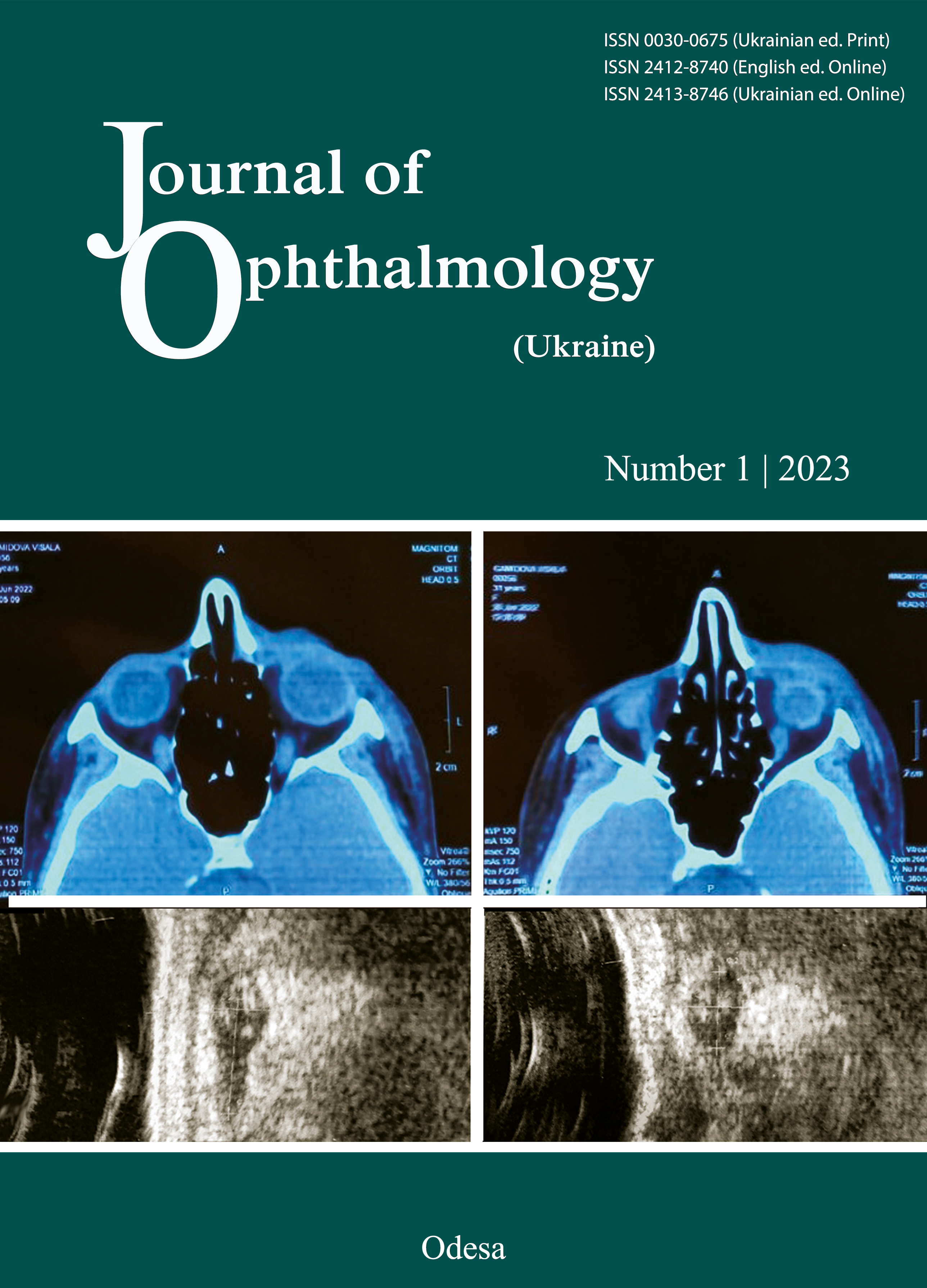
Material and Methods: Sixty-five myopic patients (130 eyes) were divided into two groups, group 1 (a LASIK group) and group 2 (a FemtoLASIK group). The control group was composed of 40 individuals (80 eyes). An examination was performed before surgery and throughout the study and included manifest and cycloplegic refraction, corneal topography, anterior segment optical coherence tomography (AS-ОСТ), tear production, tear film stability and ocular surface staining. Follow-up duration was 12 months.
Results: A myopic regression of 0.5 ± 0.1 D was observed in eyes with postoperative DES. At 6 months after ELC for myopia, study patients had a 10.7% incidence of myopic regression of 0.5 ± 0.1 D. The epithelial thickness in the central zone increased by 7.9 ± 0.25 µm, and in the peripheral zone, by 2.0 ± 0.3 µm; corneal irregularity measurement (CIM) increased to 3.01 ± 0.12 µm, and ocular surface staining score, from 0.22 ± 0.08 to 2.3 ± 0.08, over the follow-up period in patients with persistent DES. Patients without DES exhibited neither myopic regression nor ocular surface staining at 6 months, with their CIM values being in the range of 0.49–1.68 µm. An increase in the epithelial thickness in the central zone was by no more than 2.5±0.3 µm larger than that in the peripheral zone, and mean manifest refraction was +0.12 ± 0.1 D.
Conclusion: First, we found that 10.7% of our study patients had signs of persistent DES and a myopic regression of 0.5 ± 0.1 D after ELC for myopia. Second, the post-ELC increase in central corneal epithelial thickness over the follow-up period was 37.5% smaller in patients without DES than in patients with DES. Third, corneal epithelial thickness values were 6.4% lower in patients with myopia than in individuals without refractive abnormalities. Fourth, corneal topography maps of patients with DES show irregular astigmatism, which causes a decreased quality of vision associated with changes in epithelial thickness and myopic regression after ELC for myopia. Finally, the presence of corneal epithelial fluorescein staining can be considered to be evidence of DES-induced damage to the epithelium after ELC for myopia.
References
Burton, Matthew Jetal. The Lancet Global Health Commission on Global Eye Health: vision beyond 2020 The Lancet Global Health, Volume 9, Issue 4. https://doi.org/10.1016/S2214-109X(21)00138-8
World Health Organisation. Worldreportofvision. Geneva, Switzerland: WHO; 2019. p. 154.
Joffe SN. The 25th Anniversary of Laser Vision Correction in the United States. Clin Ophthalmol. 2021 Mar 17;15:1163-72. https://doi.org/10.2147/OPTH.S299752
Eydelman M, Hilmantel G, Tarver ME, Hofmeister EM, MayJ, Hammel K. et al. Symptoms and Satisfaction of Patients in the Patient-Reported Outcomes With Laser In Situ Keratomileusis (PROWL) Studies. JAMA Ophthalmol. 2017 Jan 1;135(1):13-22. https://doi.org/10.1001/jamaophthalmol.2016.4587
Sharma B, Soni D, Saxena H, Stevenson LJ, Karkhur S, Takkar B, Vajpayee RB. Impact of corneal refractive surgery on the precorneal tear film. Indian J Ophthalmol. 2020 Dec;68(12):2804-12. https://doi.org/10.4103/ijo.IJO_2296_19
Wilson SE. Laser in situ keratomileusis-induced (presumed) neurotrophic epitheliopathy. Ophthalmology. 2001 Jun;108(6):1082-7. https://doi.org/10.1016/S0161-6420(01)00587-5
Mogilevskyy SY, Zhovtoshtan M. Assessing the early and late impact of excimer laser correction for myopia on the development of dry eye syndrome. J.ophthalmol.(Ukraine) [Internet]. 2022 Nov. 15 [cited 2022 Dec. 12];(5):23-9. https://doi.org/10.31288/oftalmolzh202252329
Могілевський СЮ, Жовтоштан МЮ. Ступінь міопії та синдром сухого ока після ексимерлазерної корекції. У: Актуальні питання офтальмології; 22-23.09.2021. Миколаїв; 2021.
Могілевський СЮ, Жовтоштан МЮ. Синдром сухого ока як ускладнення ексимерлазерної корекції у ранні та віддалені терміни післяопераційного спостереження У: Матеріали науково-практичної конференції з міжнародною участю «РЕФРАКЦІЙНИЙ ПЛЕНЕР'22»; 28-30.10.2021. Київ; 2021.
Toda I. Dry Eye After LASIK. Invest Ophthalmol Vis Sci. 2018 Nov 1;59(14): DES109-DES115. https://doi.org/10.1167/iovs.17-23538
Sridhar MS. Anatomy of cornea and ocular surface. Indian J Ophthalmol. 2018 Feb;66(2):190-194. https://doi.org/10.4103/ijo.IJO_646_17
Cui X, Hong J, Wang F, et al. Assessment of corneal epithelial thickness in dry eye patients. Optom Vis Sci. 2014;91(12):1446-54. https://doi.org/10.1097/OPX.0000000000000417
Corbett M, Maycock N, Rosen E, O'Brart D. Corneal Surface Disease. In: Corneal Topography. Springer, 2019. Cham. Доступно на: https://doi.org/10.1007/978-3-030-10696-6_8
Solin S, Liam EJ, Edward EM. Effect of corneal epithelial remodeling on visual outcomes of topography-guided femtosecond LASIK. Journal of Cataract & Refractive Surgery. 2022;48(10):1155-61. https://doi.org/10.1097/j.jcrs.0000000000000940
Wolffsohn JS, Arita R, Chalmers R, Djalilian A, Dogru M, Dumbleton K. et al. Craig, TFOS DEWS II DiagnosticMethodology report. The Ocular Surface. 2017. 15 (3):539-74. https://doi.org/10.1016/j.jtos.2017.05.001
User Manual ATLAS Corneal Topography System Model 9000 and ATLAS Review Software 3.0; PathFinder II Color-CodedParameter Classification(Normal Eyes, based on Multi-Center Clinical Study), Chapter 7:147-8.
OzalpO, Atalay E. Biometric Determinants of Epithelial Thickness Profile Acrossa Wide Range of Refractive Errors. Ophthalmol Ther. 2022 Jun;11(3):1089-100. https://doi.org/10.1007/s40123-022-00489-9
Kanellopoulos AJ, AsimellisG. In vivo three-dimensional corneal epithelium imaging in normal eyes by anterior-segment optical coherence tomography: a clinical reference study. Cornea. 2013;32(11):1493-8. https://doi.org/10.1097/ICO.0b013e3182a15cee
Ma XJ, Wang L, Koch DD. Repeatability of corneal epithelial thickness measurements using Fourier-domain optical coherence tomography in normal and post-LASIK eyes. Cornea. 2013;32(12):1544-8. https://doi.org/10.1097/ICO.0b013e3182a7f39d
Maltsev DS, Kudryashova EV, Kulikov AN, Mareichev AY. Relationship between central epithelial thickness and central corneal thickness in healthy eyes and eyes after laser in situ keratomileusis. Cornea. 2018;37(8):1053-7. https://doi.org/10.1097/ICO.0000000000001568
Levy A, Georgeon C, Knoeri J, Tourabaly M, Leveziel L, Bouheraoua N, Borderie VM. Corneal Epithelial Thickness Mapping in the Diagnosis of Ocular Surface Disorders Involving the Corneal Epithelium: A Comparative Study. Cornea. 2022 Nov 1;41(11):1353-61. https://doi.org/10.1097/ICO.0000000000003012
Arba MosqueraS, Awwad ST. Theoretical analyses of the refractive implications of transepithelial PRK ablations. Br J Ophthalmol. 2013 Jul;97(7):905-11. https://doi.org/10.1136/bjophthalmol-2012-302853
Reinstein DZ, ArcherTJ, Gobbe M, et al. Epithelial thickness in the normal cornea: three-dimensional display with artemis very high-frequency digital ultrasound. J Refract Surg. 2008;24:571-81. https://doi.org/10.3928/1081597X-20080601-05
Hwang ES, Schallhorn JM, Randleman JB. Utility of regional epithelial thickness measurements in corneal evaluations. Surv Ophthalmol. 2020;65(2):187-204. https://doi.org/10.1016/j.survophthal.2019.09.003
Salomão MQ, Hofling-Lima AL, Lopes BT, et al. Role of the corneal epithelium. measurements in keratorefractive surgery. Curr Opin Ophthalmol. 2017;28(4):326-36. https://doi.org/10.1097/ICU.0000000000000379
Ryu IH, Kim WK, Nam MS, Kim JK, Kim SW. Reduction of corneal epithelial thickness during medical treatment for myopic regression following FS-LASIK. BMC Ophthalmol. 2020;20(1):296. https://doi.org/10.1186/s12886-020-01570-2
Reinstein DZ, Archer TJ, Gobbe M. Rate of change of curvature of the corneal stromal surface drives epithelial compensatory changes and remodeling. J Refract Surg. 2014;30(12):799-802. https://doi.org/10.3928/1081597X-20141113-02
Reinstein DZ, Archer TJ, Gobbe M. Change in epithelial thickness profile 24 hours and longitudinally for 1 year after myopic LASIK: three-dimensional display with Artemis very high-frequency digital ultrasound. J Refract Surg. 2012;28(3):195-201. https://doi.org/10.3928/1081597X-20120127-02
Kanellopoulos AJ, Asimellis G. Longitudinal postoperative lasik epithelial thickness profile change sin correlation with degree of myopia correction. J Refract Surg. 2014; 30(3):166-71.
Cohen E, Spierer O. Dry Eye Post-Laser-Assisted In Situ Keratomileusis: Major Reviewand Latest Updates. J Ophthalmol. 2018 Jan 28;2018:4903831. https://doi.org/10.1155/2018/4903831
Mori Y, Nejima R, Masuda A, Maruyama Y, Minami K, Miyata K, Amano S. Effect of diquafosol tetrasodium eye drop for persistent dry eye after laser in situkeratomileusis. Cornea.2014 Jul;33(7):659-62. https://doi.org/10.1097/ICO.0000000000000136
Downloads
Published
How to Cite
Issue
Section
License
Copyright (c) 2023 Mogilevskyy S.Yu., Zhovtoshtan M.Yu., Bushuyeva O.V.

This work is licensed under a Creative Commons Attribution 4.0 International License.
This work is licensed under a Creative Commons Attribution 4.0 International (CC BY 4.0) that allows users to read, download, copy, distribute, print, search, or link to the full texts of the articles, or use them for any other lawful purpose, without asking prior permission from the publisher or the author as long as they cite the source.
COPYRIGHT NOTICE
Authors who publish in this journal agree to the following terms:
- Authors hold copyright immediately after publication of their works and retain publishing rights without any restrictions.
- The copyright commencement date complies the publication date of the issue, where the article is included in.
DEPOSIT POLICY
- Authors are permitted and encouraged to post their work online (e.g., in institutional repositories or on their website) during the editorial process, as it can lead to productive exchanges, as well as earlier and greater citation of published work.
- Authors are able to enter into separate, additional contractual arrangements for the non-exclusive distribution of the journal's published version of the work with an acknowledgement of its initial publication in this journal.
- Post-print (post-refereeing manuscript version) and publisher's PDF-version self-archiving is allowed.
- Archiving the pre-print (pre-refereeing manuscript version) not allowed.


