Dirofilariasis of eyelid and orbit (clinic, diagnosis, treatment)
DOI:
https://doi.org/10.31288/oftalmolzh202312733Keywords:
ocular dirofilariasis, incidence, clinical features, treatmentAbstract
Background: Human dirofilariasis is a larval helminthiasis, and there has been an increase in its incidence in recent years, possibly due to global warmth, urbanization, and increased numbers of stray animals and their migration between settlements.
Purpose: To review the clinical features of ocular dirofilariasis in patients treated for the disease at the Filatov Institute of Eye Diseases and Tissue Therapy over 2006-2022.
Material and Methods: Medical records of 58 patients treated for dirofilariasis at the Filatov institute in 2006-2022 were retrospectively reviewed. Of these patients, 13 (22.4%) were males and 45 (77.6%) were females, and mean age was 48.8 ± 15.2 years. They underwent a routine eye examination. In addition, they underwent an ocular and orbital ultrasound, computed tomography (CT) or magnetic resonance imaging (MRI). The disease was treated by worm extraction surgery in all patients. The worm was verified at Parasitological Laboratory, and the capsule formed around the parasite was examined at Pathomorphology Laboratory of the institute. Statistics 7 software (StatSoft, Tulsa, OK) was used for statistical analysis. Data are presented as mean plus or minus standard deviation. Student t test was used for data comparison. The level of significance р ≤ 0.05 was assumed.
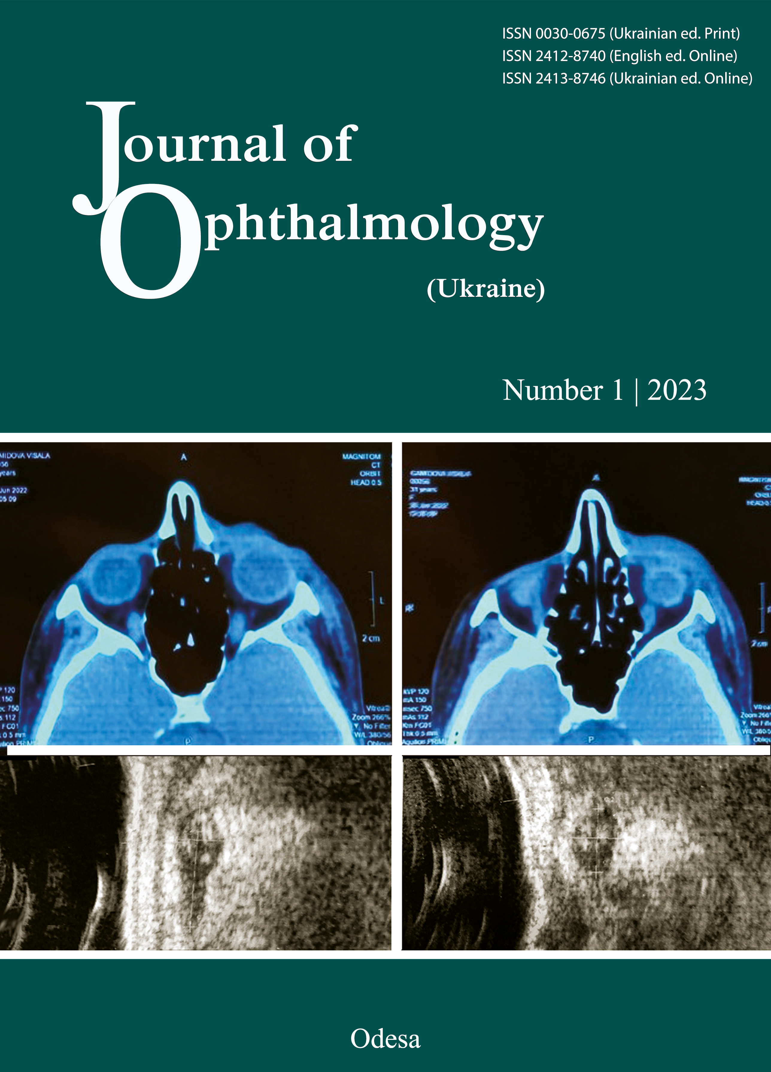
Results: Patients treated were most commonly from the city of Odesa and Odesa region (25.9% or 15 patients). Odesa-region patients most commonly were from Izmail and Bilhorod Dnistrovskyi districts. The worm was more commonly located in the orbital tissue (53 of 58 patients or 91.4%) than in the eyelids (5 patients or 8.6%). All patients complained of skin redness and swollen eyelids. With further migration of the worm deep into the orbital tissue, 15 patients (28.3%) developed exophthalmos of not more than 2-3 mm and 15 patients (28.3%) developed diplopia. The study showed evidence of D. repens infection by parasitological methods of diagnosis in all the 58 study patients, with a mature female found in 44 patients (75.9%), mature female remnants in 11 patients (19.0%), an immature female in 2 patients (3.4%), and a male in one patient (1.8%). The worms were elongated, less than or equal to 1.0-mm thick and 2.2-15 mm in length.
Conclusion: First, worm extraction surgery is an essential treatment for ocular dirofilariasis (irrespective whether the worm is located in the eyelid, orbit or eye) and must be accompanied by disinfectant, anti-inflammatory and antihistamine therapy, both topical (eyedrops and ointments) and oral. Second, an ultrasound of the orbital tissue and eyelids may become a gold standard (along with CT and MRI) for the differential diagnosis of dirofilariasis.
References
Азнабаев МТ. Редкие случаи в клинической офтальмологии. Уфа, Уфимский НИИ глазных болезней; 2001: 58 с.
Бугаев АМ. Дирофиляриоз - неизвестная опасность. [Интернет]. Доступно: meddovidka.ua/content/view/936
Голубовская ОА, редактор. Инфекционные болезни (учебник). Киев: ВСВ «Медицина» (2 изд., дополненное и переработанное); 2018. 688 с.
Майчук ЮФ. Паразитарные заболевания глаз. М., Медицина; 1988. 288 с.
Гаймутдинова РФ, Тухбатуллин МГ, Гилмуллина ФС, Нефедов ВП, Пигалова ОМ, Бикмухаметова ДА. Диагностика дирофиляроза человека. Практ. мед. 2012.1(56): 123-6.
Гончаров В. Дирофіляріоз - хвороба, з якою може зустрітися людина. [Интернет]. Доступно: ооlc.od.ua
Сакович ВН, Сердюк ВН, Тесса ЕА и др. Диагностика и лечение дирофиляриоза. Офтальмология. Восточная Европа. 2016.6(3): 378-83.
Авдюхина ТИ, Постнова ВФ, Абросимова ЛМ. и др. Дирофиляриоз (D. repens) в Российской Федерации и некоторых странах СНГ: ситуация и тенденция ее изменения. Мед. паразитол. 2003. 4: 44-8.
Журавлев АС, Калашник МВ, Пустовалова МН, Лупырь АВ, Крылова ИВ, Олейник СВ, Одарюк ИА. Дирофиляриоз, осложненный аденофлегмоной шеи у больной сахарным діабетом. Ринология. 2008. 2: 33-6.
Дирофиляриоз человека, симптомы, лечение. [Интернет]. Доступно: http://q99.it/VFihBcp
Дронова АП. Случай дирофиляриоза органа зрения. Офтальмол. журн. 1997. 5: 382.
Мельникова Л, Рулев А. Одновременное паразитирование двух личинок у одного хозяина-человека. Материалы Филатовских чтений, 2016. Одесса. 2016: 220.
Полищук Т, Клопоцкая Н, Тесса Е. Эпидемиология дирофиляриоза глаз в Днепропетровске. Материалы «Актуальные вопросы офтальмологии». Запорожье. 2015: 185-6.
Супряга ВГ, Старкова ТВ, Короткова ГИ Клинический и паразитологический диагноз дирофиляриоза человека. Мед. паразитол. 2002. 1: 53-5.
Угодіна ЛЄ, Штепа ЛФ. Остерігайтесь дирофіляріозу. [Интернет]. Доступно: www.dolc.dp.ua/wpress/?p=4260
Beden U, Hokelek М, Acici М. et al. A case of orbital dirofilariasis in Northern Turkey. Ophthalmic Plastic And Reconstructive Surgery. 2007; 23 (4): 329-31.
https://doi.org/10.1097/IOP.0b013e318073cca3
Rohela М, Jamaiah I, Hui T. et al. Dirofilaria causing eye infection in a patient from Malaysia. Southeast Asian J Trop Med. 2009. 40 (5): 914-18.
Gupta V, Sankaran P, Samantaray MJCh. Menon Bilateral intraocular dirofilariasis Indian Journal of Ophthalmology. [Internet]. 2014. 62(3). Available from: https://doi.org/10.4103/0301-4738.116252
Szénási Z, Kovács A, Pampiglione S. et al. Human dirofilariosis in Hungary: an emerging zoonosis in central Europe.Wiener Klinische Wochenschrift. 2008. 120 (3-4): 96-102. https://doi.org/10.1007/s00508-008-0928-2
Dzamić A, Colović I, Arsić-Arsenijević V et al. Human Dirofilaria repens infection in Serbia. Journal Of Helminthology. 2009; 6. 83 (2): 129-37. https://doi.org/10.1017/S0022149X09341346
Mukherjee B., Biswas J., Varde Meghana Anika, Noronha Veena. Orbital dirofilariasis. AnnTropMedPublic Health. 2012; 5: 42-3. https://doi.org/10.4103/1755-6783.92880
Pampiglione S, Rivasi F. Human dirofilaria (Nochtiella) repens: An update of world literature from 1995 to 2000. Parassitologia. 2000; 42: 231-54.
Sepideh Tavakolizadeh and Iraj Mobedi Jrbital Dirofilariasis in Iran: A Case Report // Korean J Parasitol. 47(4): 397-99, December 2009 DOJ: 10.3347/kjp. 2009. 47. 4. 397. https://doi.org/10.3347/kjp.2009.47.4.397
Nadgir S, Tallur S, Mangoli V [et al.] Subconjunctival dirofilariasis in India. Southeast Asian J. Trop. Med. 2001. 6. 32 (2): 244-6.
Velev V, Vutova K, Pelov T, Tsachev I. Human dirofilariasis in Bulgaria between 2009 and 2018. [Internet]. Helminthologia. 2019; 56(3). Available from: https://doi.org/10.2478/helm-2019-0016
Que AT, Nguyen DN, Do NA, Le TA. Dirofilariasis in Vietnam: A case report and brief review. [Internet]. Tropical Biomedicine. 2019; 36(2). Available from: https://www.researchgate.net/publication/336702990_Dirofilariasis_in_Vietnam_A_case_report_and_brief_review
Downloads
Published
How to Cite
Issue
Section
License
Copyright (c) 2023 Poliakova S.I., Karliuga I.A., Moloda A.L., Linchevska O.G.

This work is licensed under a Creative Commons Attribution 4.0 International License.
This work is licensed under a Creative Commons Attribution 4.0 International (CC BY 4.0) that allows users to read, download, copy, distribute, print, search, or link to the full texts of the articles, or use them for any other lawful purpose, without asking prior permission from the publisher or the author as long as they cite the source.
COPYRIGHT NOTICE
Authors who publish in this journal agree to the following terms:
- Authors hold copyright immediately after publication of their works and retain publishing rights without any restrictions.
- The copyright commencement date complies the publication date of the issue, where the article is included in.
DEPOSIT POLICY
- Authors are permitted and encouraged to post their work online (e.g., in institutional repositories or on their website) during the editorial process, as it can lead to productive exchanges, as well as earlier and greater citation of published work.
- Authors are able to enter into separate, additional contractual arrangements for the non-exclusive distribution of the journal's published version of the work with an acknowledgement of its initial publication in this journal.
- Post-print (post-refereeing manuscript version) and publisher's PDF-version self-archiving is allowed.
- Archiving the pre-print (pre-refereeing manuscript version) not allowed.


