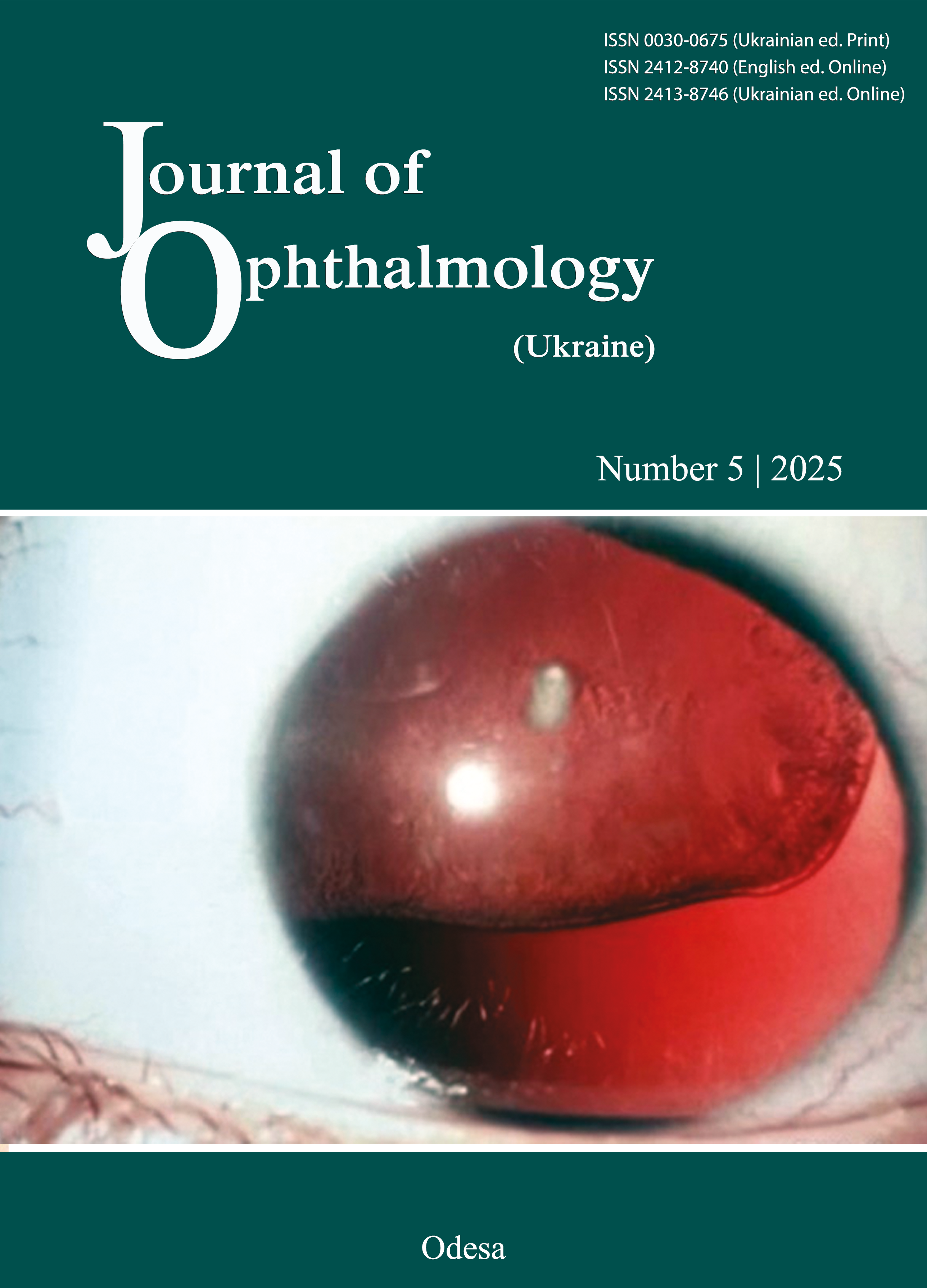
Features of corneal reparation and postoperative complications after various types of excimer laser correction of myopia
DOI:
https://doi.org/10.31288/oftalmolzh20255313Keywords:
myopia, excimer laser correction, photorefractive keratectomy, transepithelial PRK, LASEK, corneal reparation, complicationsAbstract
Purpose: To determine the features of corneal reparation and postoperative complications after various types of excimer laser correction (ELC) of myopia.
Material and Methods: This was a multicenter, prospective, observational clinical study including 255 patients (510 eyes). They were stratified into three equal groups, based on the particular type of ELC: photorefractive keratectomy (PRK), transepithelial PRK (trans-PRK) and laser-assisted subepithelial keratectomy (LASEK). Corneal re-epithelization rate, epithelial and stromal thicknesses (as assessed by anterior segment spectral domain optical coherence tomography), corneal irregularity measurement (CIM), uncorrected visual acuity (UCVA) and incidence of haze were assessed over 3 months postoperatively. Correlation analysis and Principal Component Analysis clustering were used for assessing relationships.
Results: There were statistically significant between-groups differences early after surgery. The trans-PRK group showed the best results in terms of re-epithelialization dynamics (with an incidence of delayed re-epithelialization at day 4-5 of 28% vs 38% PRK), maximum epithelial thickness in the central 5-mm map zone (64 µm vs 74 µm LASEK) and the size of stromal edema (444 µm vs 460 µm PRK). At month 3, UCVA was higher in the trans-PRK group (1.00 vs 0.90 PRK, p = 0.0059). Haze strongly correlated with stromal edema (r = 0.96, p < 0.001) and delayed re-epithelization (r = 0.93). Cluster analysis revealed subgroups with shared signs of pathological reparation irrespective of the ELC technique. The percentage of patients with a pathological cluster profile was 16.5% in the PRK group, 14.7% in the LASEK group, and 10.6% in the trans-PRK group, which confirmed independent morphometric patterns of complicated healing.
Conclusion: Among the three types of ELC, trans-PRK was the best in terms of the dynamics of corneal reparation and visual recovery. Between-groups differences in morphometric and functional parameters almost disappeared at 3 months after surgery. The PRK and trans-PRK groups had the highest and lowest incidences of haze, respectively. Haze had the strongest correlations with corneal edema and delayed re-epithelialization. Cluster analysis revealed subgroups with pathological healing irrespective of the ELC technique. Morphometric parameters may be considered as predictors of complicated corneal reparation after ELC. When comprehensively assessed, they may be used for predicting the risk of complications and in further research on the new etiological factors of complicated corneal reparation.
References
Burton MJ, Ramke J, Marques AP, Bourne RRA, Congdon N, Jones I, et al. The Lancet Global Health Commission on Global Eye Health: vision beyond 2020. Lancet Glob Health. 2021;9(4):e489-e551.
Azar DT, Chang JH, Han KY. Wound healing after keratorefractive surgery: review of biological and optical considerations. Cornea. 2012;31 Suppl 1:S9-S19. https://doi.org/10.1097/ICO.0b013e31826ab0a7
Wilson SE, Sampaio LP, Shiju TM, Hilgert GSL, de Oliveira RC. Corneal opacity: cell biological determinants of the transition from transparency to transient haze to scarring fibrosis, and resolution, after injury. Invest Ophthalmol Vis Sci. 2022;63(1):22.https://doi.org/10.1167/iovs.63.1.22
Wilson SE, Marino GK, Medeiros CS, Santhiago MR. Phototherapeutic keratectomy: science and art. J Refract Surg. 2017;33(3):203-10.https://doi.org/10.3928/1081597X-20161123-01
Abdel-Radi M, Shehata M, Mostafa MM, Aly MOM. Transepithelial photorefractive keratectomy: a prospective randomized comparative study between the two-step and the single-step techniques. Eye (Lond). 2023;37(8):1545-52. https://doi.org/10.1038/s41433-022-02174-4.https://doi.org/10.1038/s41433-022-02174-4
Moshirfar M, Wang Q, Theis J, Porter KC, Stoakes IM, Payne CJ, et al. Management of corneal haze after photorefractive keratectomy. Ophthalmol Ther. 2023;12(6):2841-62.https://doi.org/10.1007/s40123-023-00782-1
Tomás-Juan J, Murueta-Goyena Larrañaga A, Hanneken L. Corneal regeneration after photorefractive keratectomy: a review. J Optom. 2015;8(3):149-69.https://doi.org/10.1016/j.optom.2014.09.001
Kobayashi R, Hashida N. Overview of cytomegalovirus ocular diseases: retinitis, corneal endotheliitis, and iridocyclitis. Viruses. 2024;16(7):1110.https://doi.org/10.3390/v16071110
Koizumi N, Inatomi T, Suzuki T. Japan Corneal Endotheliitis Study Group. Clinical features and management of cytomegalovirus corneal endotheliitis: analysis of 106 cases from the Japan corneal endotheliitis study. Br J Ophthalmol. 2015;99(1):54-8.https://doi.org/10.1136/bjophthalmol-2013-304625
Edelhauser HF. The resiliency of the corneal endothelium to refractive and intraocular surgery. Cornea. 2000;19(3):263-73.https://doi.org/10.1097/00003226-200005000-00002
Ding K, Nataneli N. Cytomegalovirus corneal endotheliitis. In: StatPearls [Internet]. Treasure Island (FL): StatPearls Publishing; 2025 Jan. Aviable from: https://www.ncbi.nlm.nih.gov/books/NBK578174.
Kobayashi R, Hashida N, Maruyama K, Nishida K. Clinical findings of specular microscopy images in cytomegalovirus corneal endotheliitis. Asia Pac J Ophthalmol (Phila). 2022;11(3):273-8.https://doi.org/10.1097/APO.0000000000000522
Yoo WS, Kwon LH, Eom Y, Thng ZX, Or C, Nguyen QD, et al. Cytomegalovirus corneal endotheliitis: a comprehensive review. Ocul Immunol Inflamm. 2024;32(9):2228-37.https://doi.org/10.1080/09273948.2024.2320704
Bitton K, Zéboulon P, Ghazal W, Rizk M, Elahi S, Gatinel D. Deep learning model for the detection of corneal edema before Descemet membrane endothelial keratoplasty on optical coherence tomography images. Transl Vis Sci Technol. 2022;11(12):19.https://doi.org/10.1167/tvst.11.12.19
Carl Zeiss Meditec. User Manual ATLAS Corneal Topography System Model 9000 and ATLAS Review Software 3.0; PathFinder II Color-Coded Parameter Classification (Normal Eyes, based on Multi-Center Clinical Study). 2007. p.147-8.
Janiszewska-Bil D, Grabarek BO, Lyssek-Boroń A, Kiełbasińska A, Kuraszewska B, Wylęgała E, Krysik K. Comparative Analysis of Corneal Wound Healing: Differential Molecular Responses in Tears Following PRK, FS-LASIK, and SMILE Procedures. Biomedicines. 2024 Oct 9;12(10):2289. https://doi.org/10.3390/biomedicines12102289
Mohan RR, Kempuraj D, D'Souza S, Ghosh A. Corneal stromal repair and regeneration. Prog Retin Eye Res. 2022 Nov;91:101090. https://doi.org/10.1016/j.preteyeres.2022.101090
Ljubimov AV, Saghizadeh M. Progress in corneal wound healing. Prog Retin Eye Res. 2015 Nov;49:17-45. https://doi.org/10.1016/j.preteyeres.2015.07.002
Ljubimov AV. Diabetic complications in the cornea. Vision Res. 2017 Oct;139:138-152. https://doi.org/10.1016/j.visres.2017.03.002
Kundu G, D'Souza S, Lalgudi VG, Arora V, Chhabra A, Deshpande K, Shetty R. Photorefractive keratectomy (PRK) Prediction, Examination, tReatment, Follow-up, Evaluation, Chronic Treatment (PERFECT) protocol - A new algorithmic approach for managing post PRK haze. Indian J Ophthalmol. 2020 Dec;68(12):2950-2955. https://doi.org/10.4103/ijo.IJO_2623_20
Rocha-de-Lossada C, Rachwani-Anil R, Colmenero-Reina E, Borroni D, Sánchez-González JM. Laser refractive surgery in corneal dystrophies. J Cataract Refract Surg. 2021 May 1;47(5):662-670. https://doi.org/10.1097/j.jcrs.0000000000000468
Adib-Moghaddam S, Soleyman-Jahi S, Sanjari Moghaddam A, Hoorshad N, Tefagh G, Haydar AA, et al. Efficacy and safety of transepithelial photorefractive keratectomy. J Cataract Refract Surg. 2018;44(10):1267-79.https://doi.org/10.1016/j.jcrs.2018.07.021
Saad A, Saad A, Frings A. Refractive results of photorefractive keratectomy comparing trans-PRK and PTK-PRK for correction of myopia and myopic astigmatism. Int Ophthalmol. 2024;44(1):111-9.https://doi.org/10.1007/s10792-024-02999-w
Akram S, Moazzum W, Abid K. Efficacy of single-step transepithelial photorefractive keratectomy in myopia, hyperopia and astigmatism: a systematic review. BMC Ophthalmol. 2025;25(1):93.https://doi.org/10.1186/s12886-024-03830-x
Gadde AK, Srirampur A, Katta KR, Mansoori T, Armah SM. Comparison of single-step transepithelial photorefractive keratectomy and conventional photorefractive keratectomy in low to high myopic eyes. Indian J Ophthalmol. 2020;68(5):755-61.https://doi.org/10.4103/ijo.IJO_1126_19
Gharieb HM, Awad-Allah MAA, Ahmed AA, Othman IS. Transepithelial laser versus alcohol-assisted photorefractive keratectomy safety and efficacy: 1-year follow-up of a contralateral eye study. Korean J Ophthalmol. 2021;35(2):142-52.https://doi.org/10.3341/kjo.2020.0105
Gibson CR, Mader TH, Lipsky W, Schallhorn SC, Tarver WJ, Suresh R, et al. Photorefractive keratectomy and laser-assisted in situ keratomileusis on 6-month space missions. Aerosp Med Hum Perform. 2024;95(5):278-81.https://doi.org/10.3357/AMHP.6368.2024
Chen CC, Chang JH, Lee JB, Javier J, Azar DT. Human corneal epithelial cell viability and morphology after dilute alcohol exposure. Invest Ophthalmol Vis Sci. 2002;43(8):2593-602.
Downloads
Published
How to Cite
Issue
Section
License
Copyright (c) 2025 Mogilevskyy S. Yu., Kalinichenko A. A., Gulida A. O., Zhovtoshtan M. Yu.

This work is licensed under a Creative Commons Attribution 4.0 International License.
This work is licensed under a Creative Commons Attribution 4.0 International (CC BY 4.0) that allows users to read, download, copy, distribute, print, search, or link to the full texts of the articles, or use them for any other lawful purpose, without asking prior permission from the publisher or the author as long as they cite the source.
COPYRIGHT NOTICE
Authors who publish in this journal agree to the following terms:
- Authors hold copyright immediately after publication of their works and retain publishing rights without any restrictions.
- The copyright commencement date complies the publication date of the issue, where the article is included in.
DEPOSIT POLICY
- Authors are permitted and encouraged to post their work online (e.g., in institutional repositories or on their website) during the editorial process, as it can lead to productive exchanges, as well as earlier and greater citation of published work.
- Authors are able to enter into separate, additional contractual arrangements for the non-exclusive distribution of the journal's published version of the work with an acknowledgement of its initial publication in this journal.
- Post-print (post-refereeing manuscript version) and publisher's PDF-version self-archiving is allowed.
- Archiving the pre-print (pre-refereeing manuscript version) not allowed.


