Objective optical densitometry-based evaluation of longitudinal changes in the inflammatory process in the treatment of bacterial keratitis
DOI:
https://doi.org/10.31288/oftalmolzh202531114Keywords:
bacterial keratitis, corneal topography, optical densitometry, cornea, recurrent herpetic keratitisAbstract
Purpose: To objectively determine longitudinal changes in corneal optical density (COD) in patients with bacterial keratitis (BK).
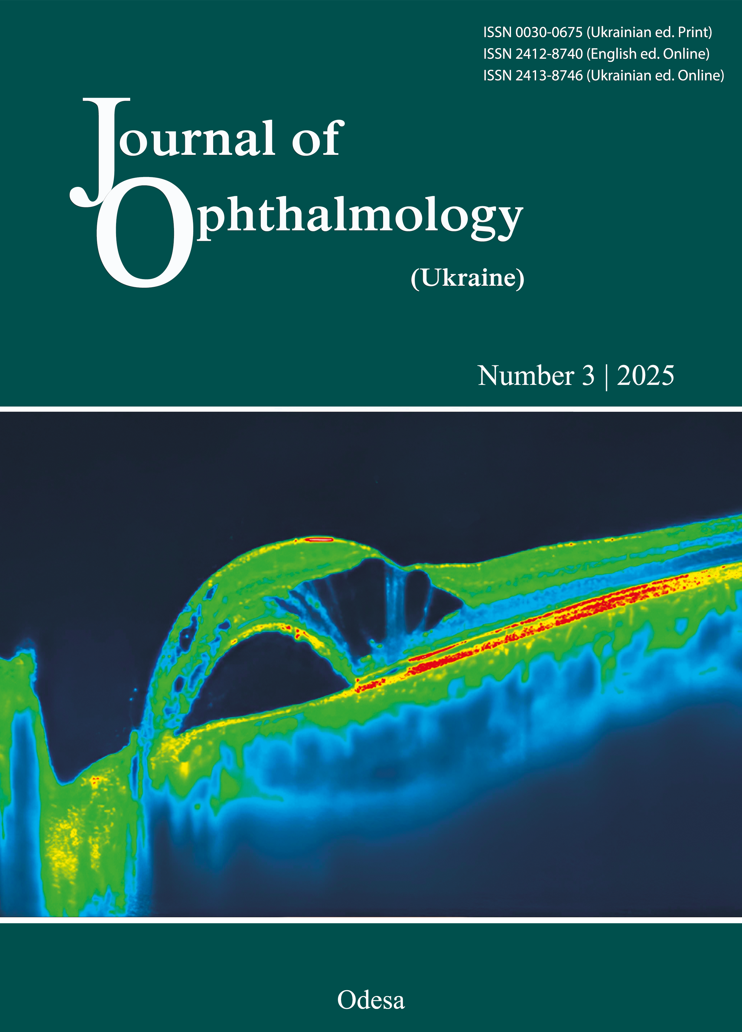
Methods: Thirty patients with unilateral BK (mean age ± standard deviation [SD], 44.7 ± 11.5 years) underwent examination. Fellow eyes were used as controls. Optical densitometry was performed using the corneal topography system Pentacam AXL at admission and days 7 and 14 of treatment.
Results: At presentation and days 7 and 14 of treatment, the average median (interquartile range [IQR]) COD value for the affected eyes was 92.8 (55.6-99.6), 89.7 (51.1-97.3) and 80.6 (32.0-93.1) grayscale units (GSU), respectively (p < 0.05), indicating a decrease in inflammatory infiltration, and the average mean (SD) COD value for the control eyes was 19.6 ± 3.0, 19.9 ± 2.8, and 19.7 ± 2.6 GSU, respectively, with no statistically significant difference.
Conclusion: The use of optical densitometry for monitoring corneal transparency in eyes treated for BK over a two-week period enabled objective recording of a gradual reduction in median (IQR) COD values in the affected eyes from 92.8 (55.6–99.6) to 80.6 (32.0–93.1) GSU, corresponding to an improvement in the clinical status of patients.
References
WHO (2019). World report on vision. Geneva: World Health Organization; 2019. Available at: https://www.who.int/publications/i/item/world-report-on-vision (accessed 30 June 2020)
World Health Organization. Causes of blindness and visual impairment. Available at: http://www.who.int/blindness/causes/en. Accessed December 7, 2016.
Teweldemedhin M, Gebreyesus H, Atsbaha AH, et al. Bacterial profile of ocular infections: a systematic review. BMC Ophthalmol. 2017 Nov 25;17(1):212. https://doi.org/10.1186/s12886-017-0612-2
Cabrera-Aguas M, Khoo P, Watson SL. Infectious keratitis: A review. Clin Experiment Ophthalmol. 2022; 50(5): 543-562. https://doi.org/10.1111/ceo.14113
He Y, Ma B-S, Zeng J-H, Ma D-J. Corneal optical density: Structural basis, measurements, influencing factors, and roles in refractive surgery. Front Bioeng Biotechnol. 2023 Apr 6:11:1144455. https://doi.org/10.3389/fbioe.2023.1144455
Kuerten D, Plange N, Koch EC, et al. Central corneal thickness determination in corneal edema using ultrasound pachymetry, a Scheimpflug camera, and anterior segment OCT. Graefes Arch Clin Exp Ophthalmol. 2015 Jul;253(7):1105-9. https://doi.org/10.1007/s00417-015-2998-y
Hsieh TH, Yu HJ, Yang IH, et al. Simultaneously Monitoring Whole Corneal Injury with Corneal Optical Density and Thickness in Patients Undergoing Cataract Surgery. Diagnostics (Basel). 2021 Sep 7;11(9):1639. https://doi.org/10.3390/diagnostics11091639
Asrar A, Ikram B, Khan H, et al. Normal values of corneal optical densitometry using pentacam scheimpflug camera. Adv Ophthalmol Vis Syst. 2016;5(1):202-9. https://doi.org/10.15406/aovs.2016.05.00142
Adran D, Vaillancourt L, Harissi-Dagher M, et al. Corneal Densitometry as a Tool to Measure Epithelial Ingrowth After Laser In Situ Keratomileusis. Cornea. 2017 Apr;36(4):406-410. https://doi.org/10.1097/ICO.0000000000001114
Wei R, Li M, Yang W, et al. Corneal Densitometry After Small Incision Lenticule Extraction (SMILE) and Femtosecond Laser-Assisted LASIK (FS-LASIK): 5-Year Prospective Comparative Study. Front. Med (Lausanne). 2020 Nov 6:7:521078. https://doi.org/10.3389/fmed.2020.521078
Alnawaiseh M, Rosentreter A, Eveslage M, et al. Changes in Corneal Transparency After Cross-linking for Progressive Keratoconus: Long-term Follow-up. J Refract Surg. 2015 Sep;31(9):614-8. https://doi.org/10.3928/1081597X-20150820-07
Böhm M, Shajari M, Remy M, Kohnen T. Corneal densitometry after accelerated corneal collagen cross-linking in progressive keratoconus. Int Ophthalmol. 2019 Apr;39(4):765-75. https://doi.org/10.1007/s10792-018-0876-4
Kobashi H, Kamiya K, Shimizu K. Impact of Forward and Backward Scattering and Corneal Higher-Order Aberrations on Visual Acuity after Penetrating Keratoplasty. Semin Ophthalmol. 2018;33(6):748-756. https://doi.org/10.1080/08820538.2018.1427767
Downloads
Published
How to Cite
Issue
Section
License
Copyright (c) 2025 Alifanov I.S., Klopotska N.G.

This work is licensed under a Creative Commons Attribution 4.0 International License.
This work is licensed under a Creative Commons Attribution 4.0 International (CC BY 4.0) that allows users to read, download, copy, distribute, print, search, or link to the full texts of the articles, or use them for any other lawful purpose, without asking prior permission from the publisher or the author as long as they cite the source.
COPYRIGHT NOTICE
Authors who publish in this journal agree to the following terms:
- Authors hold copyright immediately after publication of their works and retain publishing rights without any restrictions.
- The copyright commencement date complies the publication date of the issue, where the article is included in.
DEPOSIT POLICY
- Authors are permitted and encouraged to post their work online (e.g., in institutional repositories or on their website) during the editorial process, as it can lead to productive exchanges, as well as earlier and greater citation of published work.
- Authors are able to enter into separate, additional contractual arrangements for the non-exclusive distribution of the journal's published version of the work with an acknowledgement of its initial publication in this journal.
- Post-print (post-refereeing manuscript version) and publisher's PDF-version self-archiving is allowed.
- Archiving the pre-print (pre-refereeing manuscript version) not allowed.


