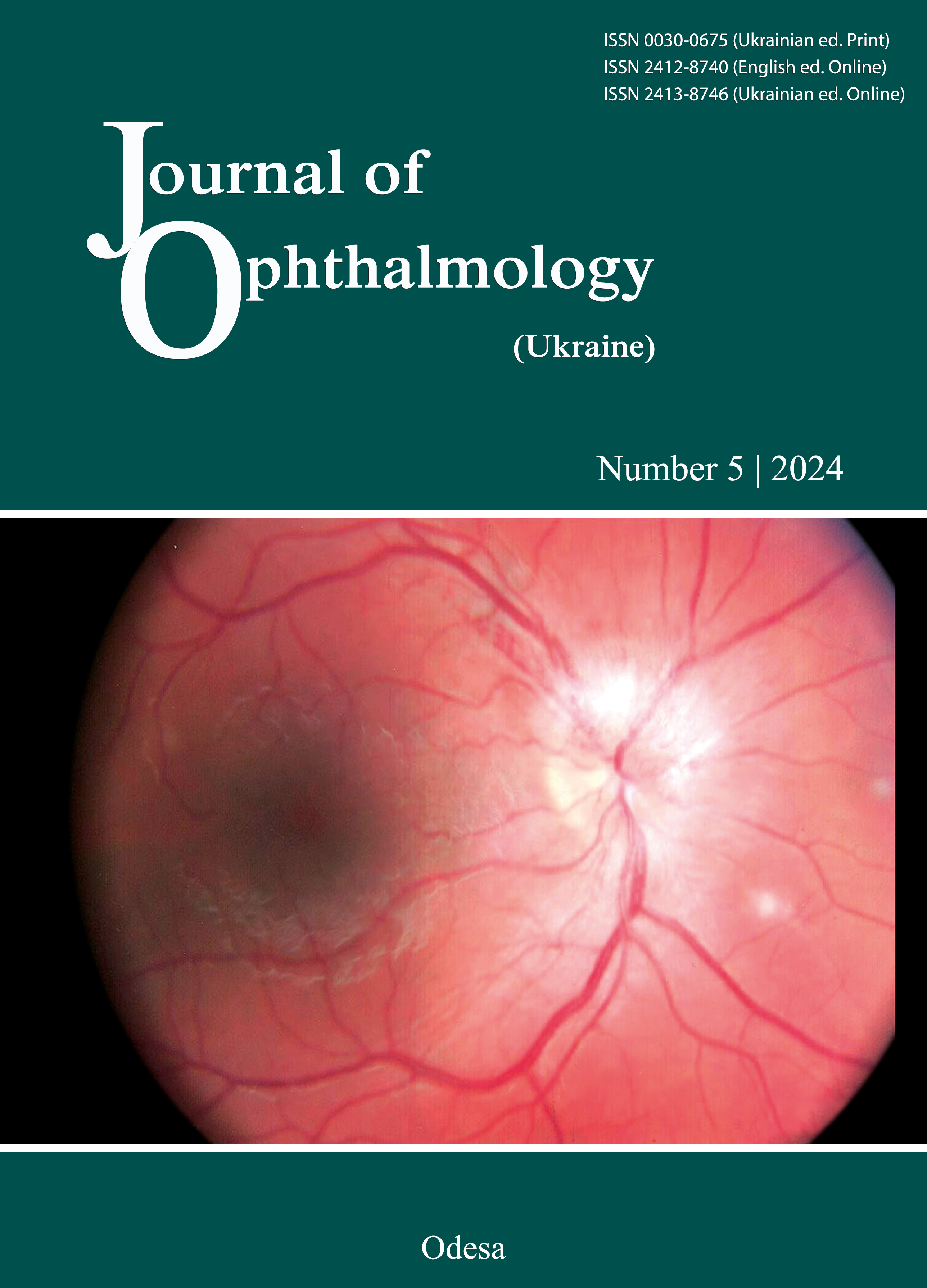
Macular thickness analysis using optical coherence tomography data, stereopsis and binocular vision in premature infants who underwent retinal laser photocoagulation due to retinopathy of prematurity in an age-related perspective
DOI:
https://doi.org/10.31288/oftalmolzh202451520Keywords:
retinopathy of prematurity, macular thickness, stereopsis, binocular visionAbstract
The purpose was to establish reference values for macular thickness, binocular vision, and stereopsis in premature infants aged 5-9 years and 10-13 years who underwent laser photocoagulation of avascular retinal zones due to retinopathy of prematurity (ROP); to assess and compare the data in an age-related perspective.
Methods. Data from 24 premature infants who underwent ophthalmological examination, including optical coherence tomography, Titmus Stereo Fly Test, and the Worth 4 Dot Test, at ages 5-9 years and again at ages 10-13 years were analyzed. All children had undergone laser photocoagulation of avascular retinal zones due to ROP in infancy.
Results. At ages 5-9 years, the mean central macular volume was 9.2 mm3, and the retinal thickness in the central fovea was 313.7 μm. At ages 10-13 years, the mean central macular volume was 9.1 mm3, and the retinal thickness in fovea was 320.8 μm. Normal binocular vision and stereopsis were observed in 79.2% and 33.3% of the children at the first time point, and in 87.5% and 45.8% at the second time point.
Conclusions. No statistically significant difference in the central macular volume and macular thickness was detected between the two time points, (p>0.05). The thickest part of the macula was identified in the inner macula, followed by the outer macula, with the nasal quadrant being the thickest. Despite the anatomical peculiarities, high rates of binocular vision were observed at both time points, along with stereopsis at the second examination.
References
Akerblom H, Larsson E, Eriksson U, Holmström G. Central macular thickness is correlated with gestational age at birth in prematurely born children. Br J Ophthalmol. 2011 Jun;95(6):799-803. https://doi.org/10.1136/bjo.2010.184747
Lee H, Proudlock FA, Gottlob I. Pediatric Optical Coherence Tomography in Clinical Practice-Recent Progress. Invest Ophthalmol Vis Sci. 2016 Jul 1;57(9):OCT69-79. https://doi.org/10.1167/iovs.15-18825
Marmor MF, Choi SS, Zawadzki RJ, Werner JS.Visual Insignificance of the foveal pit. Arch Ophthalmol 2008; 126: 907-913. https://doi.org/10.1001/archopht.126.7.907
An International Committee for the Classification of Retinopathy of Prematurity. The International Classification of Retinopathy of Prematurity Revisited // Arch. Ophthalmol. - 2005. - Vol.123. - № 7. - P. 991-999. https://doi.org/10.1001/archopht.123.7.991
Nigam B, Garg P, Ahmad L, Mullick R. OCT Based Macular Thickness in a Normal Indian Pediatric Population. J Ophthalmic Vis Res. 2018 Apr-Jun;13(2):144-148. https://doi.org/10.4103/jovr.jovr_51_17
Eriksson U, Holmström G, Alm A, Larsson E. A population-based study of macular thickness in full-term children assessed with Stratus OCT: normative data and repeatability. Acta Ophthalmol. 2009 Nov;87(7):741-5. https://doi.org/10.1111/j.1755-3768.2008.01357.x
Fieß A, Pfisterer A, Gißler S, Korb C, Mildenberger E, Urschitz MS, et al. Retinal thickness and foveal hypoplasia in adults born preterm with and without retinopathy of prematurity. The Gutenberg Prematurity Eye Study. Retina. 2022 Sep 1;42(9):1716-1728. https://doi.org/10.1097/IAE.0000000000003501
Pétursdóttir D, Åkerblom H, Holmström G, Larsson E. Central macular morphology and optic nerve fibre layer thickness in young adults born premature and screened for retinopathy of prematurity. Acta Ophthalmol. 2024 Jun;102(4):391-400. Epub 2023 Nov 22. PMID: 37991127. https://doi.org/10.1111/aos.15814
Molnar AEC, Rosén RM, Nilsson M, Larsson EKB, Holmström GE, Hellgren KM. Central macular thickness in 6.5-year-old children born extremely preterm is strongly associated with gestational age even when adjusted for risk factors. Retina. 2017 Dec;37(12):2281-2288. https://doi.org/10.1097/IAE.0000000000001469
Ecsedy M, Szamosi A, Karkó C, Zubovics L, Varsányi B, Németh J, et al. A comparison of macular structure imaged by optical coherence tomography in preterm and full-term children. Invest Ophthalmol Vis Sci. 2007 Nov;48(11):5207-11. https://doi.org/10.1167/iovs.06-1199
Mills MD. The eye in childhood. Am Fam Physician. 1999 Sep 1;60(3):907-16, 918.
Kwinta P, Herman-Sucharska I, Leśniak A, Klimek M, Karcz P, Durlak W, et al. Relationship between Stereoscopic Vision, Visual Perception, and Microstructure Changes of Corpus Callosum and Occipital White Matter in the 4-Year-Old Very Low Birth Weight Children. Biomed Res Int. 2015;2015:842143. https://doi.org/10.1155/2015/842143
Downloads
Published
How to Cite
Issue
Section
License
Copyright (c) 2024 Adakhovska A. O., Katsan S. V.

This work is licensed under a Creative Commons Attribution 4.0 International License.
This work is licensed under a Creative Commons Attribution 4.0 International (CC BY 4.0) that allows users to read, download, copy, distribute, print, search, or link to the full texts of the articles, or use them for any other lawful purpose, without asking prior permission from the publisher or the author as long as they cite the source.
COPYRIGHT NOTICE
Authors who publish in this journal agree to the following terms:
- Authors hold copyright immediately after publication of their works and retain publishing rights without any restrictions.
- The copyright commencement date complies the publication date of the issue, where the article is included in.
DEPOSIT POLICY
- Authors are permitted and encouraged to post their work online (e.g., in institutional repositories or on their website) during the editorial process, as it can lead to productive exchanges, as well as earlier and greater citation of published work.
- Authors are able to enter into separate, additional contractual arrangements for the non-exclusive distribution of the journal's published version of the work with an acknowledgement of its initial publication in this journal.
- Post-print (post-refereeing manuscript version) and publisher's PDF-version self-archiving is allowed.
- Archiving the pre-print (pre-refereeing manuscript version) not allowed.


