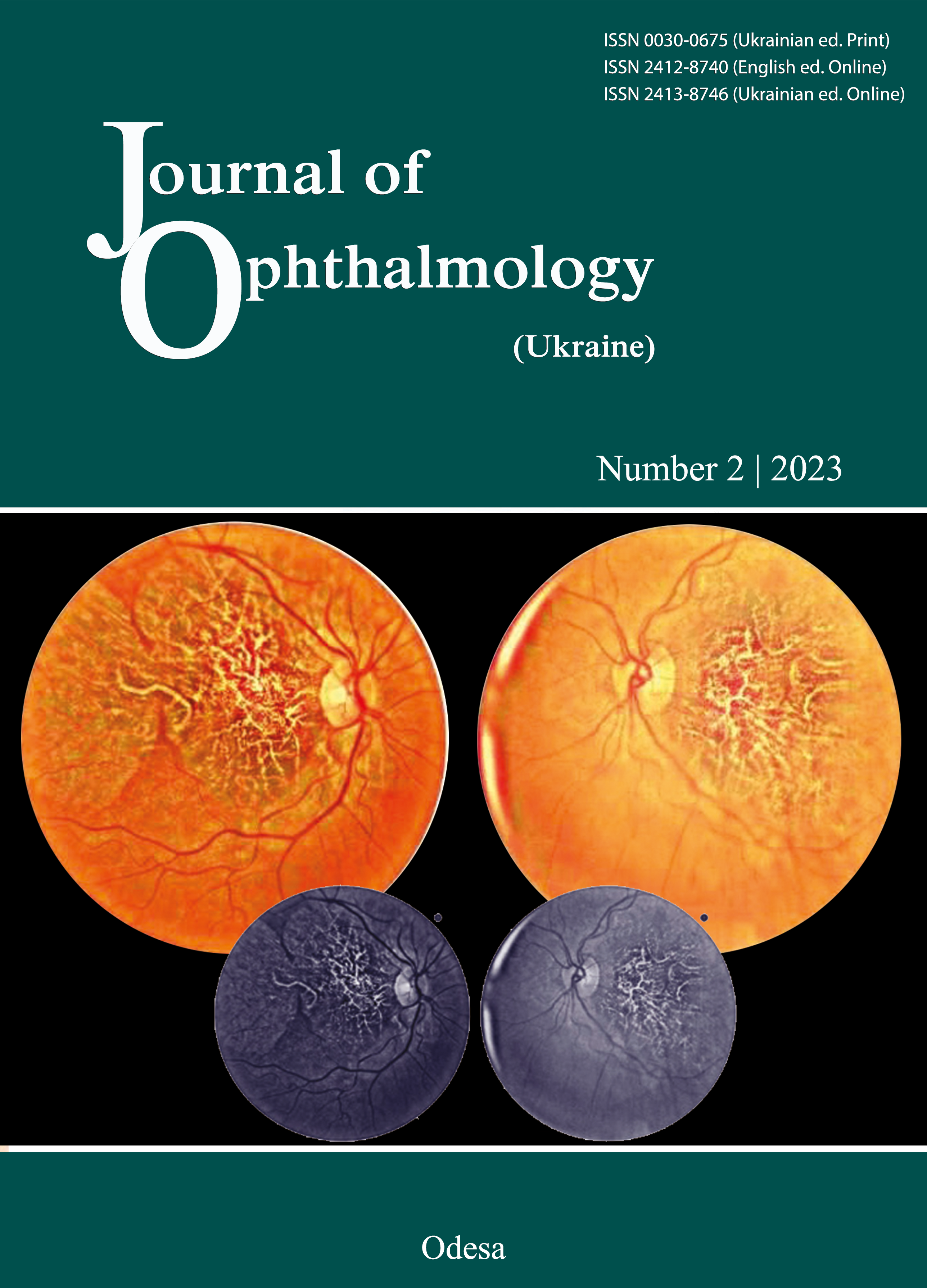
An unusual case of posterior vitreous detachment
DOI:
https://doi.org/10.31288/oftalmolzh202326062Abstract
Purpose: To describe a case of spontaneous closure of a macular microhole induced by an acute posterior vitreous detachment (PVD).
Methods: Retrospective case report and analysis of patient data.
Case Report: We present a case of spontaneous closure of a posterior vitreous detachment (PVD) induced macular microhole. In our patient, macular microhole developed a few weeks after acute PVD. The development of macular hole secondarily to acute PVD is rare, and there are reports of spontaneous closure. Reporting this case is important to alert clinicians to the potential self-resolving nature of this pathology.
Conclusion: Macular hole is a rare complication of acute PVD. Some cases resolve spontaneously without the need for surgical correction. A conservative approach may be an appropriate setting if no high-risk features are present.
References
Ahmed F, Tripathy K. Posterior Vitreous Detachment. StatPearls Publishing; 2022. Accessed December 18, 2022. https://www.ncbi.nlm.nih.gov/books/NBK563273/
Ramovecchi P, Salati C, Zeppieri M. Spontaneous posterior vitreous detachment: A glance at the current literature. World J Exp Med. 2021;11(3):30-36. https://doi.org/10.5493/wjem.v11.i3.30
Johnson MW. Posterior vitreous detachment: evolution and complications of its early stages. Am J Ophthalmol. 2010;149(3):371-382.e1. https://doi.org/10.1016/j.ajo.2009.11.022
Majumdar S, Tripathy K. Macular Hole. StatPearls Publishing; 2022. Accessed December 18, 2022. https://www.ncbi.nlm.nih.gov/books/NBK559200/
Azzolini C. Macular Hole: From Diagnosis to Therapy. J Ophthalmol. 2020;2020:1473763. https://doi.org/10.1155/2020/1473763
Cairns JD, McCombe MF. Microholes of the fovea centralis. Aust N Z J Ophthalmol. 1988;16(2):75-79. https://doi.org/10.1111/j.1442-9071.1988.tb01253.x
Zambarakji HJ, Schlottmann P, Tanner V, Assi A, Gregor ZJ. Macular microholes: pathogenesis and natural history. Br J Ophthalmol. 2005;89(2):189-193. https://doi.org/10.1136/bjo.2004.052084
Reddy CV, Folk JC, Feist RM. Microholes of the macula. Arch Ophthalmol. 1996;114(4):413-416. https://doi.org/10.1001/archopht.1996.01100130409007
Lai MM, Bressler SB, Haller JA. Spontaneous resolution of macular microhole. Am J Ophthalmol. 2006;141(1):210-212. https://doi.org/10.1016/j.ajo.2005.07.074
Downloads
Published
How to Cite
Issue
Section
License
Copyright (c) 2023 Bruno Barbosa Ribeiro, João Oliveira Leite, Angelina Meireles, Miguel Mesquita Neves

This work is licensed under a Creative Commons Attribution 4.0 International License.
This work is licensed under a Creative Commons Attribution 4.0 International (CC BY 4.0) that allows users to read, download, copy, distribute, print, search, or link to the full texts of the articles, or use them for any other lawful purpose, without asking prior permission from the publisher or the author as long as they cite the source.
COPYRIGHT NOTICE
Authors who publish in this journal agree to the following terms:
- Authors hold copyright immediately after publication of their works and retain publishing rights without any restrictions.
- The copyright commencement date complies the publication date of the issue, where the article is included in.
DEPOSIT POLICY
- Authors are permitted and encouraged to post their work online (e.g., in institutional repositories or on their website) during the editorial process, as it can lead to productive exchanges, as well as earlier and greater citation of published work.
- Authors are able to enter into separate, additional contractual arrangements for the non-exclusive distribution of the journal's published version of the work with an acknowledgement of its initial publication in this journal.
- Post-print (post-refereeing manuscript version) and publisher's PDF-version self-archiving is allowed.
- Archiving the pre-print (pre-refereeing manuscript version) not allowed.


