Histomorphologic changes after contact transscleral Nd: YAG laser cyclophotocoagulation with scleral compression
DOI:
https://doi.org/10.31288/oftalmolzh/2018/2/4144Keywords:
transscleral cyclophotocoagulation, Nd: YAG laser, ciliary body, sclera, morphological changesAbstract
Introduction. Refractive glaucoma is referred to the most severe forms of the disease. To reduce intraocular pressure and pain syndrome, transscleral laser photocoagulation of the ciliary body is currently used.
Puprose. To determine in experiment morphological changes in the sclera and ciliary body after contact transscleral cyclophotocoagulation (CTS CPC) with scleral compression (SC) using infrared Nd: YAG laser radiation.
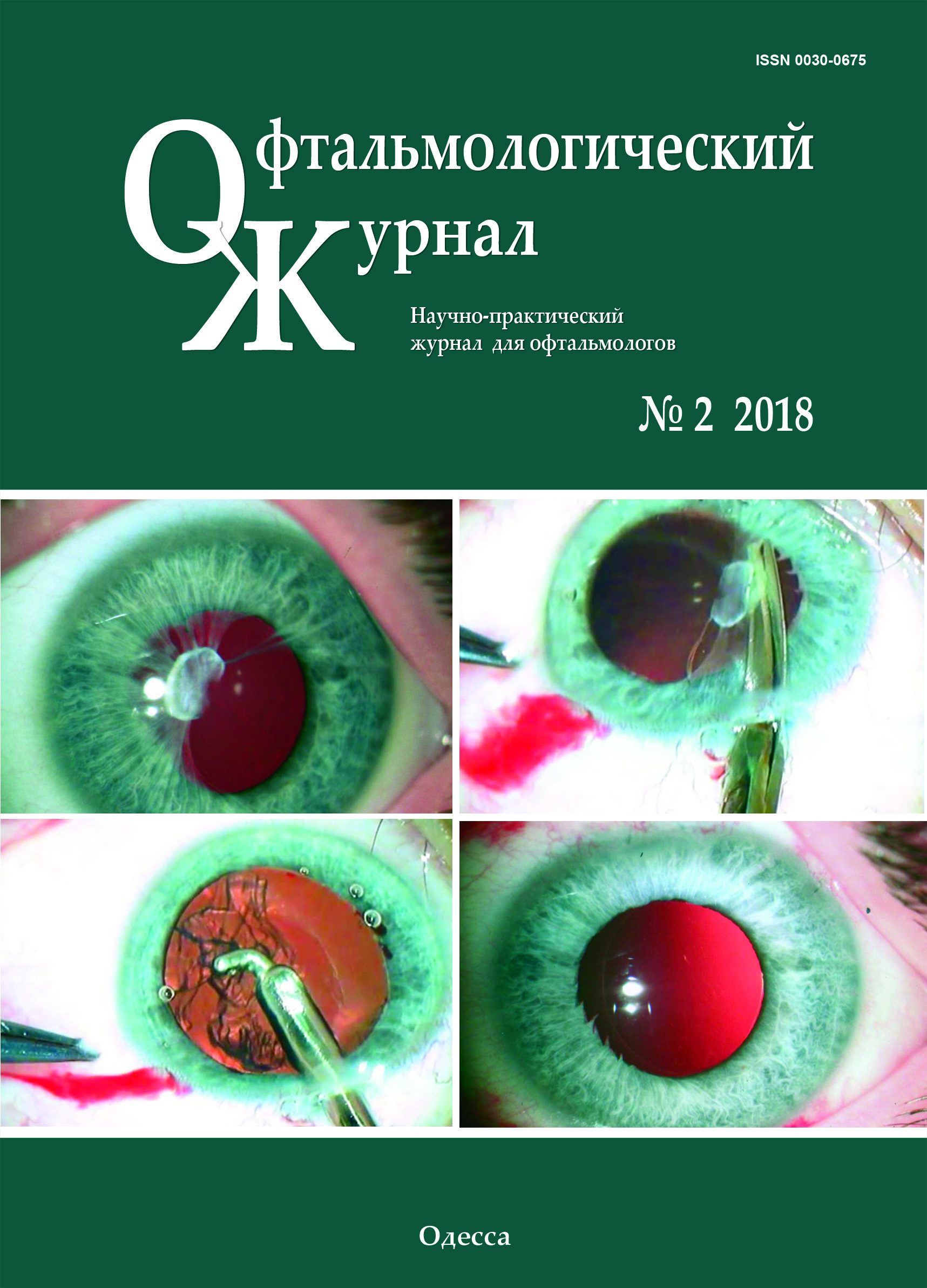
Material and Methods. Experimental studies involved two rabbits (4 eyes). Nd: YAG laser (λ=1.06 µm) was adapted to a 600 µm fiberoptic crystal probe and used for laser photocoagulation. Laser pulse was 0.7- 1.2 J; laser pulse length was 3 ms. After topical anesthesia with proxymetacaine hydrochloride (0.5%), CTS CPC SC was performed in superior and inferior eyeball quadrants, at 2-3 mm from the limbus in two rows in full circle (360°) with the sclera compressed by a 6 g endface for 5 seconds.
Results. The histological studies at Day 1 after CTS CPC SC revealed damage to the pigment epithelium of the ciliary processes and their vessels when laser pulse parameters were low (0.7-1.0 J). High-energy CTS CPC SC of the ciliary body (1.1-1.2 J) damaged the sub-conjunctival tissue and sclera. With laser radiation, collagen stromal fiber separation and channeling were noted as result of a hydrodynamic shock which can supposedly lead to increased transscleral filtration. The mechanism of antihypertensive action of CTS CPC SC using Nd:YAG laser is likely to include two components: photocoagulation and hydrodynamic cavitation.
References
Dulub LV. [Cyclodestructive surgery of glaucoma]. Meditsinskiie novosti. 2002;10:3-8. Russian.
Douglas JRI. [Diode laser transscleral cyclophotocoagulation. Ophthalmology Atlas Glaucoma]. 2010:184. Russian.
Balashevich LI, Parkhomov SD, Izmailov AS. [Comparative assessment of efficacy of diode (0.81µm) and Nd-YAG (0.523µm) lasers in treatment of open-angle glaucoma]. Oftalmol Zh. 2000;1:30-5. Russian.
Hennis HL, Stewart WC. Semiconductor diode laser transscleral cyclophotocoagulation in patients with glaucoma. Am J Ophthalmol. 1992;113:81-5. https://doi.org/10.1016/S0002-9394(14)75758-7
Brancato R, Carassa RG, Bettin Р. Contact transscleral cyclophotocoagulation with diode laser in refractory glaucoma. Eur J Ophthalmol. 1995;5:32-9.https://doi.org/10.1177/112067219500500106
Threlkeld AB, Johnson MH. Contact transscleral diode cyclophotocoagulation for refractory glaucoma. J Glaucoma. 1999;8:3-7. https://doi.org/10.1097/00061198-199902000-00003
Mistlberger A, Liebmann JM, Tschiderer H. Diode laser transscleral cyclophotocoagulation for refractory glaucoma. J Glaucoma. 2001;10:288-93.https://doi.org/10.1097/00061198-200108000-00008
Volkov VV, Kachanov AB. [Diode laser transscleral cyclophotocoagulation in treatment of secondary glaucoma and ophthalmic hypertension]. Oftalmol Zh. 1993;5-6:274-7. Russian.
Downloads
Published
How to Cite
Issue
Section
License
Copyright (c) 2025 П. П. Чечин, В. В. Вит, О. В. Гузун

This work is licensed under a Creative Commons Attribution 4.0 International License.
This work is licensed under a Creative Commons Attribution 4.0 International (CC BY 4.0) that allows users to read, download, copy, distribute, print, search, or link to the full texts of the articles, or use them for any other lawful purpose, without asking prior permission from the publisher or the author as long as they cite the source.
COPYRIGHT NOTICE
Authors who publish in this journal agree to the following terms:
- Authors hold copyright immediately after publication of their works and retain publishing rights without any restrictions.
- The copyright commencement date complies the publication date of the issue, where the article is included in.
DEPOSIT POLICY
- Authors are permitted and encouraged to post their work online (e.g., in institutional repositories or on their website) during the editorial process, as it can lead to productive exchanges, as well as earlier and greater citation of published work.
- Authors are able to enter into separate, additional contractual arrangements for the non-exclusive distribution of the journal's published version of the work with an acknowledgement of its initial publication in this journal.
- Post-print (post-refereeing manuscript version) and publisher's PDF-version self-archiving is allowed.
- Archiving the pre-print (pre-refereeing manuscript version) not allowed.


