SD OCT retinal thickness in the macula area in premature children who received laser photocoagulation of avascular retina for severe ROP
DOI:
https://doi.org/10.31288/oftalmolzh201912932Keywords:
retinopathy of prematurity, retinal thickness, SD OCT, smooth foveal depressionAbstract
Background: Immature ocular optics at birth and severe retinopathy of prematurity (ROP) may affect normal development of the retina and lead to late impairment of visual functions.
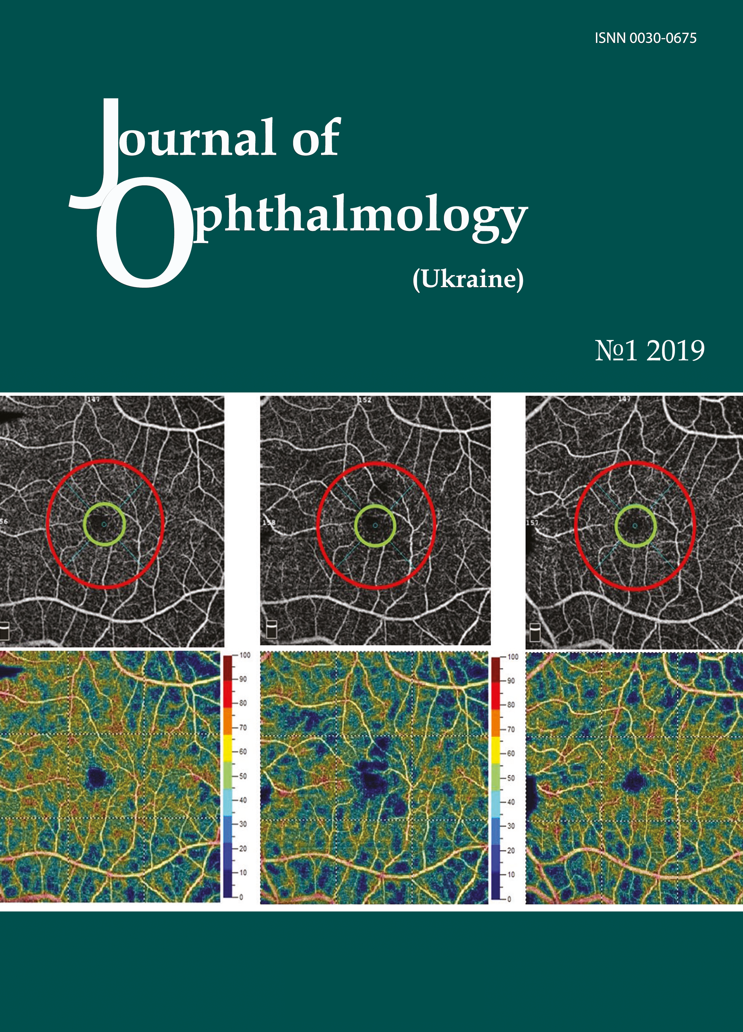
Purpose: To compare spectral-domain optical coherence tomography (SD OCT) retinal thickness in the macular area between premature children who received laser photocoagulation of avascular retina (LPCAR) and full-term children aged 4-6 years.
Materials and Methods: Twenty-five premature children (50 eyes) and 38 full-term control children (69 eyes without eye disease) aged 4-6 years received observation care at the Filatov institute between 2011 and 2017. The inclusion criterion for premature children was receiving LPCAR for severe (type 1 subthreshold, or threshold) ROP diagnosed during screening. Patients with grade 4 or 5 ROP or other ocular pathology (history of cataract, glaucoma, trauma or eye surgery) were excluded.
Results: There was a significant difference in retinal thickness in the macular area between premature and full-term children; the magnitude of the difference depended on the severity of ROP. The foveal-to-central thickness ratio was found to be increased compared to controls by at least 0.1 in 18 premature children (36 eyes, 72%) who received LPCAR (p <0.05).
Conclusion: Mean SD OCT retinal thicknesses in all macular sectors in premature children who received LPCAR for ROP were greater than in full-term children.
References
1.Larsson E, Rydberg A, Holmstrom G. Contrast sensitivity in 10 year old preterm and full term children: a population based study. Br J Ophthalmol. 2006 Jan; 90(1): 87-90. https://doi.org/10.1136/bjo.2005.081653
2.Wang J, Spencer R, Leffler JN, Birch EE. Critical period for foveal fine structure in children with regressed retinopathy of prematurity. Retina. 2012 Feb;32(2):330-9.https://doi.org/10.1097/IAE.0b013e318219e685
3.Eriksson U, Holmström G, Alm A, Larsson E. A population-based study of macular thickness in full-term children assessed with Stratus OCT: normative data and repeatability. Acta Ophthalmol. 2009 Nov;87(7):741-5.https://doi.org/10.1111/j.1755-3768.2008.01357.x
4.Akerblom H, Larsson E, Eriksson U, Holmstrom G. Central macular thickness is correlated with gestational age at birth in prematurely born children. Br J Ophthalmol. 2011 Jun;95(6):799-803.https://doi.org/10.1136/bjo.2010.184747
5.Pasyechnikova NV, Naumenko VA, Zborovska OV. [Foveal to central thickness ratio as an early sign of retinal macular edema]. Oftalmol Zh. 2004;5:4-6. Russian.
6.Ecsedy M, Szamosi A, Karkó C, et al. A comparison of macular structure imaged by optical coherence tomography in preterm and full-term children. Invest Ophthalmol Vis Sci. 2007 Nov;48(11):5207-11.https://doi.org/10.1167/iovs.06-1199
7.Chen YH, Lien R, Chiang MF, et al. Outer Retinal Structural Alternation and Segmentation Errors in Optical Coherence Tomography Imaging in Patients With a History of Retinopathy of Prematurity. Am J Ophthalmol. 2016 Jun;166:169-180.https://doi.org/10.1016/j.ajo.2016.03.030
8.Samarawickrama C, Wang JJ, Huynh SC, et al. Macular thickness, retinal thickness, and optic disk parameters in dominant compared with nondominant eyes. AAPOS. 2009 Apr;13(2):142-7.https://doi.org/10.1016/j.jaapos.2008.11.004
9.El-Dairi MA, Asrani SG, Enyedi LB, Freedman SF. Optical coherence tomography in the eyes of normal children. Arch Ophthalmol. 2009 Jan;127(1):50-8.https://doi.org/10.1001/archophthalmol.2008.553
10.Stoica F, Chirita-Ermandi A, Andreescu N, Stanciu A, Zimbru CG, Puiu M. Clinical relevance of retinal structure in children with laser-treated retinopathy of prematurity versus controls - using optical coherence tomography. Acta Ophthalmol. 2018 Mar;96(2):e222-e228. Epub 2017 Sep 19.https://doi.org/10.1111/aos.13536
11.Yanni SE, Wang J, Chan M, et al. Foveal avascular zone and foveal pit formation after preterm birth. Br J Ophthalmol. 2012 Jul;96(7):961-6.https://doi.org/10.1136/bjophthalmol-2012-301612
12.Yuodelis C, Hendrickson A. A qualitative and quantitative analysis of the human fovea during development. Vision Res. 1986;26(6):847-55.https://doi.org/10.1016/0042-6989(86)90143-4
Downloads
Published
How to Cite
Issue
Section
License
Copyright (c) 2025 Е. С. Заичко, Е. В. Иваницкая, С. В. Кацан

This work is licensed under a Creative Commons Attribution 4.0 International License.
This work is licensed under a Creative Commons Attribution 4.0 International (CC BY 4.0) that allows users to read, download, copy, distribute, print, search, or link to the full texts of the articles, or use them for any other lawful purpose, without asking prior permission from the publisher or the author as long as they cite the source.
COPYRIGHT NOTICE
Authors who publish in this journal agree to the following terms:
- Authors hold copyright immediately after publication of their works and retain publishing rights without any restrictions.
- The copyright commencement date complies the publication date of the issue, where the article is included in.
DEPOSIT POLICY
- Authors are permitted and encouraged to post their work online (e.g., in institutional repositories or on their website) during the editorial process, as it can lead to productive exchanges, as well as earlier and greater citation of published work.
- Authors are able to enter into separate, additional contractual arrangements for the non-exclusive distribution of the journal's published version of the work with an acknowledgement of its initial publication in this journal.
- Post-print (post-refereeing manuscript version) and publisher's PDF-version self-archiving is allowed.
- Archiving the pre-print (pre-refereeing manuscript version) not allowed.


