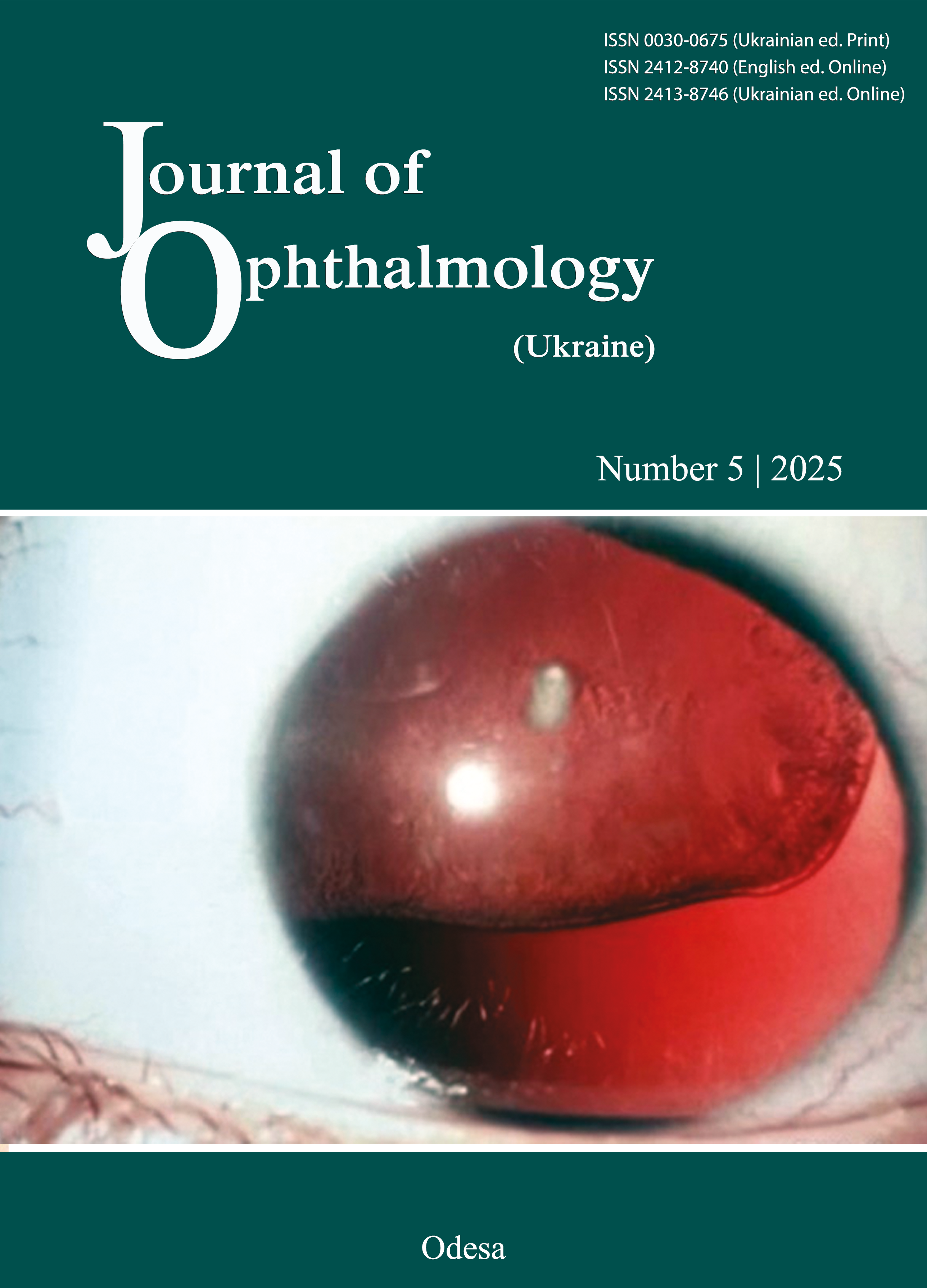
Visual evoked potentials in the diagnosis of optic neuropathies: a literature review
DOI:
https://doi.org/10.31288/oftalmolzh202556570Keywords:
optic neuropathy, visual evoked potentials, electroretinographyAbstract
This review analyzes the utility of visual evoked potentials (VEP) as a method for visual function assessment in optic neuropathies. We systematically reviewed the existing data on the application of several types of VEP (pattern-reversal, pattern-onset/offset, flash-pattern, and chromatic) for objective assessment of the visual pathway in inflammatory, demyelinating, ischemic, toxic, compressive and traumatic optic neuropathies. The review presents the typical changes in VEP parameters (latency, amplitude and morphology) for each of the pathologies considered, and their relationships with clinical manifestations and results of other neuroophthalmological examination techniques such as perimetry and optical coherence tomography. The paper highlights the diagnostic and differential diagnostic value of VEP, especially in challenging cases and for providing an objective approach for characterizing visual function deficiencies.
References
Hayreh SS. Ischemic optic neuropathy. Prog Retin Eye Res. 2009 Jan;28(1):34-62. doi: 10.1016/j.preteyeres.2008.11.002. Epub 2008 Nov 27.https://doi.org/10.1016/j.preteyeres.2008.11.002
Arteritic Anterior Ischemic Optic Neuropathy (AAION) [Інтернет]. American Academy of Ophthalmology. Aviable from: https://eyewiki.org/Arteritic_Anterior_Ischemic_Optic_Neuropathy_(AAION) .
Ischemic Optic Neuropathy: Classification and Management [Updated 2024 Dec 09]. Medscape [Internet]. Available from: https://emedicine.medscape.com/article/.
Kaur K, Margolin E. Nonarteritic Anterior Ischemic Optic Neuropathy. [Updated 2025 Sep 14]. In: StatPearls [Internet]. Treasure Island (FL): StatPearls Publishing; 2025 Jan-. Available from: https://www.ncbi.nlm.nih.gov/books/NBK559045/ .
Singla K, Agarwal P. Optic Ischemia. [Updated 2024 May 6]. In: StatPearls [Internet]. Treasure Island (FL): StatPearls Publishing; 2025 Jan-. Available from: https://www.ncbi.nlm.nih.gov/books/NBK560577/.
Fard MA, Ghahvehchian H, Subramanian PS. Optical coherence tomography in ischemic optic neuropathy. Ann Eye Sci. 2020;5:6.https://doi.org/10.21037/aes.2019.12.05
Khalili MR, Bremner F, Tabrizi R, Bashi A. Optical coherence tomography angiography (OCT angiography) in anterior ischemic optic neuropathy (AION): A systematic review and meta-analysis. Eur J Ophthalmol. 2023 Jan;33(1):530-545.https://doi.org/10.1177/11206721221113681
Rodriguez M, Siva A, Cross SA, O'Brien PC, Kurland LT. Optic neuritis: a population-based study in Olmsted County, Minnesota. Neurology. 1995 Feb;45(2):244-50. https://doi.org/10.1212/WNL.45.2.244
Balcer LJ. Clinical practice. Optic neuritis. N Engl J Med. 2006 Mar 23;354(12):1273-80. doi: 10.1056/NEJMcp053247.https://doi.org/10.1056/NEJMcp053247
Optical Coherence Tomography in Neuro-Ophthalmology [Internet]. American Academy of Ophthalmology . [Updated 2025 Sep 18]. Available from: https://eyewiki.org/Optical_Coherence_Tomography_in_Neuro-Ophthalmology
Liu A, Craver EC, Bhatti MT, Chen JJ. Population-Based Incidence and Outcomes of Compressive Optic Neuropathy. Am J Ophthalmol. 2022 Apr;236:130-135. https://doi.org/10.1016/j.ajo.2021.10.018
Biousse V, Newman NJ. Diagnosis and clinical features of common optic neuropathies. Lancet Neurol. 2016 Dec;15(13):1355-1367.
https://doi.org/10.1016/S1474-4422(16)30237-X
Karimi S, Arabi A, Ansari I, Shahraki T, Safi S. A Systematic Literature Review on Traumatic Optic Neuropathy. J Ophthalmol. 2021 Feb 26;2021:5553885. https://doi.org/10.1155/2021/5553885
Yu-Wai-Man P. Traumatic optic neuropathy-Clinical features and management issues. Taiwan J Ophthalmol. 2015 Mar 1;5(1):3-8. https://doi.org/10.1016/j.tjo.2015.01.003
Compston A. The Berger rhythm: potential changes from the occipital lobes in man, by E.D. Adrian and B.H.C. Matthews (From the Physiological Laboratory, Cambridge). Brain. 2010 Jan;133(1):3-6. https://doi.org/10.1093/brain/awp324
Marmoy OR, Viswanathan S. Clinical electrophysiology of the optic nerve and retinal ganglion cells. Eye (Lond). 2021 Sep;35(9):2386-2405.https://doi.org/10.1038/s41433-021-01614-x
Celesia GG, Kaufman D, Cone S. Effects of age and sex on pattern electroretinograms and visual evoked potentials. Electroencephalogr Clin Neurophysiol. 1987 May;68(3):161-71. https://doi.org/10.1016/0168-5597(87)90023-2
Ekayanti MS, Mahama CN, Ngantung DJ. Normative values of visual evoked potential in adults. Indian J Ophthalmol. 2021 Sep;69(9):2328-2332. https://doi.org/10.4103/ijo.IJO_2480_20
Tekavčič Pompe M, Perovšek D, Šuštar M. Chromatic visual evoked potentials indicate early dysfunction of color processing in young patients with demyelinating disease. Doc Ophthalmol. 2020 Oct;141(2):157-168. https://doi.org/10.1007/s10633-020-09761-4
Meredith JT, Celesia GG. Pattern-reversal visual evoked potentials and retinal eccentricity. Electroencephalogr Clin Neurophysiol. 1982 Mar;53(3):243-53. https://doi.org/10.1016/0013-4694(82)90082-7
Yiannikas C, Walsh JC. The variation of the pattern shift visual evoked response with the size of the stimulus field. Electroencephalogr Clin Neurophysiol. 1983 Apr;55(4):427-35. https://doi.org/10.1016/0013-4694(83)90131-1
Thompson DA, Fritsch DM, Hardy SE; POW Study Group. The changing shape of the ISCEV standard pattern onset VEP. Doc Ophthalmol. 2017 Aug;135(1):69-76. https://doi.org/10.1007/s10633-017-9596-8
Andersson L, Sjölund J, Nilsson J. Flash visual evoked potentials are unreliable as markers of ICP due to high variability in normal subjects. Acta Neurochir (Wien). 2012 Jan;154(1):121-7. https://doi.org/10.1007/s00701-011-1152-9
Mellow TB, Liasis A, Lyons R, Thompson DA. The reproducibility of binocular pattern reversal visual evoked potentials: a single subject design. Doc Ophthalmol. 2011 Jun;122(3):133-9. https://doi.org/10.1007/s10633-011-9267-0
Sarnthein J, Andersson M, Zimmermann MB, Zumsteg D. High test-retest reliability of checkerboard reversal visual evoked potentials (VEP) over 8 months. Clin Neurophysiol. 2009 Oct;120(10):1835-40. https://doi.org/10.1016/j.clinph.2009.08.014
Tandon OP, Sharma KN. Visual evoked potential in young adults: a normative study. Indian J Physiol Pharmacol. 1989 Oct-Dec;33(4):247-9.
Crognale MA, Page JW, Fuhrel A. Aging of the chromatic onset visual evoked potential. Optom Vis Sci. 2001 Jun;78(6):442-6. https://doi.org/10.1097/00006324-200106000-00018
Porciatti V, Sartucci F. Retinal and cortical evoked responses to chromatic contrast stimuli. Specific losses in both eyes of patients with multiple sclerosis and unilateral optic neuritis. Brain. 1996 Jun;119 ( Pt 3):723-40. https://doi.org/10.1093/brain/119.3.723
Sartucci F, Murri L, Orsini C, Porciatti V. Equiluminant red-green and blue-yellow VEPs in multiple sclerosis. J Clin Neurophysiol. 2001 Nov;18(6):583-91. https://doi.org/10.1097/00004691-200111000-00010
Majander A, Robson AG, João C, Holder GE, Chinnery PF, Moore AT, Votruba M, Stockman A, Yu-Wai-Man P. The pattern of retinal ganglion cell dysfunction in Leber hereditary optic neuropathy. Mitochondrion. 2017 Sep;36:138-149. https://doi.org/10.1016/j.mito.2017.07.006
Fuest M, Kieckhoefel J, Mazinani B, Kuerten D, Koutsonas A, Koch E, Walter P, Plange N. Blue-yellow and standard pattern visual evoked potentials in phakic and pseudophakic glaucoma patients and controls. Graefes Arch Clin Exp Ophthalmol. 2015 Dec;253(12):2255-61.
https://doi.org/10.1007/s00417-015-3152-6
Sartucci F, Porciatti V. Visual-evoked potentials to onset of chromatic red-green and blue-yellow gratings in Parkinson's disease never treated with L-dopa. J Clin Neurophysiol. 2006 Oct;23(5):431-5. https://doi.org/10.1097/01.wnp.0000216127.53517.4d
Carroll WM, Halliday AM, Kriss A. Improvements in the accuracy of pattern visual evoked potentials in the diagnosis of visual pathway disease. Neuro-Ophthalmology. 1982; 2(4): 237-253. https://doi.org/10.3109/01658108209009705
Odom JV, Bach M, Brigell M, Holder GE, McCulloch DL, Mizota A, Tormene AP; International Society for Clinical Electrophysiology of Vision. ISCEV standard for clinical visual evoked potentials: (2016 update). Doc Ophthalmol. 2016 Aug;133(1):1-9. https://doi.org/10.1007/s10633-016-9553-y
MacDonald DB, Dong CC, Uribe A. Intraoperative evoked potential techniques. Handb Clin Neurol. 2022;186:39-65.https://doi.org/10.1016/B978-0-12-819826-1.00012-0
Marcar VL, Battegay E, Schmidt D, Cheetham M. Parallel processing in human visual cortex revealed through the influence of their neural responses on the visual evoked potential. Vision Res. 2022 Apr;193:107994. https://doi.org/10.1016/j.visres.2021.107994
McCulloch DL, Marmor MF, Brigell MG, Hamilton R, Holder GE, Tzekov R, Bach M. ISCEV Standard for full-field clinical electroretinography (2015 update). Doc Ophthalmol. 2015 Feb;130(1):1-12. https://doi.org/10.1007/s10633-014-9473-7
Robson AG, Nilsson J, Li S, Jalali S, Fulton AB, Tormene AP, Holder GE, Brodie SE. ISCEV guide to visual electrodiagnostic procedures. Doc Ophthalmol. 2018 Feb;136(1):1-26. https://doi.org/10.1007/s10633-017-9621-y
de Seze J, Bigaut K. Multiple sclerosis diagnostic criteria: From poser to the 2017 revised McDonald criteria. Presse Med. 2021 Jun;50(2):104089. https://doi.org/10.1016/j.lpm.2021.104089
Oh J, Vidal-Jordana A, Montalban X. Multiple sclerosis: clinical aspects. Curr Opin Neurol. 2018 Dec;31(6):752-759. https://doi.org/10.1097/WCO.0000000000000622
Petzold A, Wattjes MP, Costello F, Flores-Rivera J, Fraser CL, Fujihara K, Leavitt J, Marignier R, Paul F, Schippling S, Sindic C, Villoslada P, Weinshenker B, Plant GT. The investigation of acute optic neuritis: a review and proposed protocol. Nat Rev Neurol. 2014 Aug;10(8):447-58. https://doi.org/10.1038/nrneurol.2014.108
Halliday AM, McDonald WI, Mushin J. Visual evoked response in diagnosis of multiple sclerosis. Br Med J. 1973 Dec 15;4(5893):661-4.
https://doi.org/10.1136/bmj.4.5893.661
Backner Y, Petrou P, Glick-Shames H, Raz N, Zimmermann H, Jost R, Scheel M, Paul F, Karussis D, Levin N. Vision and Vision-Related Measures in Progressive Multiple Sclerosis. Front Neurol. 2019 May 3;10:455. https://doi.org/10.3389/fneur.2019.00455
Leocani L, Guerrieri S, Comi G. Visual Evoked Potentials as a Biomarker in Multiple Sclerosis and Associated Optic Neuritis. J Neuroophthalmol. 2018 Sep;38(3):350-357. https://doi.org/10.1097/WNO.0000000000000704
Park SH, Park CY, Shin YJ, Jeong KS, Kim NH. Low Contrast Visual Evoked Potentials for Early Detection of Optic Neuritis. Front Neurol. 2022 Apr 29;13:804395. https://doi.org/10.3389/fneur.2022.804395
Jayaraman M, Gandhi RA, Ravi P, Sen P. Multifocal visual evoked potential in optic neuritis, ischemic optic neuropathy and compressive optic neuropathy. Indian J Ophthalmol. 2014 Mar;62(3):299-304. https://doi.org/10.4103/0301-4738.118452
Grzybowski A, Zülsdorff M, Wilhelm H, Tonagel F. Toxic optic neuropathies: an updated review. Acta Ophthalmol. 2015 Aug;93(5):402-410. https://doi.org/10.1111/aos.12515
Sen S, Mandal S, Banerjee M, Gk R, Saxena A, Aalok SP, Saxena R. Ethambutol-induced optic neuropathy: Functional and structural changes in the retina and optic nerve. Semin Ophthalmol. 2022 Aug;37(6):730-739. https://doi.org/10.1080/08820538.2022.2085517
Kooi KA, Yamada T, Marshall RE. Field studies of monocularly evoked cerebral potentials in bitemporal hemianopsia. Neurology. 1973 Nov;23(11):1217-25. https://doi.org/10.1212/WNL.23.11.1217
Vaughan HG Jr, Katzman R. Evoked response in visual disorders. Ann N Y Acad Sci. 1964 May 8;112:305-19. https://doi.org/10.1111/j.1749-6632.1964.tb26759.x
Brecelj J. A VEP study of the visual pathway function in compressive lesions of the optic chiasm. Full-field versus half-field stimulation. Electroencephalogr Clin Neurophysiol. 1992 May-Jun;84(3):209-18. https://doi.org/10.1016/0168-5597(92)90002-S
Dotto PF, Berezovsky A, Sacai PY, Rocha DM, Fernandes AG, Salomão SR. Visual function assessed by visually evoked potentials in adults with orbital and other primary intracranial tumors. Eur J Ophthalmol. 2021 May;31(3):1351-1360. https://doi.org/10.1177/1120672120925643
Bowman R, Walters B, Smith V, Prise KL, Handley SE, Green K, Mankad K, O'Hare P, Dahl C, Jorgensen M, Opocher E, Hargrave D, Thompson DA. Visual outcomes and predictors in optic pathway glioma: a single centre study. Eye (Lond). 2023 Apr;37(6):1178-1183. https://doi.org/10.1038/s41433-022-02096-1
Allen D, Riordan-Eva P, Paterson RW, Hadden RD. Subacute peripheral and optic neuropathy syndrome with no evidence of a toxic or nutritional cause. Clin Neurol Neurosurg. 2013 Aug;115(8):1389-93. https://doi.org/10.1016/j.clineuro.2013.01.002
Wilson WB. Visual-evoked response differentiation of ischemic optic neuritis from the optic neuritis of multiple sclerosis. Am J Ophthalmol. 1978 Oct;86(4):530-5. https://doi.org/10.1016/0002-9394(78)90302-1
Barbano L, Ziccardi L, Parisi V. Correlations between visual morphological, electrophysiological, and acuity changes in chronic non-arteritic ischemic optic neuropathy. Graefes Arch Clin Exp Ophthalmol. 2021 May;259(5):1297-1308.https://doi.org/10.1007/s00417-020-05023-w
Janáky M, Fülöp Z, Pálffy A, Benedek K, Benedek G. Electrophysiological findings in patients with nonarteritic anterior ischemic optic neuropathy. Clin Neurophysiol. 2006 May;117(5):1158-66. https://doi.org/10.1016/j.clinph.2006.01.013
Rangaswamy NV, Frishman LJ, Dorotheo EU, Schiffman JS, Bahrani HM, Tang RA. Photopic ERGs in patients with optic neuropathies: comparison with primate ERGs after pharmacologic blockade of inner retina. Invest Ophthalmol Vis Sci. 2004 Oct;45(10):3827-37.https://doi.org/10.1167/iovs.04-0458
Baiano C, Zeppieri M. Visual Evoked Potential. [Updated 2023 May 11]. In: StatPearls [Internet]. Treasure Island (FL): StatPearls Publishing; 2025 Jan-. Available from: https://www.ncbi.nlm.nih.gov/books/NBK582128/ .
Downloads
Published
How to Cite
Issue
Section
License
Copyright (c) 2025 Kyslitska M. S., Vasyuta V. A., Solonovych O. S., Chebotariova L. L., Mytsak O. I., Severenchuk Ie. I.

This work is licensed under a Creative Commons Attribution 4.0 International License.
This work is licensed under a Creative Commons Attribution 4.0 International (CC BY 4.0) that allows users to read, download, copy, distribute, print, search, or link to the full texts of the articles, or use them for any other lawful purpose, without asking prior permission from the publisher or the author as long as they cite the source.
COPYRIGHT NOTICE
Authors who publish in this journal agree to the following terms:
- Authors hold copyright immediately after publication of their works and retain publishing rights without any restrictions.
- The copyright commencement date complies the publication date of the issue, where the article is included in.
DEPOSIT POLICY
- Authors are permitted and encouraged to post their work online (e.g., in institutional repositories or on their website) during the editorial process, as it can lead to productive exchanges, as well as earlier and greater citation of published work.
- Authors are able to enter into separate, additional contractual arrangements for the non-exclusive distribution of the journal's published version of the work with an acknowledgement of its initial publication in this journal.
- Post-print (post-refereeing manuscript version) and publisher's PDF-version self-archiving is allowed.
- Archiving the pre-print (pre-refereeing manuscript version) not allowed.


