Longitudinal structural changes in the optic nerve in ischemic optic neuropathy: a case report
DOI:
https://doi.org/10.31288/oftalmolzh202535358Keywords:
non-arteritic ischemic optic neuropathy , 3-dioxygenase, optic disc, hypoperfusion, optic nerveAbstract
Purpose: To describe the case of longitudinal structural changes in the optic disc in ischemic optic neuropathy.
Methods: A 63-year-old male patient (two eyes) was under observation for three months, from August to October 2024. Informed consent to use medical records for research purposes was obtained from the patient. Ethical approval was obtained from the local ethics committee (committee meeting minutes of March 24, 2022). Visual acuity and ophthalmoscopy OU were done and optic coherence tomography (OCT) OS was performed in August and October, 2024, and OCT OD was performed in October, 2024. Automated perimetry and OCT angiography (OCTA) OU were performed in October, 2024.
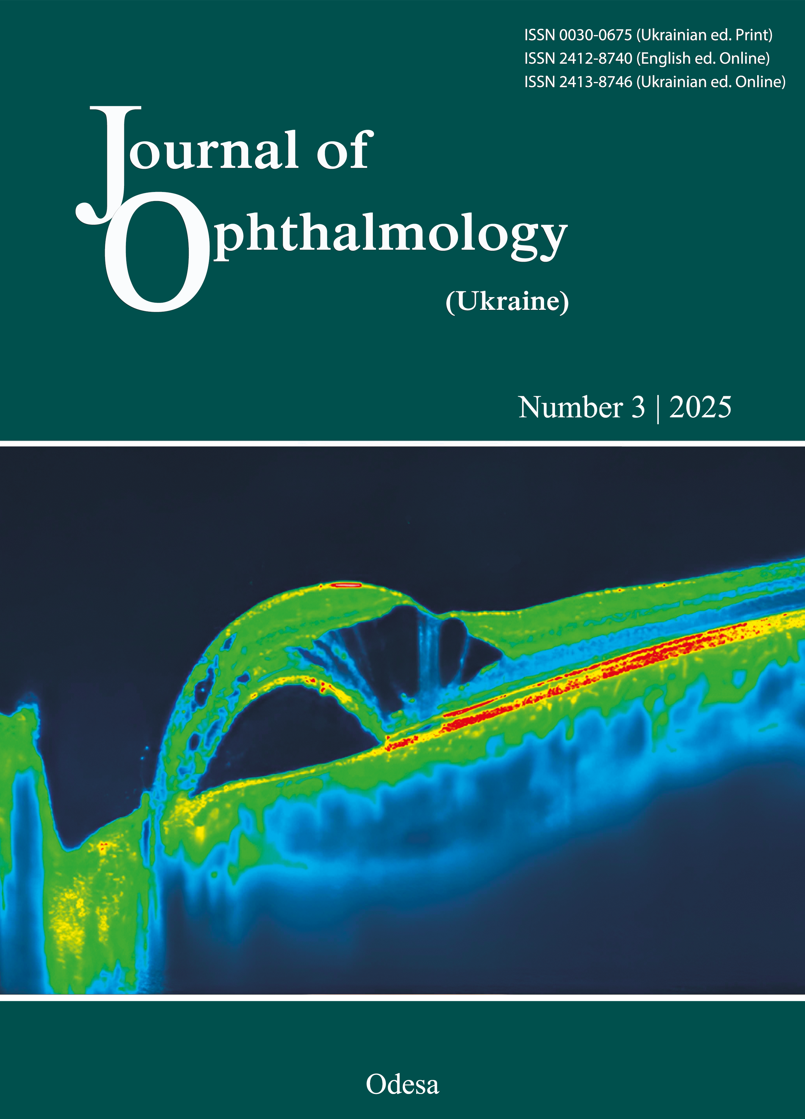
Results: The 63-year-old male patient was examined and diagnosed with “optic atrophy OS and non-arteritic ischemic optic neuropathy (NAION) OU”. The OCT study of structural components of the optic disc OS showed a 54.9% reduction in the average thickness of retinal nerve fiber layer (RNFL) of the optic disc and a 4.4% reduction in the thickness of the inner retinal complex (GCL++) in October compared to August 2024. In August 2024, RNFL thickness and GCL++ thickness in the superior and inferior segments were thicker in the left eye than in the right eye. In October 2024, the thickness of both layers in the inferior segment became thicker than the thickness of the superior segment for the left eye, likely reflecting the expansion of swelling to the inferior segment. In October 2024, it was in the inferior segment of the peripapillary region that OCTA showed a reduction in vessel density along with dilated and tortuous capillaries.
Conclusion: Studies of longitudinal structural changes in the optic disc reflect the role of localized swelling as a consequence of impaired blood perfusion in NAION, and confirm an increase in the affected area, likely due to secondary factors.
References
Biousse V, Newman NJ. Ischemic Optic Neuropathies. N Engl J Med. 2015 Oct 22;373(17):1677. https://doi.org/10.1056/NEJMc1509058
Hayreh SS. Ischemic optic neuropathies - where are we now? Graefes Arch Clin Exp Ophthalmol. 2013 Aug;251(8):1873-84. https://doi.org/10.1007/s00417-013-2399-z
Rizzo JF 3rd. Unraveling the Enigma of Nonarteritic Anterior Ischemic Optic Neuropathy. J Neuroophthalmol. 2019 Dec;39(4):529-544. https://doi.org/10.1097/WNO.0000000000000870
Suh MH, Kim SH, Park KH, Kim SJ, Kim TW, Hwang SS, Kim DM. Comparison of the correlations between optic disc rim area and retinal nerve fiber layer thickness in glaucoma and nonarteritic anterior ischemic optic neuropathy. Am J Ophthalmol. 2011 Feb;151(2):277-86.e1. https://doi.org/10.1016/j.ajo.2010.08.033
Arnold AC. Pathogenesis of nonarteritic anterior ischemic optic neuropathy. J Neuroophthalmol. 2003 Jun;23(2):157-63. https://doi.org/10.1097/00041327-200306000-00012
Raizada K, Margolin E. Non-Arteritic Anterior Ischemic Optic Neuropathy. 2022 Oct 31. In: StatPearls [Internet]. Treasure Island (FL): StatPearls Publishing; 2025 Jan-. PMID: 32644471
Tournaire-Marques E. Neuropathies optiques ischémiques [Ischemic optic neuropathies]. J Fr Ophtalmol. 2020 Jun;43(6):552-558. French. https://doi.org/10.1016/j.jfo.2019.10.020
Levin LA, Louhab A. Apoptosis of retinal ganglion cells in anterior ischemic optic neuropathy. Arch Ophthalmol. 1996 Apr;114(4):488-91. https://doi.org/10.1001/archopht.1996.01100130484027
Hashimoto H, Hata M, Kashii S, Oishi A, Suda K, Nakano E, Miyata M, Tsujikawa A. Analysis of Retinal Nerve Fibre Thickening in Progressive and Non-progressive Non-arteritic Anterior Ischaemic Optic Neuropathy Using Optical Coherence Tomography. Neuroophthalmology. 2020 Jun 25;44(5):307-314. https://doi.org/10.1080/01658107.2020.1755991
Al-Nashar HY, Hemeda S. Assessment of peripapillary vessel density in acute non-arteritic anterior ischemic optic neuropathy. Int Ophthalmol. 2020 May;40(5):1269-1276. https://doi.org/10.1007/s10792-020-01293-9
Gandhi U, Chhablani J, Badakere A, Kekunnaya R, Rasheed MA, Goud A, Chhablani PP. Optical coherence tomography angiography in acute unilateral nonarteritic anterior ischemic optic neuropathy: A comparison with the fellow eye and with eyes with papilledema. Indian J Ophthalmol. 2018 Aug;66(8):1144-1148. https://doi.org/10.4103/ijo.IJO_179_18
Downloads
Published
How to Cite
Issue
Section
License
Copyright (c) 2025 Moyseyenko N.M.

This work is licensed under a Creative Commons Attribution 4.0 International License.
This work is licensed under a Creative Commons Attribution 4.0 International (CC BY 4.0) that allows users to read, download, copy, distribute, print, search, or link to the full texts of the articles, or use them for any other lawful purpose, without asking prior permission from the publisher or the author as long as they cite the source.
COPYRIGHT NOTICE
Authors who publish in this journal agree to the following terms:
- Authors hold copyright immediately after publication of their works and retain publishing rights without any restrictions.
- The copyright commencement date complies the publication date of the issue, where the article is included in.
DEPOSIT POLICY
- Authors are permitted and encouraged to post their work online (e.g., in institutional repositories or on their website) during the editorial process, as it can lead to productive exchanges, as well as earlier and greater citation of published work.
- Authors are able to enter into separate, additional contractual arrangements for the non-exclusive distribution of the journal's published version of the work with an acknowledgement of its initial publication in this journal.
- Post-print (post-refereeing manuscript version) and publisher's PDF-version self-archiving is allowed.
- Archiving the pre-print (pre-refereeing manuscript version) not allowed.


