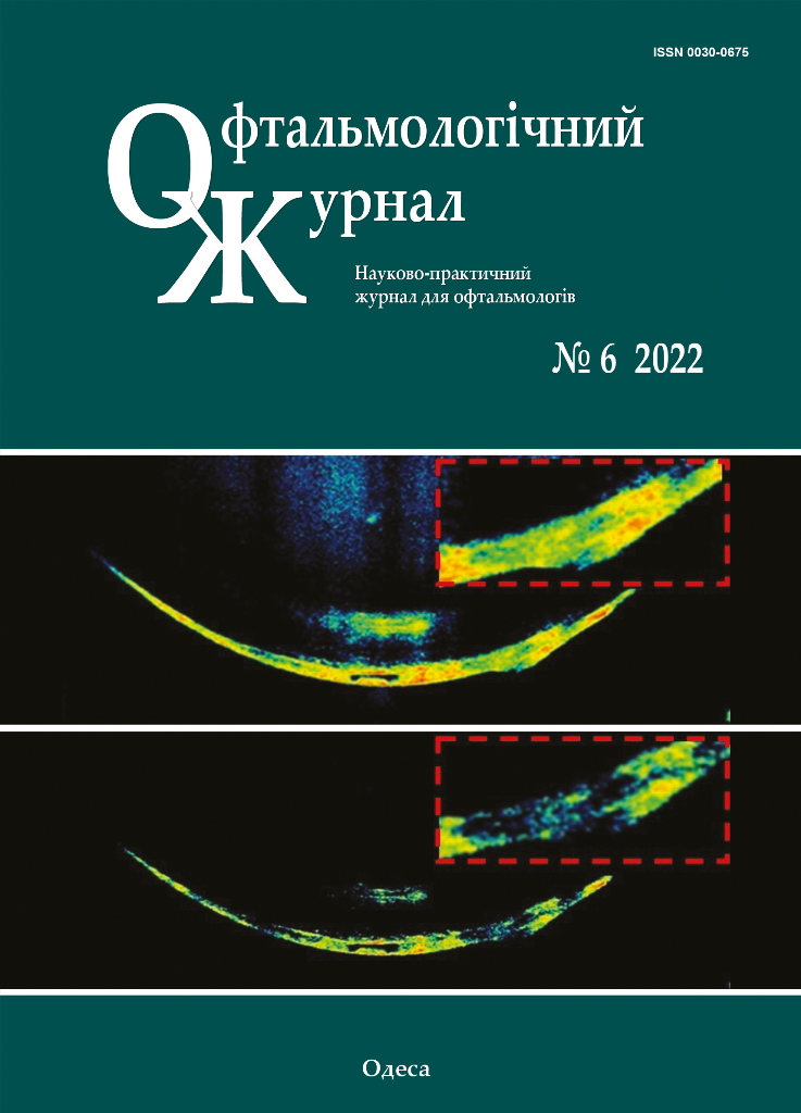
Неврит зорового нерва чи запальна оптична нейропатія? (огляд літератури)
DOI:
https://doi.org/10.31288/oftalmolzh202264449Ключові слова:
неврит зорового нерва, запальна оптична нейропатія, ОКТ, МРТ, демілієнізуючі захворюванняАнотація
Актуальність. За даними літератури зустрічаються два рівнозначних терміни «неврит зорового нерва» і «запальна оптична нейропатія». Класифікація, патогенез і клінічні прояви неоднозначні. Тим не менше, посилений інтерес до проблем демієлінізуючих та інфекційних захворювань сприяє деталізації даних про механізми окремих форм запалення зорового нерва.
Мета. Дослідити сучасні погляди та перспективи їх розвитку щодо патогенезу запалення зорового нерва.
Методи. Літературний пошук джерел української та зарубіжної науки.
Результати. Сучасні методики діагностики, такі як оптична когерентна томографія (ОКТ), магнітно-резонансна томографія (МРТ), імунологічні та вірусологічні тести дозволяють вивчити роль окремих факторів, а також їх комбінації у розвитку невриту, динаміку зорових функцій, пошкодження окремих структурних елементів зорової системи та відновність після отриманих ушкоджень.
Висновок. Аналіз літературних даних щодо невриту зорового нерва та/чи запальної оптичної нейропатії показав високий актуальний інтерес наукової громадськості. Подальші поглиблені дослідження, імовірно дозволять удосконалити чинні класифікації запалень зорового нерва та розробити диференційований підхід щодо лікування запалень зорового нерва.
Посилання
1.Günay Ç, Yardım E, Yaşar E, Hız-Kurul AS, Uzan GS, Öztürk T et al. Optic neuritis in CD59 deficiency: an extremely rare presentation. Turk J Pediatr. 2022; 64(4): 787-94. https://doi.org/10.24953/turkjped.2021.1405
2.Eslami M, Lichtman-Mikol S, Razmjou, S, Bernitsas E. Optical Coherence Tomography in Chronic Relapsing Inflammatory Optic Neuropathy, Neuromyelitis Optica and Multiple Sclerosis: A Comparative Study. Brain Sci. 2022; 12: 1140. https://doi.org/10.3390/brainsci12091140
3.Protocol of medical care for patients with optic neuritis (papillitis, retrobulbar neuritis). Annex to the Order of the Ministry of Health №117 dated 15-03-2007.
4.Critical review: Typical and atypical optic neuritis. Anne Abel, Collin McClelland, Michael S. Lee. J. Survophthal. 2019. 64 (6). P. 1-10. https://doi.org/10.1016/j.survophthal.2019.06.001
5.Liang, J., et al. Comparing evolvement of visual field defect in neuromyelitis optica spectrum disorder-optic neuritis and idiopathic optic neuritis: a prospective study. BMC Ophthalmol 22, 338 (2022). https://doi.org/10.1186/s12886-022-02510-y
6.Kitchens N., Nichols L., Hope T. Educational Case: Neuromyelitis optica. Acad Pathol. 2022 Aug 16;9(1):100041. https://doi.org/10.1016/j.acpath.2022.100041
7.Kahloun R, Abroug N, Ksiaa I, Mahmoud A, Zeghidi H. et al. Infectious optic neuropathies: a clinical update. Eye Brain. 2015 Sep 28; 7: 59-81. https://doi.org/10.2147/EB.S69173
8.Trebst C., Jarius S., Berthele A, et al. Update on the diagnosisand treatment of neuromyelitis optica: Recommendations of the Neuromyelitis Optica Study Group (NEMOS). J Neurol. 2014; 261(1): 1e16. https://doi.org/10.1007/s00415-013-7169-7
9.Vanda A. Lennon. at al. A serum autoantibody marker of neuromyelitis optica: distinction from multiple sclerosis. The Lancet. 2004; 364 (9451): 2106-2112. https://doi.org/10.1016/S0140-6736(04)17551-X
10.Roed H, Frederiksen J, Langkilde A, Sørensen TL, Lauritzen M, Sellebjerg F. Systemic T-cell activation in acute clinically isolated optic neuritis. J. Neuroimmunol. 2005; 162(1-2): 165. https://doi.org/10.1016/j.jneuroim.2005.02.002
11.Söderström M, Link H, Xu Z, Fredriksson S. Optic neuritis and multiple sclerosis: anti-MBP and anti-MBP peptide antibody-secreting cells are accumulated in CSF. Neurology. 1993; 43(6): 1215. https://doi.org/10.1212/WNL.43.6.1215
12.Crotty S. Follicular helper CD4 T cells (TFH). Annu Rev. Immunol. 2011; 29: 621-663. https://doi.org/10.1146/annurev-immunol-031210-101400
13.Shetty A. et al. Immunodominant T-cell epitopes of MOG reside in its transmembrane and cytoplasmic domains in EAE. Neurol. Neuroimmunol. Neuroinflamm. 2014; 1: e22. https://doi.org/10.1212/NXI.0000000000000022
14.Francis PJ, Jackson H, Stanford MR, Graham EM. Inflammatory optic neuropathy as the presenting feature of herpes simplex acute retinal necrosis. Br J Ophthalmol. 2003 Apr; 87(4): 512-4. https://doi.org/10.1136/bjo.87.4.512-a
15.Melancia D, Fernandes A, Manita M, Cordeiro IM. Cytomegalovirus optic neuropathy in a young immunocompetent patient. J. Neurovirol. 2021 Apr; 27(2): 364-366. https://doi.org/10.1007/s13365-021-00963-3
16.Sarkar P, Mehtani A, Gandhi HC, Dubey V, Tembhurde PM, Gupta MK. Atypical optic neuritis: An overview. Indian J. Ophthalmol. 2021; 69(1): 27-35. https://doi.org/10.4103/ijo.IJO_451_20
17.Rolak LA, Beck RW, Paty DW, Tourtellotte WW, Whitaker JN, Rudick RA. Cerebrospinal fluid in acute optic neuritis: experience of the optic neuritis treatment trial. Neurology. 1996 Feb; 46(2): 368-72. https://doi.org/10.1212/WNL.46.2.368
18.Skov AG, Skov T, Frederiksen JL. Oligoclonal bands predict multiple sclerosis after optic neuritis: a literature survey. Mult Scler. 2011 Apr; 17(4): 404-10. https://doi.org/10.1177/1352458510391340
19.Olesen MN, Soelberg K, Debrabant B, Nilsson AC, Lillevang ST, Grauslund J et al. Cerebrospinal fluid biomarkers for predicting development of multiple sclerosis in acute optic neuritis: a population-based prospective cohort study. J Neuroinflammation. 2019 Mar 11; 16(1): 59. https://doi.org/10.1186/s12974-019-1440-5
20.Rodríguez-Rodríguez MS, Romero-Castro RM, Alvarado-de la Barrera C, González-Cannata MG, García-Morales AK, Ávila-Ríos S. Optic neuritis following SARS-CoV-2 infection. J. Neurovirol. 2021; 27(2): 359-363. https://doi.org/10.1007/s13365-021-00959-z
21.Kahloun R, Abroug N, Ksiaa I, Mahmoud A, Zeghidi H, Zaouali S. et al. Infectious optic neuropathies: a clinical update. Eye Brain. 2015; 7: 59-81. https://doi.org/10.2147/EB.S69173
22.Varrin-Doyer M. et al. MOG transmembrane and cytoplasmic domains contain highly stimulatory T-cell epitopes in MS. Neurol Neuroimmunol Neuroinflamm. 2014; 1: e20. https://doi.org/10.1212/NXI.0000000000000020
23.Zamvil SS, Slavin AJ. Does MOG Ig-positive AQP4-seronegative opticospinal inflammatory disease justify a diagnosis of NMO spectrum disorder? Neurol Neuroimmunol Neuroinflamm. Feb 2015; 2(1): e62. https://doi.org/10.1212/NXI.0000000000000062
24.D'Haeseleer M, Cambron M, Vanopdenbosch L, De Keyser J. Vascular aspects of multiple sclerosis. The Lancet Neurology. 2011; 10(7): 657-666. https://doi.org/10.1016/S1474-4422(11)70105-3
25.Saindane AM, Law M, Ge Y, Johnson G, Babb JS, Grossman RI. Correlation of diffusion tensor and dynamic perfusion MR imaging metrics in normal-appearing corpus callosum: support for primary hypoperfusion in multiple sclerosis. AJNR Am Journal of Neuroradiology. 2007; 28(4): 767-772.
26.Cambron M, D'Haeseleer M, Laureys G, Clinckers R, Debruyne J, De Keyser J. White-matter astrocytes, axonal energy metabolism, and axonal degeneration in multiple sclerosis. Journal of cerebral blood flow and metabolism. 2012; 32(3): 413-424. https://doi.org/10.1038/jcbfm.2011.193
27.Pache M. et al. Extraocular blood flow and endothelin-1 plasma levels in patients with multiple sclerosis. European neurology. 2003; 49(3): 164-168. https://doi.org/10.1159/000069085
28.Chen T-C. et al. Vascular hypoperfusion in acute optic neuritis is a potentially new neurovascular model for demyelinating diseases. PLoS ONE. 2017; 12(9): e0184927. https://doi.org/10.1371/journal.pone.0184927
29.Nguyen AQ, Cherry BH, Scott GF, Ryou MG, Mallet RT. Erythropoietin: powerful protection of ischemic and post-ischemic brain. Experimental biology and medicine (Maywood, NJ). 2014; 239(11):1461-1475. https://doi.org/10.1177/1535370214523703
30.Eslami M, Lichtman-Mikol S, Razmjou S, Bernitsas E. Optical Coherence Tomography in Chronic Relapsing Inflammatory Optic Neuropathy, Neuromyelitis Optica and Multiple Sclerosis: A Comparative Study. Brain Sci. 2022; 12: 1140. https://doi.org/10.3390/brainsci12091140
31.Chen JJ, Sotirchos ES, Henderson AD, Vasileiou ES, Flanagan EP, Bhatti MT. et al. OCT retinal nerve fiber layer thickness differentiates acute optic neuritis from MOG antibody-associated disease and Multiple Sclerosis: RNFL thickening in acute optic neuritis from MOGAD vs. MS. Mult. Scler. Relat. Disord. 2022; 58: 103525. https://doi.org/10.1016/j.msard.2022.103525
32.Kaushik M, Wang CY, Barnett MH, Garrick R, Parratt J, Graham SL. et al. Inner nuclear layer thickening is inversely proportional to retinal ganglion cell loss in optic neuritis. PLoS ONE. 2013; 8: e78341. https://doi.org/10.1371/journal.pone.0078341
33.Al-Louzi OA et al. Outer retinal changes following acute optic neuritis. Mult. Scler. 2016; 22: 362-372. https://doi.org/10.1177/1352458515590646
##submission.downloads##
Опубліковано
Як цитувати
Номер
Розділ
Ліцензія
Авторське право (c) 2025 Н. М. Мойсеєнко

Ця робота ліцензується відповідно до Creative Commons Attribution 4.0 International License.
Ця робота ліцензується відповідно до ліцензії Creative Commons Attribution 4.0 International (CC BY). Ця ліцензія дозволяє повторно використовувати, поширювати, переробляти, адаптувати та будувати на основі матеріалу на будь-якому носії або в будь-якому форматі за умови обов'язкового посилання на авторів робіт і первинну публікацію у цьому журналі. Ліцензія дозволяє комерційне використання.
ПОЛОЖЕННЯ ПРО АВТОРСЬКІ ПРАВА
Автори, які подають матеріали до цього журналу, погоджуються з наступними положеннями:
- Автори отримують право на авторство своєї роботи одразу після її публікації та назавжди зберігають це право за собою без жодних обмежень.
- Дата початку дії авторського права на статтю відповідає даті публікації випуску, до якого вона включена.
ПОЛІТИКА ДЕПОНУВАННЯ
- Редакція журналу заохочує розміщення авторами рукопису статті в мережі Інтернет (наприклад, у сховищах установ або на особистих веб-сайтах), оскільки це сприяє виникненню продуктивної наукової дискусії та позитивно позначається на оперативності і динаміці цитування.
- Автори мають право укладати самостійні додаткові угоди щодо неексклюзивного розповсюдження статті у тому вигляді, в якому вона була опублікована цим журналом за умови збереження посилання на первинну публікацію у цьому журналі.
- Дозволяється самоархівування постпринтів (версій рукописів, схвалених до друку в процесі рецензування) під час їх редакційного опрацювання або опублікованих видавцем PDF-версій.
- Самоархівування препринтів (версій рукописів до рецензування) не дозволяється.


