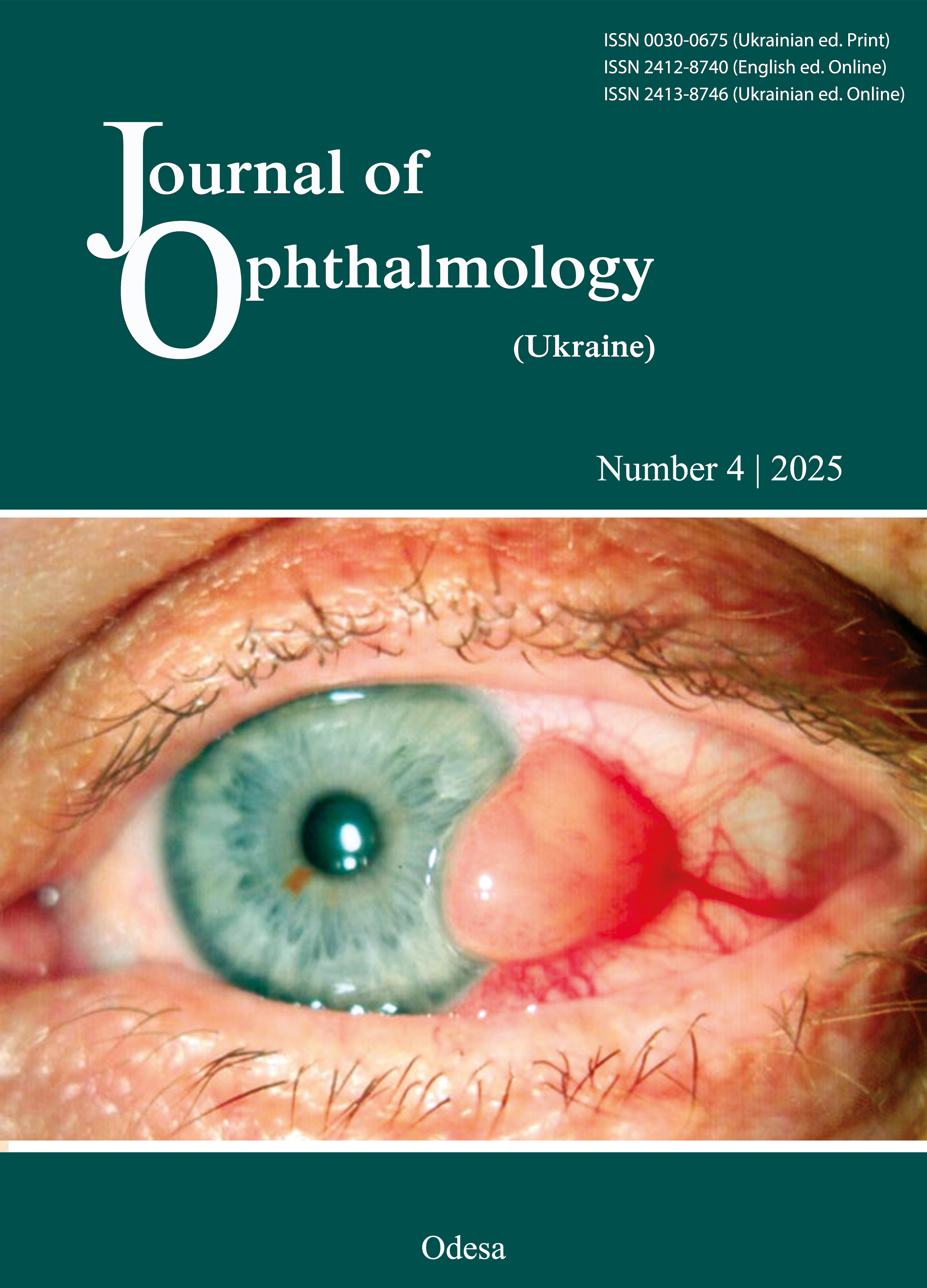
Features of the human eye microbiota in normal and pathological conditions
DOI:
https://doi.org/10.31288/oftalmolzh202545565Keywords:
eye microbiome, ocular surface, ophthalmic diseases, ocular dysbiosisAbstract
Microorganisms isolated from the ocular surface and conjunctiva have been primarily associated with ocular surface diseases such as blepharitis, trachoma, and dry eye syndrome, and have also suggested a role for the ocular and nonocular microbiome in retinal diseases including age-related macular degeneration, glaucoma, uveitis, and diabetic retinopathy. However, relatively little attention is paid to understanding that microorganisms are an integral part of the structure of the eye. Colonizer pioneers interacting with the receptors of eye cells form local immunity, activating pro-inflammatory cytokines. During human ontogenesis, the qualitative and quantitative composition of the ocular microbiome changes depending on the environment.
In 2008, when the "human microbiome" project was launched, the eye was excluded from this study due to the low biomass of the environment.
The purpose of this review is to summarize current data on the microbiota of the human eye in normal conditions and its changes in ophthalmological diseases. We summarized research results from around the world and found that this contradicts conventional wisdom. So, at first glance, the area for settlement is quite small, however, the ability of microorganisms to form structurally specific colonies, using various adhesive factors, creates not just a surface biofilm, but a unique individual landscape of the eye. However, not all of these microorganisms can be isolated using culture methods, so perhaps this is what influenced the formation of the theory about the low biomass of the eye microbiome.
Conclusion: The healthy ocular surface is characterized by a relatively stable microbiome with a relatively low diversity of microorganisms of a certain species. Biofilm landscapes are an integral anatomical unit of the eye, as this structure plays a role in maintaining its homeostasis by activating or inhibiting local cytokines.
References
Michels TC, Ivan O. Glaucoma: Diagnosis and Management. Am Fam Physician. 2023 Mar;107(3):253-262.
Damato B, Coupland SE. Conjunctival melanoma and melanosis a reappraisal of terminology, classification and staging. Clin. Experiment Ophthalmol. 2008; V.36: 785-795. https://doi.org/10.1111/j.1442-9071.2008.01888.x
Safronenkova IO. Epidemiological profile of patients with conjunctival malignancy based on the data on patients presenting to SI «The Filatov Institute of Eye Diseases and Tissue Therapy of the NAMS of Ukraine» Ophthalmol Journal. 2025; 2(523): 42-48. https://doi.org/10.31288/oftalmolzh202524248
Bai X, Xu Q, Zhang W, Wang C. The Gut-Eye Axis: Correlation Between the Gut Microbiota and Autoimmune Dry Eye in Individuals With Sjögren Syndrome. Eye Contact Lens. 2023 Jan 1;49(1):1-7. https://doi.org/10.1097/ICL.0000000000000953
Biscarini F, Masetti G, Muller I, Verhasselt HL, Covelli D, Colucci G, et al. Gut Microbiome Associated With Graves Disease and Graves Orbitopathy: The INDIGO Multicenter European Study. J Clin Endocrinol Metab. 2023 Jul 14;108(8):2065-2077. https://doi.org/10.1210/clinem/dgad030
Turnbaugh PJ, Ley RE, Hamady M, Fraser-Liggett CM, Knight R, Gordon JI. The human microbiome project. Nature. 2007 Oct 18;449(7164):804-10. https://doi.org/10.1038/nature06244
Chen Z, Jia Y, Xiao Y, Lin Q, Qian Y, Xiang Z, et al. Microbiological Characteristics of Ocular Surface Associated With Dry Eye in Children and Adolescents With Diabetes Mellitus. Invest Ophthalmol Vis Sci. 2022 Dec 1;63(13):20. https://doi.org/10.1167/iovs.63.13.20
Petrillo F, Pignataro D, Lavano MA, Santella B, Folliero V, Zannella C, et al. Current Evidence on the Ocular Surface Microbiota and Related Diseases. Microorganisms. 2020 Jul 13;8(7):1033. https://doi.org/10.3390/microorganisms8071033
Willcox M. Characterization of the normal microbiota of the ocular surface. Experimental eye research. Exp Eye Res. 2013 Dec:117:99-105.https://doi.org/10.1016/j.exer.2013.06.003
Trojacka E, Izdebska J, Szaflik J, Przybek-Skrzypecka J. The Ocular Microbiome: Micro-Steps Towards Macro-Shift in Targeted Treatment? A Comprehensive Review. Microorganisms. 2024 Nov 4;12(11):2232. https://doi.org/10.3390/microorganisms12112232
Sharma S, Mohler J, Mahajan SD, Schwartz SA, Bruggemann L, Aalinkeel R. Microbial Biofilm: A Review on Formation, Infection, Antibiotic Resistance, Control Measures, and Innovative Treatment. Microorganisms. 2023 Jun 19;11(6):1614. doi: 10.3390/microorganisms11061614. Erratum in: Microorganisms. 2024 Sep 27;12(10):1961. https://doi.org/10.3390/microorganisms12101961
Wang Y, Chen H, Xia T, Huang Y. Characterization of fungal microbiota on normal ocular surface of humans. Clin Microbiol Infect.. 2020 Jan;26(1):123.e9-123.e13. https://doi.org/10.1016/j.cmi.2019.05.011
Nash AK, Auchtung TA, Wong MC, Smith DP, Gesell JR, Ross MC, et al. The gut mycobiome of the Human Microbiome Project healthy cohort. Microbiome. 2017 Nov 25;5(1):153. https://doi.org/10.1186/s40168-017-0373-4
Zimmerman AB, Nixon AD, Rueff EM. Contact lens associated microbial keratitis: practical considerations for the optometrist. Clin Optom (Auckl). 2016 Jan 29;8:1-12. https://doi.org/10.2147/OPTO.S66424
Shin H, Price K, Albert L, Dodick J, Park L, Dominguez-Bello MG. Changes in the Eye Microbiota Associated with Contact Lens Wearing. mBio. 2016 Mar 22;7(2):e00198. https://doi.org/10.1128/mBio.00198-16
Haikun Zhang, Fuxin Zhao, Hutchinson DS, Wenfeng Sun, Ajami NJ, Shujuan Lai, et al. Conjunctival Microbiome Changes Associated With Soft Contact Lens and Orthokeratology Lens Wearing. Invest. Ophthalmol. Vis. Sci. 2017;58(1):128-136.https://doi.org/10.1167/iovs.16-20231
Szczotka-Flynn LB, Pearlman E, Ghannoum M. Microbial contamination of contact lenses, lens care solutions, and their accessories: a literature review. Eye Contact Lens. 2010 Mar;36(2):116-29. https://doi.org/10.1097/ICL.0b013e3181d20cae
Wang Y, Ding Y, Jiang X, Yang J, Li X. Bacteria and Dry Eye: A Narrative Review. J Clin Med. 2022 Jul 12;11(14):4019. https://doi.org/10.3390/jcm11144019
Barabino S. Is dry eye disease the same in young and old patients? A narrative review of the literature. BMC Ophthalmol. 2022 Feb 22;22(1):85. https://doi.org/10.1186/s12886-022-02269-2
Naqvi M, Utheim TP, Charnock C. Whole genome sequencing and characterization of Corynebacterium isolated from the healthy and dry eye ocular surface. BMC Microbiol. 2024; 24, 368. https://doi.org/10.1186/s12866-024-03517-9
Severn MM, Horswill AR. Staphylococcus epidermidis and its dual lifestyle in skin health and infection. Nat Rev Microbiol. 2023 Feb;21(2):97-111. https://doi.org/10.1038/s41579-022-00780-3
Kumar NG, Contaifer D Jr, Wijesinghe DS, Jefferson KK. Staphylococcus aureus Lipase 3 (SAL3) is a surface-associated lipase that hydrolyzes short chain fatty acids. PLoS One. 2021 Oct 7;16(10):e0258106. https://doi.org/10.1371/journal.pone.0258106
Chhadva P, Goldhardt R, Galor A. Meibomian Gland Disease: The Role of Gland Dysfunction in Dry Eye Disease. Ophthalmology. 2017 Nov;124(11S):S20-S26. https://doi.org/10.1016/j.ophtha.2017.05.031
Iqbal F, Stapleton F, Masoudi S, Papas EB, Tan J. Meibomian Gland Shortening Is Associated With Altered Meibum Composition. Invest Ophthalmol Vis Sci. 2024 Jul 1;65(8):49. https://doi.org/10.1167/iovs.65.8.49
Foulks GN, Bron AJ. Meibomian gland dysfunction: a clinical scheme for description, diagnosis, classification, and grading. Ocul Surf. 2003 Jul;1(3):107-26. https://doi.org/10.1016/S1542-0124(12)70139-8
Chen KJ, Sun MH, Hou CH, Chen HC, Chen YP, Wang NK, et al. Susceptibility of bacterial endophthalmitis isolates to vancomycin, ceftazidime, and amikacin. Sci Rep. 2021 Aug 5;11(1):15878. https://doi.org/10.1038/s41598-021-95458-w
Chen KJ, Hou CH, Sun MH, Lai CC, Sun CC, Hsiao CH. Endophthalmitis caused by Acinetobacter baumannii: report of two cases. J Clin Microbiol. 2008 Mar;46(3):1148-50. https://doi.org/10.1128/JCM.01604-07
Chen KJ, Hwang YS, Chen YP, Lai CC, Chen TL, Wang NK. Endogenous Klebsiella endophthalmitis associated with Klebsiella pneumoniae pneumonia. Ocul Immunol Inflamm. 2009 May-Jun;17(3):153-9. https://doi.org/10.1080/09273940902752250
Ji Y, Jiang C, Ji J et al. Post-cataract endophthalmitis caused by multidrug-resistant Stenotrophomonas maltophilia: clinical features and risk factors. BMC Ophthalmol 2015; 15, 14. https://doi.org/10.1186/1471-2415-15-14
https://ua.ozhurnal.com/index.php/files/article/view/297/version/297
Arunasri K, Mahesh M, Sai Prashanthi G, Jayasudha R, Kalyana Chakravarthy S, Tyagi M, et al. Comparison of the Vitreous Fluid Bacterial Microbiomes between Individuals with Post Fever Retinitis and Healthy Controls. Microorganisms. 2020 May 17;8(5):751.https://doi.org/10.3390/microorganisms8050751
Patel AS, Hemady RK, Rodrigues M, Rajagopalan S, Elman MJ. Endogenous Fusarium endophthalmitis in a patient with acute lymphocytic leukemia. Am J Ophthalmol. 1994 Mar 15;117(3):363-8. https://doi.org/10.1016/S0002-9394(14)73147-2
Mukherjee S, Weir C, Hammer H. Presumed fungal retinitis associated with pseudomyxoma peritonei. BMJ Case Rep. 2009;2009:bcr06.2008.0345. https://doi.org/10.1136/bcr.06.2008.0345
Menia NK, Sharma SP, Bansal R. Fungal retinitis following influenza virus type A (H1N1) infection. Indian J Ophthalmol. 2019 Sep;67(9):1483-1484. https://doi.org/10.4103/ijo.IJO_1691_18
Gomes JÁP, Frizon L, Demeda VF. Ocular Surface Microbiome in Health and Disease. Asia Pac J Ophthalmol (Phila). 2020 Dec;9(6):505-511.https://doi.org/10.1097/APO.0000000000000330
Xue W, Li JJ, Zou Y, Zou B, Wei L. Microbiota and Ocular Diseases. Frontiers in cellular and infection microbiology. 2021; 11: 759333. https://doi.org/10.3389/fcimb.2021.759333
Ozkan J, Majzoub ME, Coroneo M, Thomas T, Willcox M. Ocular microbiome changes in dry eye disease and meibomian gland dysfunction. Exp Eye Res. 2023 Oct;235:109615. https://doi.org/10.1016/j.exer.2023.109615
Song J, Dong H, Wang T, Yu H, Yu J, Ma S, et al. What is the impact of microbiota on dry eye: a literature review of the gut-eye axis. BMC Ophthalmol. 2024 Jun 19;24(1):262. https://doi.org/10.1186/s12886-024-03526-2
Petrillo F, Pignataro D, Lavano MA, Santella B, Folliero V, Zannella C, et al. Current Evidence on the Ocular Surface Microbiota and Related Diseases. Microorganisms. 2020 Jul 13;8(7):1033. https://doi.org/10.3390/microorganisms8071033
Traish A, Bolanos J, Nair S, Saad F, Morgentaler A. Do Androgens Modulate the Pathophysiological Pathways of Inflammation? Appraising the Contemporary Evidence. Journal of clinical medicine. 2018; 7(12): 549. https://doi.org/10.3390/jcm7120549
Ueta M. Innate immunity of the ocular surface and ocular surface inflammatory disorders. Cornea. 2008 Sep;27 Suppl 1:S31-40.
https://doi.org/10.1097/ICO.0b013e31817f2a7f
Ma B, Barathan M, Ng MH, Law JX. Oxidative Stress, Gut Microbiota, and Extracellular Vesicles: Interconnected Pathways and Therapeutic Potentials. Int J Mol Sci. 2025 Mar 28;26(7):3148.https://doi.org/10.3390/ijms26073148
Flohé L. Looking Back at the Early Stages of Redox Biology. Antioxidants (Basel). 2020 Dec 9;9(12):1254. https://doi.org/10.3390/antiox9121254
Samalia PD, Solanki J, Kam J, Angelo L, Niederer RL. From Dysbiosis to Disease: The Microbiome's Influence on Uveitis Pathogenesis. Microorganisms. 2025 Jan 25;13(2):271. https://doi.org/10.3390/microorganisms13020271
Zemlicková H, Urbásková P, Adámková V, Motlová J, Lebedová V, Procházka B. Characteristics of Streptococcus pneumoniae, Haemophilus influenzae, Moraxella catarrhalis and Staphylococcus aureus isolated from the nasopharynx of healthy children attending day-care centres in the Czech Republic. Epidemiol Infect. 2006 Dec;134(6):1179-87. https://doi.org/10.1017/S0950268806006157
Leck AK, Thomas PA, Hagan M, Kaliamurthy J, Ackuaku E, John M, et al. Aetiology of suppurative corneal ulcers in Ghana and south India, and epidemiology of fungal keratitis. Br J Ophthalmol. 2002 Nov;86(11):1211-5. https://doi.org/10.1136/bjo.86.11.1211
Lee SH, Oh DH, Jung JY, Kim JC, Jeon CO. Comparative ocular microbial communities in humans with and without blepharitis. Invest Ophthalmol Vis Sci. 2012 Aug 15;53(9):5585-93. https://doi.org/10.1167/iovs.12-9922
Shin JH, Lee JW, Lim SH, Yoon BW, Lee Y, Seo JH. The microbiomes of the eyelid and buccal area of patients with uveitic glaucoma. BMC Ophthalmol. 2022 Apr 14;22(1):170.https://doi.org/10.1186/s12886-022-02395-x
Alqawlaq S, Flanagan JG, Sivak JM. All roads lead to glaucoma: Induced retinal injury cascades contribute to a common neurodegenerative outcome. Exp Eye Res. 2019 Jun;183:88-97. https://doi.org/10.1016/j.exer.2018.11.005
Awad R, Ghaith AA, Awad K, Mamdouh Saad M, Elmassry AA. Fungal Keratitis: Diagnosis, Management, and Recent Advances. Clin Ophthalmol. 2024 Jan 10;18:85-106. https://doi.org/10.2147/OPTH.S447138
Bourcier T, Sauer A, Dory A, Denis J, Sabou M. Fungal keratitis. J Fr Ophtalmol. 2017 Nov;40(9):e307-e313. https://doi.org/10.1016/j.jfo.2017.08.001
Downloads
Published
How to Cite
Issue
Section
License
Copyright (c) 2025 Melnyk O.V., Hural A.R., Lavryk H.S., Shykula R.G.

This work is licensed under a Creative Commons Attribution 4.0 International License.
This work is licensed under a Creative Commons Attribution 4.0 International (CC BY 4.0) that allows users to read, download, copy, distribute, print, search, or link to the full texts of the articles, or use them for any other lawful purpose, without asking prior permission from the publisher or the author as long as they cite the source.
COPYRIGHT NOTICE
Authors who publish in this journal agree to the following terms:
- Authors hold copyright immediately after publication of their works and retain publishing rights without any restrictions.
- The copyright commencement date complies the publication date of the issue, where the article is included in.
DEPOSIT POLICY
- Authors are permitted and encouraged to post their work online (e.g., in institutional repositories or on their website) during the editorial process, as it can lead to productive exchanges, as well as earlier and greater citation of published work.
- Authors are able to enter into separate, additional contractual arrangements for the non-exclusive distribution of the journal's published version of the work with an acknowledgement of its initial publication in this journal.
- Post-print (post-refereeing manuscript version) and publisher's PDF-version self-archiving is allowed.
- Archiving the pre-print (pre-refereeing manuscript version) not allowed.


