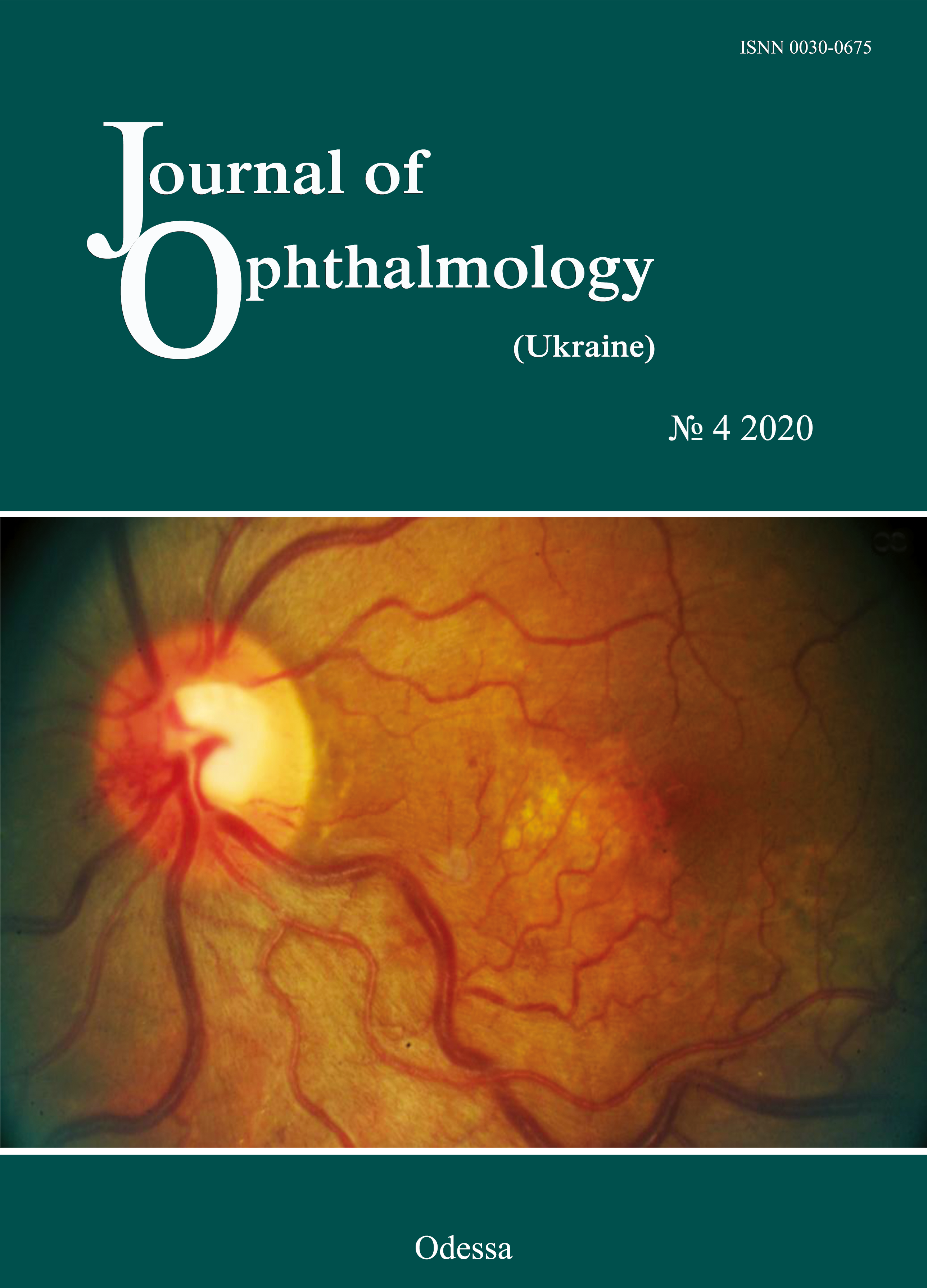
Clinical course of compressive optic neuropathy in skull-base tumors
DOI:
https://doi.org/10.31288/oftalmolzh202042327Keywords:
skull-base tumors, chiasmal syndrome, compressive optic atrophyAbstract
Background: Skull-base tumors (SBTs) of the middle and anterior fossae typically cause mass effect on the optic nerve/chiasm complex. The most common of these neoplasms are pituitary adenomas, meningiomas and craniopharyngiomas. Clinical manifestations of SBTs vary and depend on tumor involvement, nature of growth pattern, and rate of growth. Chiasmal compression is accompanied by a gradual decrease in visual acuity, bitemporal visual field defects and development of primary descending optic atrophy (OA).
Purpose: To investigate neuro-ophthalmological symptoms in patients with SBTs.
Material and Methods: This retrospective study included the records of 500 patients who received treatment for SBT and loss of visual acuity and/or visual fields at the Romodanov Neurosurgery Institute during the period from 2017 through 2019. Patients underwent clinical and neurological, eye, and otoneurological examination (a routine otoneurological examination with assessment of cranial nerve function).
Results: In patients with pituitary adenoma, chiasmal syndrome most commonly was symmetric (63.8%), and developed either gradually or (in a stroke-like course of tumor progression) abruptly. In addition, it was accompanied by a symmetric loss of visual acuity (77.1%), visual field defects (89.8%) and development of OA (50.5%). In patients with sella turcica meningioma, chiasmal syndrome most commonly was markedly asymmetric (65.7%), and accompanied by mild loss of visual acuity and visual fields in one eye, and severe or very severe visual acuity loss, residual visual field and OA in the fellow eye, as well as a particular compression of the anterior chiasm. Patients with supradiaphragmatic craniopharyngiomas had various amounts of loss of visual acuity and/or visual fields, and, most commonly, symmetric chiasmal syndrome and bilateral optic atrophy. In addition, the posterior chiasm (especially, the papillomacular bundle) was affected, which was manifested by bitemporal central scotoma (33.3%).
Conclusion: We identified ophthalmological features of disease course in various histologic types of skull-base tumors. Loss of visual acuity and/or visual fields was an early and major symptom in the clinical picture of disease.
References
1.Abouaf L, Vighetto A, Lebas M. Neuro-ophthalmologic exploration in non-functioning pituitary adenoma. Ann Endocrinol (Paris). 2015; 76(3):210-9.https://doi.org/10.1016/j.ando.2015.04.006
2.Predoi D, Badiu C, Alexandrescu D, Agarbiceanu C, Stangu C, Ogrezeanu I, Ciubotaru V, Dumitrascu A, Constantinescu Al. Assessment of compressive optic neuropathy in long-standing pituitary macroadenomas. Acta Endocrinologica (Buc). 2008; 4(1):11-22.https://doi.org/10.4183/aeb.2008.11
3.Ekpene U, Ametefe M, Akoto H, Bankah P, Totimeh T, Wepeba G, Dakurah T. Ekpene U, et al. Ghana Med J. 2018; 52(6):79-83.https://doi.org/10.4314/gmj.v52i2.3
4.Masaya-anon P, Lorpattanakasem J. Intracranial tumors affecting visual system: 5-year review in Prasat Neurological Institute. J Med Assoc Thai. 2008; 91(4):515-19.
5.Foroozan R. Chiasmal syndromes. Curr Opin Ophthalmol. 2003; 14(6):325-31.https://doi.org/10.1097/00055735-200312000-00002
6.Astorga-Carballo A, Serna-Ojeda JC, Camargo-Suarez MF. Chiasmal syndrome: Clinical characteristics in patients attending an ophthalmological center. Saudi J Ophthalmol. 2017; 31(24):229-33.https://doi.org/10.1016/j.sjopt.2017.08.004
7.Kitthaweesin K, Ployprasith C. Ocular manifestations of suprasellar tumors. J Med Assoc Thai. 2008; 91(5):711-5.
8.Wadud SA, Ahmed S, Choudhury N, Chowdhury D. Evaluation of ophthalmic manifestations in patients with intracranial tumours. Mymensingh Med J. 2014 Apr;23(2):268-71.
9.Sefi-Yurdakul N. Sefi-Yurdakul N. Int J Ophthalmol. 2015 Aug 18;8(4):800-3. doi: 10.3980/j.issn.2222-3959.2015.04.28.
10.Tagoe NN, Essuman VA., Fordjuor G, Akpalu G, Bankah P, Ndanu T. Neuro-ophthalmic and clinical characteristics of brain tumours in a tertiary hospital in Ghana. Ghana Med J. 2015; 49(3):181-6.https://doi.org/10.4314/gmj.v49i3.9
11.Guk MO. [Diagnosis and comprehensive treatment of hormonally-inactive pituitary adenomas]. Dissertation for Doctor of Medical Sciences. Kyiv. 2017.
12.Serova NK. [Clinical neuroophthalmology. Neurosurgery aspects]. Tver': Triada; 2011. Russian.
Downloads
Published
How to Cite
Issue
Section
License
Copyright (c) 2025 К. С. Єгорова, Л. В. Задояний, М. О. Гук, А. А. Чуков, О. В. Українець, Л. О. Даневич, А. О. Мумлєв

This work is licensed under a Creative Commons Attribution 4.0 International License.
This work is licensed under a Creative Commons Attribution 4.0 International (CC BY 4.0) that allows users to read, download, copy, distribute, print, search, or link to the full texts of the articles, or use them for any other lawful purpose, without asking prior permission from the publisher or the author as long as they cite the source.
COPYRIGHT NOTICE
Authors who publish in this journal agree to the following terms:
- Authors hold copyright immediately after publication of their works and retain publishing rights without any restrictions.
- The copyright commencement date complies the publication date of the issue, where the article is included in.
DEPOSIT POLICY
- Authors are permitted and encouraged to post their work online (e.g., in institutional repositories or on their website) during the editorial process, as it can lead to productive exchanges, as well as earlier and greater citation of published work.
- Authors are able to enter into separate, additional contractual arrangements for the non-exclusive distribution of the journal's published version of the work with an acknowledgement of its initial publication in this journal.
- Post-print (post-refereeing manuscript version) and publisher's PDF-version self-archiving is allowed.
- Archiving the pre-print (pre-refereeing manuscript version) not allowed.


