Клинические особенности течения неврита зрительного нерва как осложнения переднего идиопатического увеита
DOI:
https://doi.org/10.31288/oftalmolzh202154146Ключові слова:
передний идиопатический увеит, осложнения, неврит зрительного нерва, клинические особенности теченияАнотація
Цель. Выявить особенности клинического течения неврита зрительного нерва как осложнения переднего идиопатического увеита.
Материал и методы. Исследования выполнены у 150 человек с идиопатическим монолатеральным передним увеитом (114 – без признаков неврита зрительного нерва, 36 – с невритом на фоне увеита) в отделе воспалительной патологии глаза ГУ «Институт глазных болезней и тканевой терапии им. В. П. Филатова НАМН Украины». Обследование включало офтальмоскопию, биомикроскопию, Хамфри периметрию, определение внутриглазного давления, остроты зрения). Лечение проводили согласно протоколу (антибиотики, кортикостероиды, нестероидные противовоспалительные средства, иммуносупрессанты, модуляторы иммунного ответа).
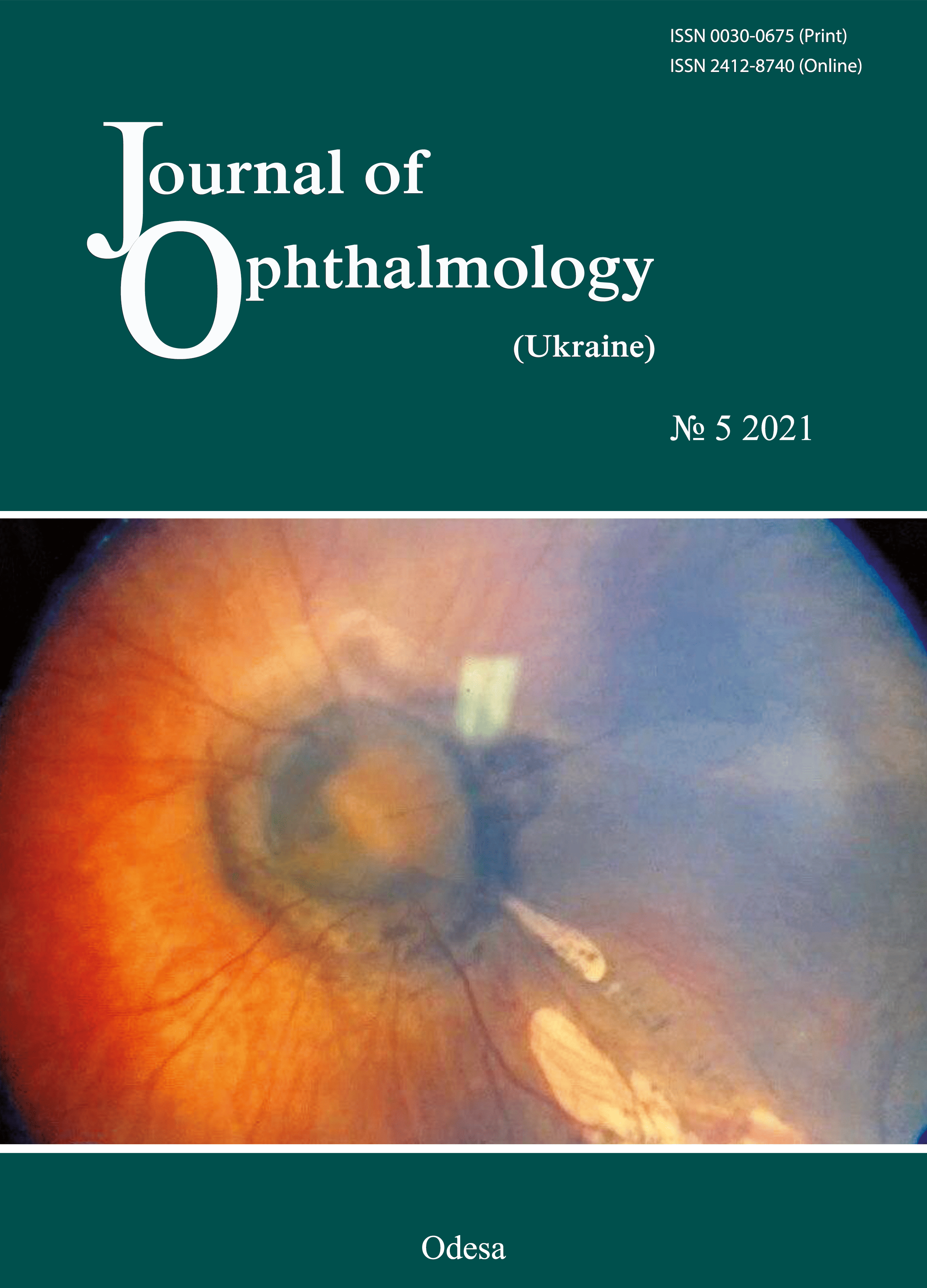
Результаты. Выявлено, что для пациентов с невритом зрительного нерва как осложнением переднего увеита характерна более выраженная воспалительная реакция (множественные преципитаты в 69,7%, единичные – не обнаружены вообще, гипопион более, чем у двух третей пациентов, интенсивность помутнений стекловидного тела максимальна у всех больных). У лиц с увеитом без неврита единичные преципитаты в 19,3%, множественные – не выявлены, гипопион отмечен в одном случае, помутнения стекловидного тела в – 94,7% случаев средней выраженности. Между интенсивностью признаков воспаления у больных передним увеитом и развитием неврита нерва выявлена положительная сильная связь (r Спирмена 0,566, p<0,05). У пациентов с передним увеитом как без, так и с наличием неврита в подавляющем большинстве случаев имелись отоларингологические и одонтогенные заболевания (82,5 и 63,9%). Выявлена достоверная связь между сопутствующими воспалительными заболеваниями у лиц с передним увеитом и развитием неврита зрительного нерва (χ2 Пирсона = 5,50, p=0,0191).
Посилання
1. Fardeau C, Champion E, Massamba N, Lehoang P. Uveitic macular edema. Eye. 2016; 30(10):1277-92. https://doi.org/10.1038/eye.2016.115
2. Anesi SD, Foster CS. Anterior uveitis: etiology and treatment. Advanced Ocular Care. 2011;2(1):32-4.
3. Miserocchi E, Fogliato G, Modorati M, Bandello F. Review on the worldwide epidemiology of uveitis. Eur J Ophthalmol. 2013; 23: 705- 717.https://doi.org/10.5301/ejo.5000278
4. Arbenyeva NS, Chekhova TA, Bratko GV, Chernykh VV. [Comparative analysis of the incidence of patients with uveitis]. In: [Current issues of ophthalmology: a collection of science works. Proceedings of the 7th National Russian Conference of Young Scientists]. Editor, B.E. Malyugin. Moscow: Ophthalmology; 2012. p. 28-9. Russian.
5. McCannel CA, Holland GN, Helm CJ, et al. Causes of uveitis in the general practice of ophthalmology. UCLA Community-Based Uveitis Study Group. Am J Ophthalmol. 1996; 121 (1): 35-46. https://doi.org/10.1016/S0002-9394(14)70532-X
6. Panova IE, Drozdova IE. [Uveitides: a manual for physicians]. Moscow: Meditsinskoie informatsionnoie agenstvo; 2014. Russian.
7. Nussenblatt RB, Whitcup SM, editors. Uveitis: fundamental and clinical practice. 4th ed. Elsevier/Mosby; 2012.
8. Emmett T Cunningham, Zierhut M. Uveitic Macular Edema. Ocul Immunol Inflamm. 2018;26(7):987-90,https://doi.org/10.1080/09273948.2018.1529466
9. Khramenko NI, Konovalova NV. Findings of ocular and brain hemodynamics in patients with anterior uveitis complicated by macular edema. J Ophthalmol (Ukraine). 2020; 4:14-22.
10. Panchenko NV, Samofalova MN, Gonchar EN, Litvishchenko AV, Friantseva MV. [Thinning of the peripapillary nerve fiber layer in uveitis complicated by optic nerve inflammation]. Archive of Ukrainian ophthalmology. 2016;3(1):50-3. Russian.
11. Penkov MA, Shpak NI, Avrushchenko NM. [Endogenous uveitis]. Kyiv: Zdorov'ia; 1979. Russian.
12. Jabs DA, Nussenblatt RB, Rosenbaum JT. Standardization of Uveitis Nomenclature (SUN) Working Group. Standardization of uveitis nomenclature for reporting clinical data. Results of the First International Workshop. Am J Ophthalmol. 2005; 140(3): 509- 516.https://doi.org/10.1016/j.ajo.2005.03.057
13. Deschenes J, Murray PI, Rao NA, Nussenblatt RB. & International Uveitis Study Group. International Uveitis Study Group (IUSG): clinical classification of uveitis. Ocul Immunol Inflamm. 2008; 16: 1-2.https://doi.org/10.1080/09273940801899822
14. McNeil R. Grading of ocular inflammation in uveitis: an overview. Eye news. 2016; 22 (5): Fabruary/March; Available at: http://www.eyenews.uk.com.
15. Glanz S. [Biomedical statistics]. Moscow: Praktika;1998. Russian.
16. Cimino L, Auer C, Herbort CP. Sensitivity of indocyanine green angiography for the follow-up of active inflammatory choriocapillaropathies. Ocul Immunol Inflamm. 2000; 8(4): 275-83.https://doi.org/10.1076/ocii.8.4.275.6462
17. Ioyleva ЕЕ, Krivosheeva MS, Smirnova MA. [Unilateral optic disc edema: features of the differential diagnosis]. Tavricheskii medikobiologicheskii vestnik. 2013;(3):166-70. Russian.
18. Ioyleva E, Krivosheeva M. Microperimetry in the diagnosis of the first manifestation of optic neuritis in multiple sclerosis. J Neurol Sci. 2015; 357: 47.https://doi.org/10.1016/j.jns.2015.08.196
19. Trusko B, Thort J, Jabs D et al. The Standardization of Uveitis Nomenclature (SUN) Project. Development of clinical evidence base utilizing informatics tools and techniques. Methods Inf Med. 2013; 7. 52 (3): 259-265.https://doi.org/10.3414/ME12-01-0063
20. Bennett JL. Optic Neuritis. Continuum (Minneap Minn). 2019; 25(5): 1236-64.https://doi.org/10.1212/CON.0000000000000768
21. Shantha GJ, Crozier I, Hayek BR, Bruce BB, Gargu C, Brown J, Fankhauser J, Yeh S. Ophthalmic Manifestations and Causes of Vision Impairment in Ebola Virus Disease Survivors in Monrovia, Liberia. Ophthalmology. 2017; 124(2): 170-177. doi:10.1016/j.ophtha.2016.10.011.HHS Public Access.https://doi.org/10.1016/j.ophtha.2016.10.011
22. Smit RL, Baarsma GS, de Vries J. Classification of 750 consecutive uveitis patients in the Rotterdam Eye Hospital. J Int Ophthalmol. 1993; 17(2):71-76.https://doi.org/10.1007/BF00942778
23. Beck RW, Cleary PA, Anderson MM et al. A randomized, controlled trial of corticosteroids in the treatment of acute optic neuritis. The Optic Neuritis Study Group. N Engl J Med. 1992; 326(9): 581-8.https://doi.org/10.1056/NEJM199202273260901
24. Whitley W, Sheppard J. The basics of uveitis. Rev Optom. 2011; Available at: http://www.reviewofoptometry.com/continuingeducation/tabviewtest/lessoni.
##submission.downloads##
Опубліковано
Як цитувати
Номер
Розділ
Ліцензія
Авторське право (c) 2025 Л. В. Венгер , В. В. Савко , А. В. Ковтун , В. Н. Соколов

Ця робота ліцензується відповідно до Creative Commons Attribution 4.0 International License.
Ця робота ліцензується відповідно до ліцензії Creative Commons Attribution 4.0 International (CC BY). Ця ліцензія дозволяє повторно використовувати, поширювати, переробляти, адаптувати та будувати на основі матеріалу на будь-якому носії або в будь-якому форматі за умови обов'язкового посилання на авторів робіт і первинну публікацію у цьому журналі. Ліцензія дозволяє комерційне використання.
ПОЛОЖЕННЯ ПРО АВТОРСЬКІ ПРАВА
Автори, які подають матеріали до цього журналу, погоджуються з наступними положеннями:
- Автори отримують право на авторство своєї роботи одразу після її публікації та назавжди зберігають це право за собою без жодних обмежень.
- Дата початку дії авторського права на статтю відповідає даті публікації випуску, до якого вона включена.
ПОЛІТИКА ДЕПОНУВАННЯ
- Редакція журналу заохочує розміщення авторами рукопису статті в мережі Інтернет (наприклад, у сховищах установ або на особистих веб-сайтах), оскільки це сприяє виникненню продуктивної наукової дискусії та позитивно позначається на оперативності і динаміці цитування.
- Автори мають право укладати самостійні додаткові угоди щодо неексклюзивного розповсюдження статті у тому вигляді, в якому вона була опублікована цим журналом за умови збереження посилання на первинну публікацію у цьому журналі.
- Дозволяється самоархівування постпринтів (версій рукописів, схвалених до друку в процесі рецензування) під час їх редакційного опрацювання або опублікованих видавцем PDF-версій.
- Самоархівування препринтів (версій рукописів до рецензування) не дозволяється.


