Ultrasound features of epibulbar malignancies of various histogenesis (melanoma and carcinoma)
DOI:
https://doi.org/10.31288/oftalmolzh202532530Keywords:
ocular oncology, conjunctival melanoma, conjunctival carcinoma, ultrasound examination, radiation therapy, cryodestruction, radiation therapy plus cryosurgery, sclera, epibulbar neoplasmAbstract
Purpose: To assess the ultrasound features of epibulbar malignancies of various histogenesis (melanoma and carcinoma), including the thickness of adjacent sclera and the sonographic density and dimensions of the neoplasm before and at various time points after radiation therapy plus cryosurgery.
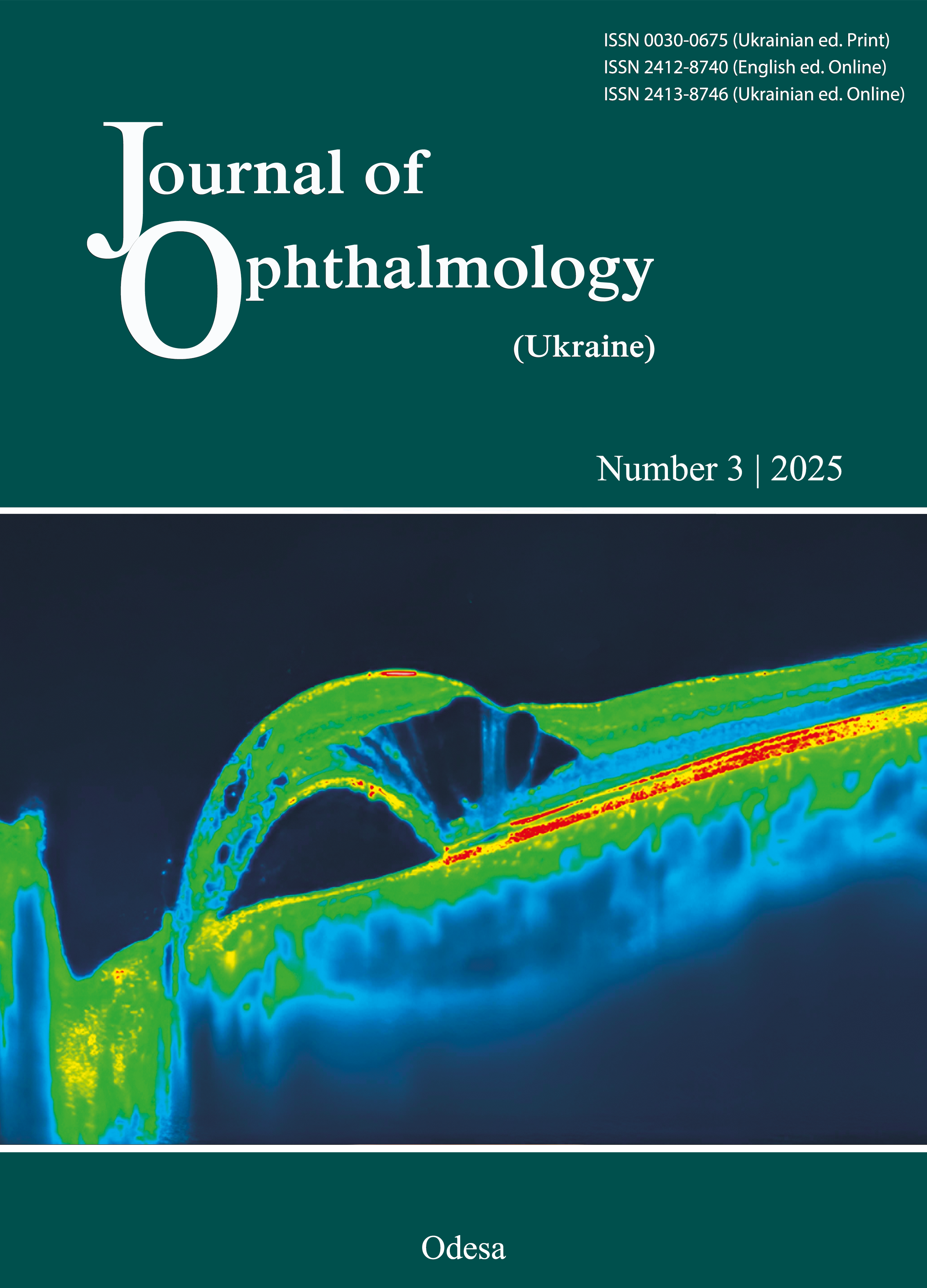
Material and Methods: Ultrasound (US) examination was performed in 75 patients with conjunctival melanoma (CM) or conjunctival carcinoma (CC) who underwent treatment at SI “The Filatov Institute of Eye Diseases and Tissue Therapy of the NAMS of Ukraine” in 2004-2021. Of these, 58 (77.3%) had CM, and 17 (24%) had CC. Of the patients with CM, 30 (51.7%) were men aged 18 to 88 years (median, 51.3 years), and 28 (48.3%) were women aged 26 to 87 years (median, 57.3 years). Of the patients with CC, 12 (70.6%) were men aged 28 to 82 years (median, 66.3 years), and 5 (29.4 %) were women aged 35 to 74 years (median, 57.0 years). US biomicroscopy images were obtained using the 50-MHz Aviso Ultrasound Platform. Dimensions and sonographic density of the tumor, thickness of the sclera underlying the tumor and scleral thickness on the opposite side were assessed at baseline and each 3 months over a one-year follow-up. All patients received radiation therapy (RT) plus cryosurgery (alias, cryodestruction of the tumor).
Results: For the total study sample, the thickness of the sclera beneath the tumor was, on average, 1.9 times thinner compared to scleral thickness on the opposite side, and this difference was significant (p < 0.01). Eyes with partial resorption of CM exhibited a thickness of the underlying sclera (0.42 mm) that was 1.3 times and 1.2 times thinner compared to eyes with complete resorption of CM (χ2 = 3.13, р = 0.006) and eyes with partial resorption of CC (χ2 = 4.31, р = 0.01), respectively. A middle sonographic density was the most common category of sonographic density for CM as well as CC (χ2 = 3.1, р = 0.004; χ2 = 4.3, р = 0.006, respectively). There was a conjugation between the category of sonographic density and the presence of tumor recurrence in patients with CM or CC. Recurrence of CM or CC was observed only in tumors with a middle sonographic density (χ2 = 6.01, р = 0.004). A thickness of the sclera underlying the tumor not exceeding 0.30 mm may be considered as a risk factor for the development of scleromalacia (odds ratio (OR) = 7.7, 95% confidence interval (CI) 3.2-53.8) (Copyright Registration Certificate for a Work No. 134003 issued on March 3, 2025).
Conclusion: The thickness of the sclera beneath the tumor as assessed by US was thinner compared to scleral thickness on the opposite side. A middle sonographic density was the most common category of sonographic density for CM. There was a conjugation between the category of sonographic density and the presence of tumor recurrence in patients with CM and those with CC. Recurrence of CM or CC was observed only in tumors with a middle sonographic density. A thickness of the sclera underlying the tumor not exceeding 0.30 mm may be considered as a risk factor for the development of scleromalacia (OR = 7.7, 95% CI 3.2-53.8).
References
Shields CL, Shields JA, Gunduz K, et al. Conjunctival melanoma: risk factors for recurrence, exenteration, metastasis, and death in 150 consecutive patients. Arch Ophthalmol. 2000; 118 (11): 1497-1507. https://doi.org/10.1001/archopht.118.11.1497
Brownstein S. Malignant melanoma of the conjunctiva. Cancer Control. 2004; 11: 310-316. https://doi.org/10.1177/107327480401100505
Damato B, Coupland SE. Conjunctival melanoma and melanosis: a reappraisal of terminology, classification and staging. Clin Experiment Ophthalmol. 2008; 36(8):786-95. https://doi.org/10.1111/j.1442-9071.2008.01888.x
Tunc M, Char DH, Crawford B, Miller T. Intraepithelial and invasive squamous cell carcinoma of the conjunctiva: Analysis of 60 cases. 1999;83(1): 98-103. https://doi.org/10.1136/bjo.83.1.98
Wong JR, Nanji AA, Galor A, Karp CL. Management of conjunctival malignant melanoma: a review and update. Expert Rev Ophthalmol. 2014;9(3):185-204. https://doi.org/10.1586/17469899.2014.921119
Buchwald HJ, Müller A, Kampmeier J, Lang GK. [Optical coherence tomography versus ultrasound biomicroscopy of conjunctival and eyelid lesions]. Klin Monatsbl Augenheilkd. 2003; 220(12):822-829. German. https://doi.org/10.1055/s-2003-812563
Finger PT, Tran H, Turbin RE, et al. High-frequency ultrasonographic evaluation of conjunctival intra-epithelial neoplasia and squamous cell carcinoma. Arch Ophthalmol. 2003; 121 (2): 168-172. https://doi.org/10.1001/archopht.121.2.168
Damato B, Coupland SE. Management of conjunctival melanoma. Expert Rev Anticancer Ther. 2009;9 (9):1227-1239. https://doi.org/10.1586/era.09.85
Damato B, Coupland SE. An audit of conjunctival melanoma treatment in Liverpool. Eye (Lond) 2009;23 (4):801-809. https://doi.org/10.1038/eye.2008.154
Damato B, Coupland SE. Clinical mapping of conjunctival melanomas. Br J Ophthalmol. 2008;92 (11):1545-1549. doi: 10.1136.
https://doi.org/10.1136/bjo.2007.129882
Faul F, Erdfelder E, Lang AG, Buchner A. G*Power 3: A flexible statistical power analysis program for the social, behavioral, and biomedical sciences. Behav Res Methods. 2007; 39: 175-191. https://doi.org/10.3758/BF03193146
Kanellopoulos AJ, Asimellis G. Comparison of high-resolution Scheimpflug and high-frequency ultrasound biomicroscopy to anterior-segment OCT corneal thickness measurements. Clin Ophthalmol. 2013; 20(7): 2239-2247. https://doi.org/10.2147/OPTH.S53718
Buchwald HJ, Spraul CW, Kampmeier J, Lang GK. Ultrasound biomicroscopy in eyelid lesions - a clinical study on 30 patients. Klin Monbl Augenheilkd. 2002; 219(3): 95-100. https://doi.org/10.1055/s-2002-26715
Downloads
Published
How to Cite
Issue
Section
License
Copyright (c) 2025 Safronenkova I.O., Buiko O.S., Yelagina V.A.

This work is licensed under a Creative Commons Attribution 4.0 International License.
This work is licensed under a Creative Commons Attribution 4.0 International (CC BY 4.0) that allows users to read, download, copy, distribute, print, search, or link to the full texts of the articles, or use them for any other lawful purpose, without asking prior permission from the publisher or the author as long as they cite the source.
COPYRIGHT NOTICE
Authors who publish in this journal agree to the following terms:
- Authors hold copyright immediately after publication of their works and retain publishing rights without any restrictions.
- The copyright commencement date complies the publication date of the issue, where the article is included in.
DEPOSIT POLICY
- Authors are permitted and encouraged to post their work online (e.g., in institutional repositories or on their website) during the editorial process, as it can lead to productive exchanges, as well as earlier and greater citation of published work.
- Authors are able to enter into separate, additional contractual arrangements for the non-exclusive distribution of the journal's published version of the work with an acknowledgement of its initial publication in this journal.
- Post-print (post-refereeing manuscript version) and publisher's PDF-version self-archiving is allowed.
- Archiving the pre-print (pre-refereeing manuscript version) not allowed.


