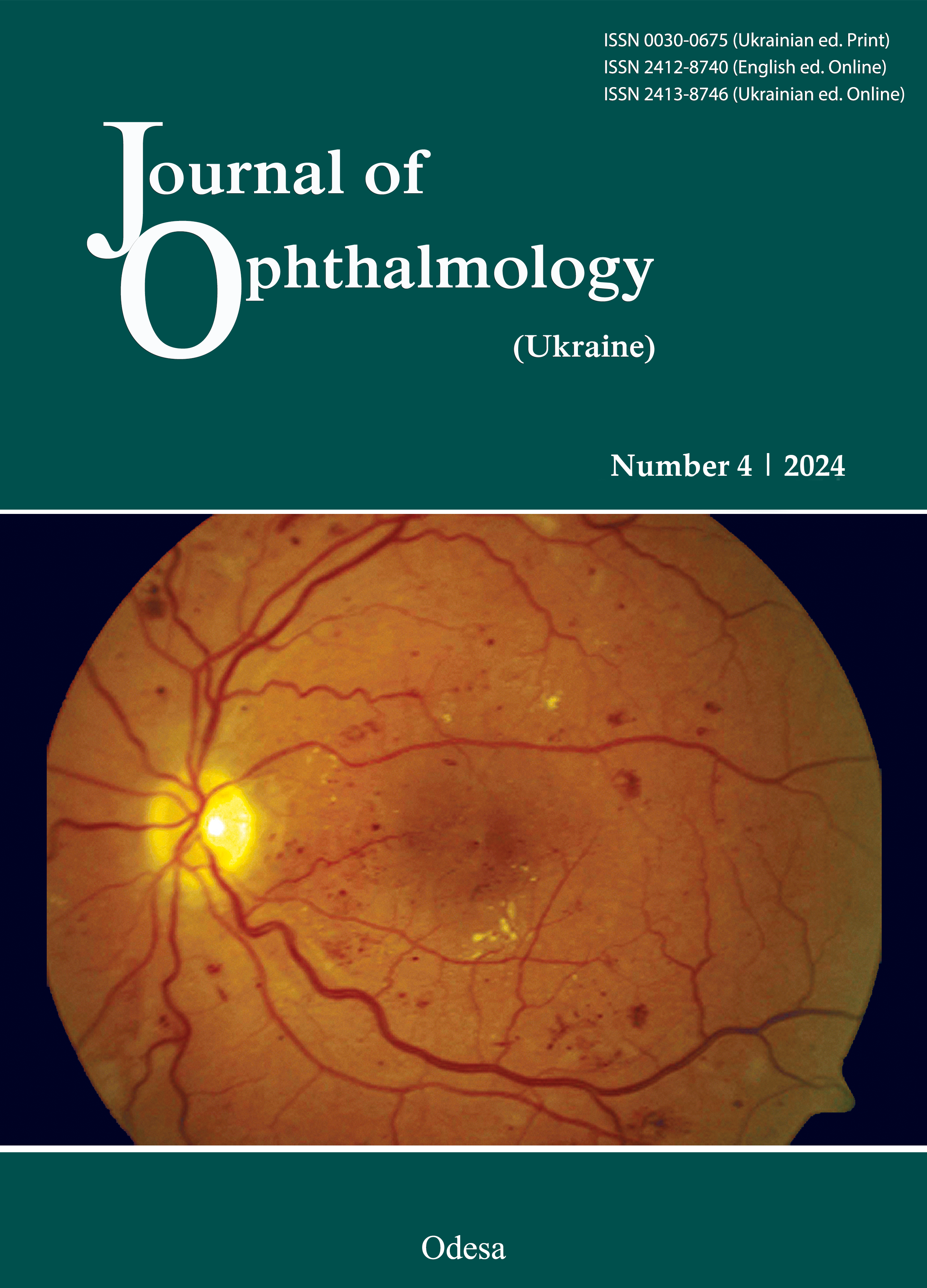
Prognostic biomarkers of non-proliferative diabetic retinopathy progression in type 2 diabetes mellitus
DOI:
https://doi.org/10.31288/oftalmolzh202443845Keywords:
type 2 diabetes mellitus, diabetic retinopathy, prognosis, modeling, precision medicine, biomarkers, HbA1c, lipoproteins, blood coagulationAbstract
Purpose: To establish prognostic biomarkers of non-prolefarative diabetic retinopathy (NPDR) progression in T2DM on the basis of examination of clinical, ophthalmological and laboratory (carbohydrate and lipid metabolism and coagulation and hemostasis) characteristics.
Methods: This study included 358 T2DM2 patients with diabetic retinopathy (DR); for each patient, the eye with more severe DR was included in the study. Cases were dichotomized into three groups based on DR severity: group 1 (NPDR; 189 eyes), group 2 (preproliferative DR or PPDR; 96 eyes), and group 3 (proliferative DR or PDR; 73 eyes). Central retinal thickness (CRT; µm) and central retinal volume (CRV) were assessed by optical coherence tomography (OCT; mm3). Serum samples were taken to assess fasting serum glucose, glycosylated hemoglobin (HbA1c), total cholesterol, high-density lipoprotein (HDL), low-density lipoprotein (LDL), very low-density lipoprotein (VLDL), triglycerides, and fibrinogen. Coagulation and hemostasis parameters were assessed. Statistical analyses were performed with EZR version 1.54.
Results: Associations of the variables with the risk of NPDR progression were confirmed by a univariate logistic regression analysis. The area under curve (AUC) was largest for HbA1c (AUC = 0.84), followed by CRT and CRV (AUC = 0.79). In addition, the AUC ranged from 0.56 to 0.67 for the rest of variables. Age, gender, diabetes duration, and blood HDL level were not associated with NPDR progression. Our multivatiate logistic regression analysis found six biomarkers (age, HbA1c, LDL, VLDL, prothrombin index and CRT) directly associated with the risk of NPDR progression (AUC = 0.91; p < 0.001). The established association was mostly due to the three independent variables, HbA1c, VLDL and CRT (AUC = 0.90; p < 0.001).
Conclusion: Independent effects on the risk of NPDR progression were confirmed for HbA1c, VLDL and CRT. The built model was found to be of very high predictive quality, which allowed recommending it for confirming high risk for NPDR progression in equivocal clinical cases or as a criterion for conclusive prognosis in the relevant expert systems.
References
International Diabetes Federation Diabetes Facts and Figures. [(accessed on 9 July 2023)]. Available online: https://idf.org/aboutdiabetes/what-is-diabetes/facts-figures.html.
Saeedi P, Petersohn I, Salpea P, Malanda B, Karuranga S, Unwin N, , et al. IDF Diabetes Atlas Committee. Global and regional diabetes prevalence estimates for 2019 and projections for 2030 and 2045: Results from the International Diabetes Federation Diabetes Atlas, 9th edition. Diabetes Res Clin Pract. 2019 Nov;157:107843. https://doi.org/10.1016/j.diabres.2019.107843
Wong TY, Sabanayagam C. Strategies to tackle the global burden of diabetic retinopathy: From epidemiology to artificial intelligence. Ophthalmologica. 2020;243(1):9-20. https://doi.org/10.1159/000502387
Teo ZL, Tham YC, Yu M, Chee ML, Rim TH, Cheung N, et al. Global prevalence of diabetic retinopathy and projection of burden through 2045: Systematic review and meta-analysis. Ophthalmology. 2021 Nov;128(11):1580-1591. https://doi.org/10.1016/j.ophtha.2021.04.027
Schiborn C, Schulze MB. Precision prognostics for the development of complications in diabetes. Diabetologia. 2022 Nov;65(11):1867-1882. https://doi.org/10.1007/s00125-022-05731-4
Dai L, Wu L, Li H, Cai C, Wu Q, Kong H, et al. A deep learning system for detecting diabetic retinopathy across the disease spectrum. Nat Commun. 2021 May 28;12(1):3242. https://doi.org/10.1038/s41467-021-23458-5
Liu Y, Yang J, Tao L, Lv H, Jiang X, Zhang M, Li X. Risk factors of diabetic retinopathy and sight-threatening diabetic retinopathy: a cross-sectional study of 13 473 patients with type 2 diabetes mellitus in mainland China. BMJ Open. 2017 Sep 1;7(9):e016280. https://doi.org/10.1136/bmjopen-2017-016280
Ghamdi AHA. Clinical predictors of diabetic retinopathy progression; A systematic review. Curr Diabetes Rev. 2020;16(3):242-247. https://doi.org/10.2174/1573399815666190215120435
Cardoso CRL, Leite NC, Dib E, Salles GF. Predictors of development and progression of retinopathy in patients with type 2 diabetes: Importance of blood pressure parameters. Sci Rep. 2017 Jul 7;7(1):4867. https://doi.org/10.1038/s41598-017-05159-6
Austen DEG. A laboratory manual of blood coagulation. Hardcover. 1975:109.
Bennett ST, Lehman CM, Rodgers GM. Laboratory Hemostasis. A Practical Guide for Pathologists. Second Edition. Springer. 2015:210. https://doi.org/10.1007/978-3-319-08924-9
Kanda Y. Investigation of the freely available easy-to-use software 'EZR' for medical statistics. Bone Marrow Transplant. 2013;48:452-8. https://doi.org/10.1038/bmt.2012.244
Gur'yanov VG, Lyakh YuE, Parii VD, Korotky OV, Chalyy OV, Chalyy KO, Tsekhmister YaV. [Handbook of biostatistics. Analysis of the results of medical research in the EZR (R-statistics) package]. Kyiv: Vistka; 2018. Ukrainain.
Chung WK, Erion K, Florez JC, Hattersley AT, Hivert MF, Lee CG, at al. Precision medicine in diabetes: a Consensus Report from the American Diabetes Association (ADA) and the European Association for the Study of Diabetes (EASD). Diabetologia. 2020 Sep;63(9):1671-1693. https://doi.org/10.1007/s00125-020-05181-w
Seid MA, Akalu Y, Gela YY, Belsti Y, Diress M, Fekadu SA, et al. Microvascular complications and its predictors among type 2 diabetes mellitus patients at Dessie town hospitals, Ethiopia. Diabetol Metab Syndr. 2021 Aug 17;13(1):86. https://doi.org/10.1186/s13098-021-00704-w
American Diabetes Association. 2. Classification and Diagnosis of Diabetes: Standards of Medical Care in Diabetes-2020. Diabetes Care. 2020 Jan;43(Suppl 1):S14-S31. https://doi.org/10.2337/dc20-S002
Cho A, Park HC, Lee YK, Shin YJ, Bae SH, Kim H. Progression of diabetic retinopathy and declining renal function in patients with type 2 diabetes. J Diabetes Res. 2020 Jul 26;2020:8784139. https://doi.org/10.1155/2020/8784139
Lee CC, Hsing SC, Lin YT, Lin C, Chen JT, Chen YH, Fang WH. The importance of close follow-up in patients with early-grade diabetic retinopathy: a Taiwan population-based study grading via deep learning model. Int J Environ Res Public Health. 2021 Sep 16;18(18):9768. https://doi.org/10.3390/ijerph18189768
Liu Y, Li J, Ma J, Tong N. The threshold of the severity of diabetic retinopathy below which intensive glycemic control is beneficial in diabetic patients: Estimation using data from large randomized clinical trials. J Diabetes Res. 2020 Jan 17;2020:8765139. https://doi.org/10.1155/2020/8765139
Sivaprasad S, Pearce E. The unmet need for better risk stratification of non-proliferative diabetic retinopathy. Diabet Med. 2019 Apr;36(4):424-433. https://doi.org/10.1111/dme.13868
Srinivasan S, Raman R, Kulothungan V, Swaminathan G, Sharma T. Influence of serum lipids on the incidence and progression of diabetic retinopathy and macular oedema: Sankara Nethralaya Diabetic Retinopathy Epidemiology And Molecular genetics Study-II. Clin Exp Ophthalmol. 2017 Dec;45(9):894-900. https://doi.org/10.1111/ceo.12990
Alattas K, Alsulami DW, Alem RH, Alotaibi FS, Alghamdi BA, Baeesa LS. Relation between lipid profile, blood pressure and retinopathy in diabetic patients in King Abdulaziz University hospital: A retrospective record review study. Int J Retina Vitreous. 2022 Mar 9;8(1):20. https://doi.org/10.1186/s40942-022-00366-4
Callaghan BC, Xia R, Banerjee M, de Rekeneire N, Harris TB, Newman AB, et al. Health ABC Study. Metabolic syndrome components are associated with symptomatic polyneuropathy independent of glycemic status. Diabetes Care. 2016 May;39(5):801-7. https://doi.org/10.2337/dc16-0081
Goldberg IJ. Clinical review 124: Diabetic dyslipidemia: causes and consequences. J Clin Endocrinol Metab. 2001 Mar;86(3):965-71. https://doi.org/10.1210/jcem.86.3.7304
Eid S, Sas KM, Abcouwer SF, Feldman EL, Gardner TW, Pennathur S, Fort PE. New insights into the mechanisms of diabetic complications: role of lipids and lipid metabolism. Diabetologia. 2019 Sep;62(9):1539-1549. https://doi.org/10.1007/s00125-019-4959-1
Zhou Y, Wang C, Shi K, Yin X. Relationship between dyslipidemia and diabetic retinopathy: A systematic review and meta-analysis. Medicine (Baltimore). 2018 Sep;97(36):e12283. https://doi.org/10.1097/MD.0000000000012283
Vincent AM, Hayes JM, McLean LL, Vivekanandan-Giri A, Pennathur S, Feldman EL. Dyslipidemia-induced neuropathy in mice: the role of oxLDL/LOX-1. Diabetes. 2009 Oct;58(10):2376-85. https://doi.org/10.2337/db09-0047
Huang JK, Lee HC. Emerging evidence of pathological roles of Very-Low-Density Lipoprotein (VLDL). Int J Mol Sci. 2022 Apr 13;23(8):4300. https://doi.org/10.3390/ijms23084300
Ouweneel AB, Van Eck M. Lipoproteins as modulators of atherothrombosis: From endothelial function to primary and secondary coagulation. Vascul Pharmacol. 2016 Jul;82:1-10. https://doi.org/10.1016/j.vph.2015.10.009
Li Z, Yuan Y, Qi Q, Wang Q, Feng L. Relationship between dyslipidemia and diabetic retinopathy in patients with type 2 diabetes mellitus: a systematic review and meta-analysis. Syst Rev. 2023 Aug 24;12(1):148. https://doi.org/10.1186/s13643-023-02321-2
Behl T, Velpandian T, Kotwani A. Role of altered coagulation-fibrinolytic system in the pathophysiology of diabetic retinopathy. Vascul Pharmacol. 2017 May;92:1-5. https://doi.org/10.1016/j.vph.2017.03.005
Mogilevskyy S Y, Panchenko Y O, Ziablittsev SV. [The role of treatment of thrombocyte reactivity in the progress of diabetic makulopathy and the initialization of the diabetic macular edema in diabetes mellitus 2 type]. Bulletin of problems in biology and medicine. 2018;4,1(146):107-11. Ukrainian https://doi.org/10.29254/2077-4214-2018-4-1-146-107-111
Zhao H, Zhang LD, Liu LF, Li CQ, Song WL, Pang YY, et al. Blood levels of glycated hemoglobin, D-dimer, and fibrinogen in diabetic retinopathy. Diabetes Metab Syndr Obes. 2021 Jun 2;14:2483-2488. https://doi.org/10.2147/DMSO.S309068
Huang Q, Wu H, Wo M, Ma J, Song Y, Fei X. Clinical and predictive significance of plasma fibrinogen concentrations combined monocyte-lymphocyte ratio in patients with diabetic retinopathy. Int J Med Sci. 2021 Jan 26;18(6):1390-1398. https://doi.org/10.7150/ijms.51533
Pokhrel U, Pradhan E, Thakuri RS, Pokhrel K, Paudyal G. Comparison of central macular thickness between diabetic patients without clinical retinopathy and non-diabetic patients. Nepal J Ophthalmol. 2022 Jul;14(28):41-48. https://doi.org/10.3126/nepjoph.v14i2.40259
Saxena S, Caprnda M, Ruia S, Prasad S, Ankita, Fedotova J, et al. Spectral domain optical coherence tomography based imaging biomarkers for diabetic retinopathy. Endocrine. 2019 Dec;66(3):509-516. https://doi.org/10.1007/s12020-019-02093-7
Downloads
Published
How to Cite
Issue
Section
License
Copyright (c) 2024 Mogilevskyy S. Iu., Serdiuk A. V., Ziablitsev S. V.

This work is licensed under a Creative Commons Attribution 4.0 International License.
This work is licensed under a Creative Commons Attribution 4.0 International (CC BY 4.0) that allows users to read, download, copy, distribute, print, search, or link to the full texts of the articles, or use them for any other lawful purpose, without asking prior permission from the publisher or the author as long as they cite the source.
COPYRIGHT NOTICE
Authors who publish in this journal agree to the following terms:
- Authors hold copyright immediately after publication of their works and retain publishing rights without any restrictions.
- The copyright commencement date complies the publication date of the issue, where the article is included in.
DEPOSIT POLICY
- Authors are permitted and encouraged to post their work online (e.g., in institutional repositories or on their website) during the editorial process, as it can lead to productive exchanges, as well as earlier and greater citation of published work.
- Authors are able to enter into separate, additional contractual arrangements for the non-exclusive distribution of the journal's published version of the work with an acknowledgement of its initial publication in this journal.
- Post-print (post-refereeing manuscript version) and publisher's PDF-version self-archiving is allowed.
- Archiving the pre-print (pre-refereeing manuscript version) not allowed.


