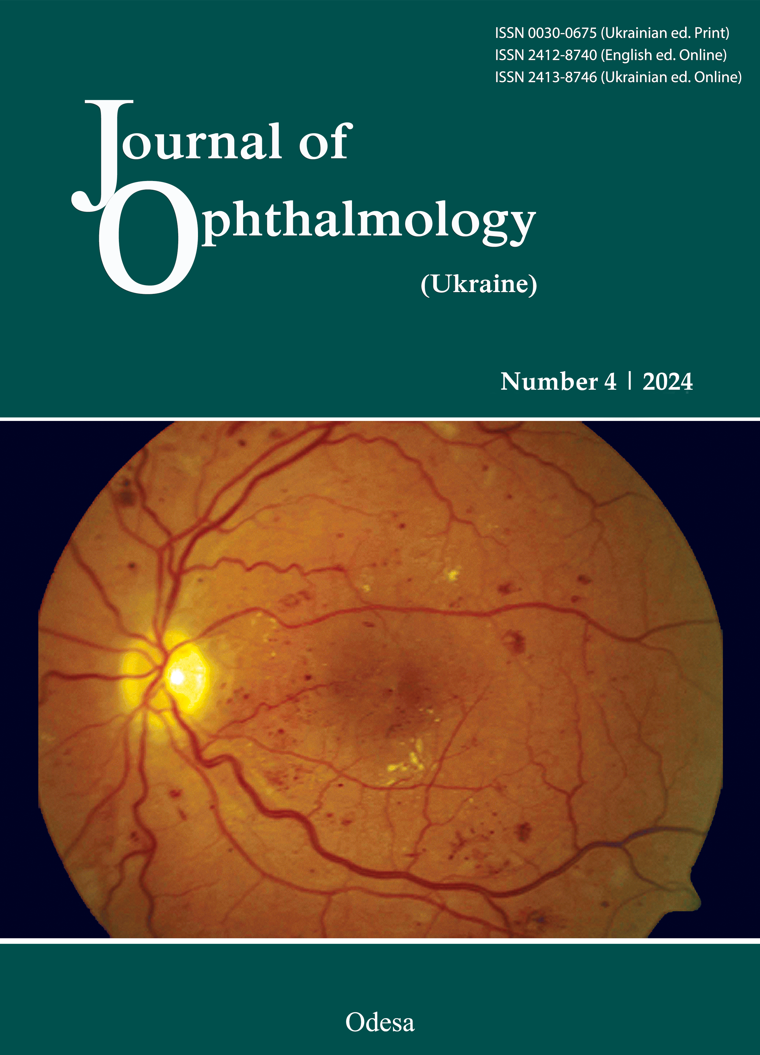
Levels of neuroinflammation markers in type 2 diabetes mellitus patients with diabetic retinopathy and genetically determined hyperhomocysteinemia
DOI:
https://doi.org/10.31288/oftalmolzh202442837Keywords:
cytokines, non-neuronal enolase, retinaAbstract
Background: Hyperhomocysteinemia determined by polymorphisms of genes encoding folate cycle enzymes is closely correlated with markers of systemic inflammation. Prolonged excessive cytokine production in the neural tissue in patients with type 2 diabetes mellitus (T2DM) causes gliosis, posing a threat of neuroinflammation, a potential pathogenetic component of diabetic retinopathy (DR).
Purpose: To assess plasma levels of neuroinflammation markers (IL-1β and IL-10) and gliosis marker, non-neuronal enolase (NNE), in T2DM patients with DR and genetically determined hyperhomocysteinemia.
Methods: One hundred and six T2DM patients with DR were involved in the study. DR severity was classified according to ETDRS DR severity system (DRSS) as non-proliferatve (NPDR; DRSS level 47–53), proliferative (NPDR; DRSS level 47–53) and advanced (APDR; DRSS level 81-85). The control group was composed of 64 individuals. Cases and controls were comparable for age, sex and way of life. MTHFR C677T (rs1801133), MTHFR A1298C (rs1801131), and MTR A2756G (rs1805087) genotypes were determined using the TaqMan® SNP Genotyping Assay (Applied Biosystems, Foster City, CA) and the ABI 7500 real-time PCR system (Applied Biosystem). Enzyme-linked immunosorbent assay (ELISA) kits were used to assess plasma levels of L-homocystein, cytokines and NNE.
Results: Plasma IL-1β levels were 1.7 times higher in patients with diabetes duration shorter than 15 years compared to those with longer diabetes duration. In addition, plasma NNE levels were higher in the former patients, but the difference was not significant. There was correlation of plasma L-homocystein levels with plasma IL-10 (R = 0.357, p < 0.01), IL-1b (R = 0.320, p < 0.01) and NNE (R = 0.286, p < 0.01) levels. Plasma NNE levels correlated with plasma IL-10 (R = 0.279, p < 0.01) and IL-1beta (R = 0.368, p < 0.01).
Conclusion: Elevated plasma levels of pro-inflammatory cytokines in patients indicate an important role of neuroinflammation in the pathogenesis of DR. The rs1801131 CC, rs1805087 GG and rs1801131 CC genotypes may be considered risk factors for the development of DR in patients with T2DM.
References
Mikheytseva IM. Current View On Pathogenic Mechanisms Of Diabetic Retinopathy. Fiziol Zh. 2023; 69(3): 106-114. https://doi.org/10.15407/fz69.03.106
Gardner TW, Davila JR. The neurovascular unit and the pathophysiologic basis of diabetic retinopathy. Graefes Arch. Clin. Exp. Ophthalmol. 2017; 255: 1-6. https://doi.org/10.1007/s00417-016-3548-y
Nian S, Lo ACY, Mi Y, Ren K, Yang D. Neurovascular unit in diabetic retinopathy: Pathophysiological roles and potential therapeutical targets. Eye Vis. 2021; 8:15. https://doi.org/10.1186/s40662-021-00239-1
Bianco L, Arrigo A, Aragona E, Antropoli A, Berni A, Saladino A, et al. Neuroinflammation and neurodegeneration in diabetic retinopathy. Front Aging Neurosci. 2022; Aug 16;14:937999. https://doi.org/10.3389/fnagi.2022.937999
Solomon SD, Chew E, Duh EJ, Sobrin L, Sun JK, VanderBeek B., et al. Diabetic retinopathy: A position statement by the American Diabetes Association. Diabetes Care. 2017; 40: 412-418.
https://doi.org/10.2337/dc16-2641
Hawkins BT, Davis TP. The blood-brain barrier/neurovascular unit in health and disease. Pharmacol. Rev. 2005; 57: 173-185. https://doi.org/10.1124/pr.57.2.4
Antonetti DA, Klein R, Gardner TW. (Diabetic retinopathy. N. Engl. J. Med. 2012; 366: 1227-1239. https://doi.org/10.1056/NEJMra1005073
Luzzi S, Cherubini V, Falsetti L, Viticchi G, Silvestrini M, Toraldo A. Homocysteine, Cognitive Functions, and Degenerative Dementias: State of the Art. Biomedicines. 2022; Oct 28;10(11):2741. https://doi.org/10.3390/biomedicines10112741
Ansari R, Mahta A, Mallack E, Luo JJ. Hyperhomocysteinemia and neurologic disorders: a review. J Clin Neurol. 2014 Oct; 10(4):281-8. https://doi.org/10.3988/jcn.2014.10.4.281
Quan Y, Xu J, Xu Q, Guo Z, Ou R, Shang H, Wei Q. Association between the risk and severity of Parkinson's disease and plasma homocysteine, vitamin B12 and folate levels: a systematic review and meta-analysis. Front Aging Neurosci. 2023 Oct 24;15:1254824. https://doi.org/10.3389/fnagi.2023.1254824
Kowluru RA, Mohammad G, Sahajpal N. Faulty homocysteine recycling in diabetic retinopathy. Eye Vis (London). 2020;7:4. https://doi.org/10.1186/s40662-019-0167-9
Gu J, Lei C, Zhang M. Folate and retinal vascular diseases. BMC Ophthalmol. 2023 Oct 13;23(1):413. https://doi.org/10.1186/s12886-023-03149-z
Rykov SO, Prokopenko IuV, Natrus LV, Panchenko IuO. Role of polymorphisms of folate-cycle enzymes in diabetic retinopathy progression in patients with type 2 diabetic mellitus. Journal of Ophthalmology (Ukraine). 2022; 5:3-11. https://doi.org/10.31288/oftalmolzh20225311
Malaguarnera G, Gagliano C, Salomone S, Giordano M, Bucolo C, Pappalardo A, Drago F, Caraci F, Avitabile T, Motta M. Folate status in type 2 diabetic patients with and without retinopathy. Clin Ophthalmol. 2015 Aug 7;9:1437-42. https://doi.org/10.2147/OPTH.S77538
Maltsev D, Kurchenko A, Marushko Y, Yuriev S. Biochemical profile of children with autism spectrum disorders associated with genetic deficiency of the folate cycle. Biochimica Clinica. 2023; 47(2): 132-140.
Marangos PJ, Schmechel D, Zis AP, Goodwin FK. The existence and neurobiological significance of neuronal and glial forms of the glycolytic enzyme enolase. Biol Psychiatry. 1979, Aug;14(4):563-79.
Davis M, Fisher M, Gangnon R, et al. Risk factors for high-risk proliferative diabetic retinopathy and severe visual loss: Early Treatment Diabetic Retinopathy Study Report 18. Invest Ophthalmol Vis Sci. 1998; 39: 233-52.
Downloads
Published
How to Cite
Issue
Section
License
Copyright (c) 2024 Panchenko Iu. O., Tsybulskyi V. S., Natrus L. V., Zakharevych H. Ie.

This work is licensed under a Creative Commons Attribution 4.0 International License.
This work is licensed under a Creative Commons Attribution 4.0 International (CC BY 4.0) that allows users to read, download, copy, distribute, print, search, or link to the full texts of the articles, or use them for any other lawful purpose, without asking prior permission from the publisher or the author as long as they cite the source.
COPYRIGHT NOTICE
Authors who publish in this journal agree to the following terms:
- Authors hold copyright immediately after publication of their works and retain publishing rights without any restrictions.
- The copyright commencement date complies the publication date of the issue, where the article is included in.
DEPOSIT POLICY
- Authors are permitted and encouraged to post their work online (e.g., in institutional repositories or on their website) during the editorial process, as it can lead to productive exchanges, as well as earlier and greater citation of published work.
- Authors are able to enter into separate, additional contractual arrangements for the non-exclusive distribution of the journal's published version of the work with an acknowledgement of its initial publication in this journal.
- Post-print (post-refereeing manuscript version) and publisher's PDF-version self-archiving is allowed.
- Archiving the pre-print (pre-refereeing manuscript version) not allowed.


