OCT angiography for diagnosing and postoperative monitoring of serous maculopathy associated with optic disc pit
DOI:
https://doi.org/10.31288/oftalmolzh201915660Keywords:
optic disc pit, serous maculopathy, optical coherence tomography angiographyAbstract
Background: Optic disc pits (ODP) usually present with the serous maculopathy found in 30% to 75% of these patients.
Purpose: To demonstrate the potential of optical coherence tomography angiography (OCTA) for monitoring the natural and postoperative course of ODP maculopathy.
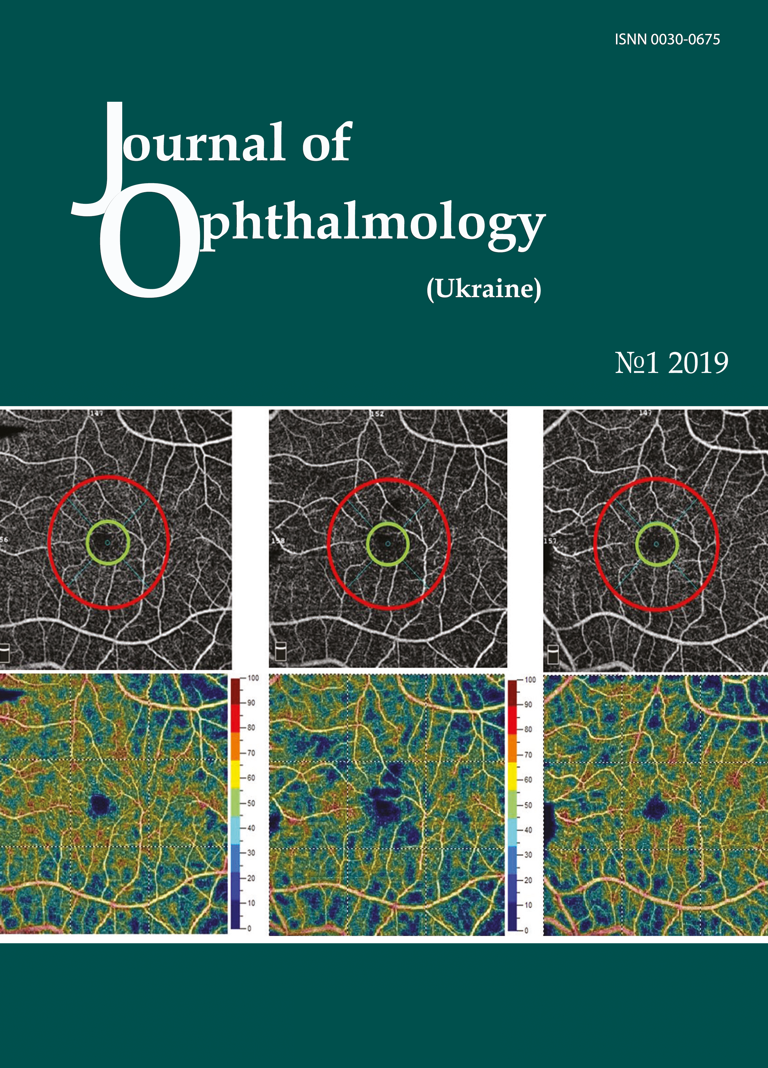
Materials and Methods: We present a case of a 27-year-old female patient with serous maculopathy associated with optic disc pit. OCTA scans were obtained using RTVue XR Avanti (Optovue, Fremont, CA). The OCTA-based vessel density was assessed. The follow-up after presentation was 10 months.
Results: The development of maculopathy in this case was found to be associated with penetration of liquor from the subarachnoidal space through the intermembranous spaces of the optic nerve. It is important to monitor the pathological areas found with the use of OCT and OCTA not only intraoperatively, but also postoperatively. Complete blockage of fluid tracts was achieved, evidencing the efficacy of treatment measures.
A decrease in vessel densities of the superficial retinal plexus in the foveal and parafoveal regions of the affected eye was accompanied by deterioration of visual functions, and guided the choice of treatment strategy. OCTA enables monitoring of morphological changes before and after surgical treatment and assist in making a prognosis for visual function.
References
1.Bayborodov YV, Izmailov AS. [The detachment of the retina caused by the pit of the optic nerve disk and its surgical treatment]. Fyodorov Journal of Ophthalmic Surgery. 2017;(4):20-5. Russian
2.Ganichenko IN. [Photo and laser coagulation for management of optic disc pit and its complications]. Oftalmol Zh. 1986;4:199. Russian.
3.Moisseiev E, Moisseiev J, Loewenstein A. Optic disc pit maculopathy: when and how to treat? A review of the pathogenesis and treatment options. Int J Retina Vitreous. 2015 Aug 7;1:13. https://doi.org/10.1186/s40942-015-0013-8
4. Apple DJ, Rabb MF, Walsh PM. Congenital anomalies of the optic disc. Surv Ophthalmol. 1982 Jul-Aug;27(1):3-41.https://doi.org/10.1016/0039-6257(82)90111-4
5.Brown GC, Shields JA, Goldberg RE. Congenital pits of the optic nerve head II. Clinical studies in humans. Ophthalmology. 1980 Jan;87(1):51-65.https://doi.org/10.1016/S0161-6420(80)35278-0
6.Gass JDM. Serous detachment of the macula secondary to congenital pit of the optic nerve head. Am J Ophthalmol.1969;67:821-49.https://doi.org/10.1016/0002-9394(69)90075-0
7.Theodossiadis GP, Ladas ID, Panagiotidis DN, Kollia AC, Voudouri AN, Theodossiadis PG. Fluorescein and indocyanine green angiographic findings in congenital optic disk pit associated with macular detachment. 1999;19(1):6-11.https://doi.org/10.1097/00006982-199901000-00002
8.Lee KJ, Peyman GA. Surgical management of retinal detachment associated with optic nerve pit. Int Ophthalmol. 1993 Apr;17(2):105-7.https://doi.org/10.1007/BF00942784
9.Georgalas I, Papaconstantinou D, Koutsandrea C, et al. Optic disc pit maculopathy: the value of small-gauge vitrectomy, peeling, laser treatment, and gas tamponade. Eur J Ophthalmol. 2013;23:275.https://doi.org/10.5301/ejo.5000216
10.Ghosh YK, Banerjee S, Konstantinidis A, Athanasiadis I, Kirkby GR, Tyagi AK. Surgical management of optic disc pit associated maculopathy. Eur J Ophthalmol. 2008 Jan-Feb;18:142-6.https://doi.org/10.1177/112067210801800126
11.Hirakata A, Inoue M, Hiraoka T, McCuen BW 2nd. Vitrectomy without laser treatment or gas tamponade for macular detachment associated with an optic disc pit. Ophthalmology. 2012 Apr;119(4):810-8.https://doi.org/10.1016/j.ophtha.2011.09.026
12.Rizzo S, Belting C, Genovesi-Ebert F, et al. Optic disc pit maculopathy: the value of small-gauge vitrectomy, peeling, laser treatment, and gas tamponade. Eur J Ophthalmol. 2012;22:620-5.https://doi.org/10.5301/ejo.5000083
13.Sandali O, Barale PO, Bui Quoc E, et al. [Long-term results of the treatment of optic disc pit associated with serous macular detachment: a review of 20 cases]. J Fr Ophtalmol. 2011 Oct;34(8):532-8.
14.Ohno-Matsui K, Hirakata A, Inoue M, Akiba M, Ishibashi T. Evaluation of congenital optic disc pit and optic disc colobomas by Swept-Source optical coherence tomography. Invest Ophthalmol Vis Sci. 2013 Nov 25;54(12):7769-78.https://doi.org/10.1167/iovs.13-12901
16.Schatz H, McDonald HR. Treatment of sensory retinal detachment associated with optic nerve pit or coloboma. 1988 Feb;95(2):178-86.https://doi.org/10.1016/S0161-6420(88)33197-0
17.Lumbroso B, Huang D, Chen CJ, et al., editors. Clinical OCT Angiography Atlas. New Delhi: Jaypee Brothers Medical Publishers; 2015.
Downloads
Published
How to Cite
Issue
Section
License
Copyright (c) 2025 Н. С. Луценко, О. А. Рудычева, О. А. Исакова, А. Н. Сергиенко

This work is licensed under a Creative Commons Attribution 4.0 International License.
This work is licensed under a Creative Commons Attribution 4.0 International (CC BY 4.0) that allows users to read, download, copy, distribute, print, search, or link to the full texts of the articles, or use them for any other lawful purpose, without asking prior permission from the publisher or the author as long as they cite the source.
COPYRIGHT NOTICE
Authors who publish in this journal agree to the following terms:
- Authors hold copyright immediately after publication of their works and retain publishing rights without any restrictions.
- The copyright commencement date complies the publication date of the issue, where the article is included in.
DEPOSIT POLICY
- Authors are permitted and encouraged to post their work online (e.g., in institutional repositories or on their website) during the editorial process, as it can lead to productive exchanges, as well as earlier and greater citation of published work.
- Authors are able to enter into separate, additional contractual arrangements for the non-exclusive distribution of the journal's published version of the work with an acknowledgement of its initial publication in this journal.
- Post-print (post-refereeing manuscript version) and publisher's PDF-version self-archiving is allowed.
- Archiving the pre-print (pre-refereeing manuscript version) not allowed.


