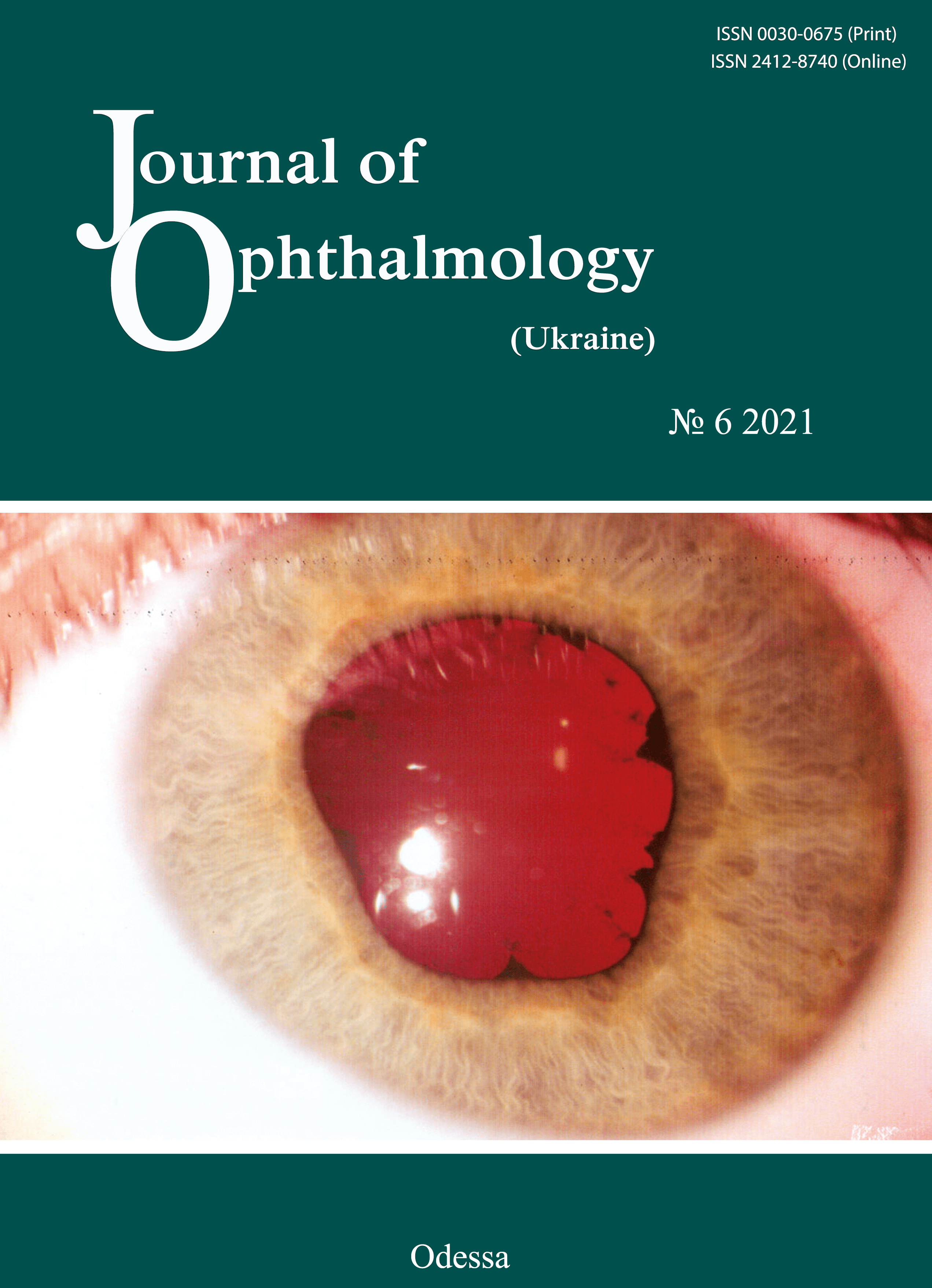
Report on the clinical study of glautan and original travoptost in patients with primary open angle glaucoma
DOI:
https://doi.org/10.31288/oftalmolzh202167783Abstract
Open angle glaucoma is characterized by specific lesions of the optic disc and is accompanied by visual field loss in the presence of elevated intraocular pressure (IOP). The management of glaucoma is challenging, the disease has become a matter of rising social and economic concern and a major cause of the deterioration in quality of life in patients [1, 2]. It is important that glaucoma is highly prevalent among economically-active working-age individuals and is the most common cause of certified visual disability [3, 4]. Glaucoma incidence increases with age. Primary open angle glaucoma (POAG) is more common in individuals older than 40 years of age.
References
1. Sommer A, Quigley HA, Robin AL, et al. Evaluation of nerve fiber layer assessment. Arch Ophthalmol. 1984 Dec;102(12):1766-71.https://doi.org/10.1001/archopht.1984.01040031430017
2. Sommer A, Katz J, Quigley HA, et al. Clinically detectable nerve fiber atrophy precedes the onset of glaucomatous field loss. Arch Ophthalmol. 1991 Jan;109(1):77-83. https://doi.org/10.1001/archopht.1991.01080010079037
3. Airaksinen PJ, Drance SM, Douglas GR, et al. Diffuse and localized nerve fiber loss in glaucoma. Am J Ophthalmol. 1984 Nov;98(5):566-71. https://doi.org/10.1016/0002-9394(84)90242-3
4. Kurysheva NI. [Glaucomatous optic neuropathy]. Moscow: Medpressinform; 2006. Russian.
5. Curcio CA, Allen KA. Topography of ganglion cells in human retina. J Comp Neurol. 1990 Oct 1;300(1):5-25. https://doi.org/10.1002/cne.903000103
6. Munemasa Y, Kitaoka Y. Molecular mechanisms of retinal ganglion cell degeneration in glaucoma and future prospects for cell body and axonal protection. Front Cell Neurosci. 2013 Jan 9;6:60. https://doi.org/10.3389/fncel.2012.00060
7. Zeimer R, Shahidi M, Mori M, et al. A new method for rapid mapping of the retinal thickness at the posterior pole. Invest Ophthalmol Vis Sci. 1996 Sep;37(10):1994-2001.
8. Duker JC, Waheed MK, Goldman DR. Handbook of Retinal OCT. London: Elsevier. 2014.
9. Nesterov AP. [Primary open angle glaucoma: pathogenesis and principles of management]. Rossiiskii meditsinskii zhurnal. Klinicheskaia Oftalmologiia. 2000; 1:4-5. Russian.
10. Zhaboiedov GD, Petrenko OV, Parkhomenko EG. [Comparing tear nitric oxide levels in healthy individuals and patients with glaucoma]. In: [Proceedings of Fyodorov Memorial Lectures-2006]. p. 201-3. Russian.
11. Bagga H, Greenfield DS, Knighton RW. Macular symmetry testing for glaucoma detection. J Glaucoma. 2005 Oct;14(5):358-63.https://doi.org/10.1097/01.ijg.0000176930.21853.04
12. Tan O, Chopra V, Lu AT, et al. Detection of macular ganglion cell loss in glaucoma by Fourier-domain optical coherence tomography. Ophthalmology 2009; 116: 2305-14. https://doi.org/10.1016/j.ophtha.2009.05.025
Downloads
Published
How to Cite
Issue
Section
License
Copyright (c) 2025 І. А. Соболєва

This work is licensed under a Creative Commons Attribution 4.0 International License.
This work is licensed under a Creative Commons Attribution 4.0 International (CC BY 4.0) that allows users to read, download, copy, distribute, print, search, or link to the full texts of the articles, or use them for any other lawful purpose, without asking prior permission from the publisher or the author as long as they cite the source.
COPYRIGHT NOTICE
Authors who publish in this journal agree to the following terms:
- Authors hold copyright immediately after publication of their works and retain publishing rights without any restrictions.
- The copyright commencement date complies the publication date of the issue, where the article is included in.
DEPOSIT POLICY
- Authors are permitted and encouraged to post their work online (e.g., in institutional repositories or on their website) during the editorial process, as it can lead to productive exchanges, as well as earlier and greater citation of published work.
- Authors are able to enter into separate, additional contractual arrangements for the non-exclusive distribution of the journal's published version of the work with an acknowledgement of its initial publication in this journal.
- Post-print (post-refereeing manuscript version) and publisher's PDF-version self-archiving is allowed.
- Archiving the pre-print (pre-refereeing manuscript version) not allowed.


