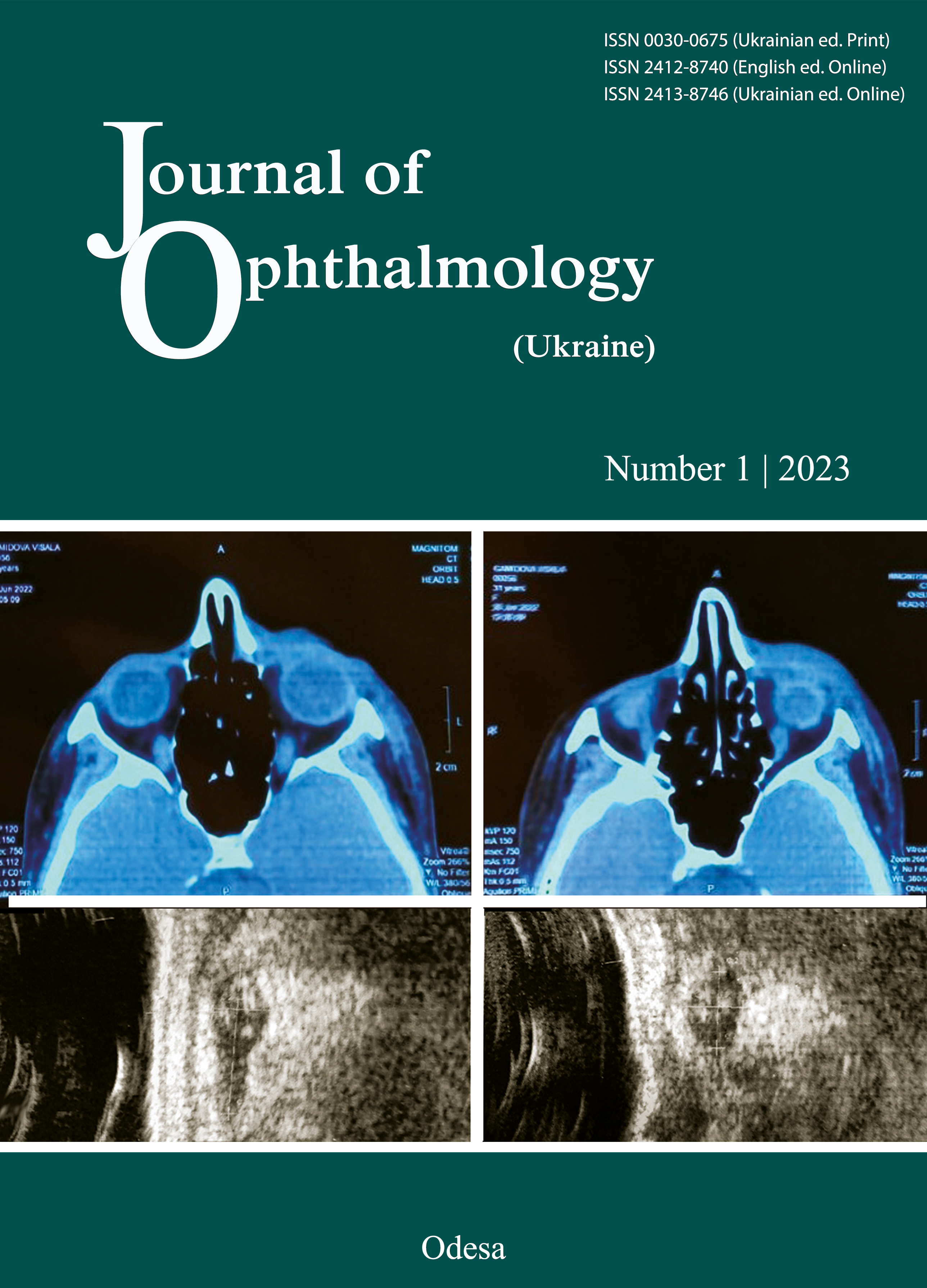
Features of regression of different stages of retinoblastoma after primary combined (intravitreal plus systemic) polychemotherapy and additional consolidation therapy
DOI:
https://doi.org/10.31288/oftalmolzh202311418Abstract
Background: Complete retinoblastoma (RB) regression is defined as final changes undergone by the tumor in the course of eye-salvage treatment. Studies of RB regression patterns following various types of eye-salvage treatment are important for assessing the outcomes of this treatment.
Purpose: To study the regression patterns in different stages of RB after primary combined polychemotherapy (PCPC) and additional consolidation therapy.
Material and Methods: We reviewed RB regression patterns in 89 children (119 eyes) aged 1.5 to 77 months after PCPC and various types of consolidation therapy. Of these children, 37 had unilateral retinoblastoma, and 52, bilateral retinoblastoma. At presentation, T3 RB was the most prevalent (67.2%), followed by T2 (23.6%). T1 stage was observed only in 9.2% - most often it was diagnosed on a fellow "healthy" eye at a spreading T3 stage of the countralateral eye. Sixteen eyes (13%) had multifocal tumors type of tumor growth. Because the number of tumor foci per eye ranged from one to three, the total number of foci (124) exceeded the total number of affected eyes. Treatment was carried out according to the developed PСPС method, which invсcluded the intravitral injection of melfalan at the dose of 10-30 mcg, depending on the tumor stage, followed by intravenous systemic therapy (VEC protocol).
Results: We found different RB regression patterns after PCPC. After the first cycle of PCPC, type 2 regression pattern was typical for small T1 tumors, whereas type 3 regression pattern was most prevalent (60%) for T2 and T3 tumors. This is likely to indicate tumor mosaicism with the presence of less differentiated and, consequently, more malignant cell types, which faster reacted to PCPC by calcification than surrounding more differentiated and, consequently, less malignant cells which reacted weaker. After the completion of PCPC, type 1 regression pattern was seen in 29%, which indicated complete regression, whereas type 3 regression pattern persisted in 33% foci. The features of tumor regression following PCPC included (a) fragmentation of a large RB (59.3%) after first 1-2 PCPC cycles with appearance of necrotic foci which resolved or calcified finally; (b) presence of various types of regression in one eye in the multifocal growth; and (c) transformation of a regression type into another regression type: most commonly, transformation of type 2 into type 3 and type 3 into type 1.
References
Боброва НФ, Науменко ВА, Сорочинская ТА, Братишко АЮ, Комарницкая ТИ. Комбинация локальных методов воздействия на ретинобластому. Мат. Научно-практич. конф. c междунар. участ. «Филатовские чтения-2019»: 186-187.
Боброва НФ. Ретинобластома: монография. Одесса: Издательский центр, 2020. 324 с.
Боброва НФ, Сорочинская ТА. Комбинированная (интравитреальная и внутривенная) полихимиотерапия в системе органосохранного лечения ретинобластомы. Офтальмол. журн. 2011; 2: 38-44. https://doi.org/10.31288/oftalmolzh201123844
Боброва НФ, Сорочинська ТА, Троніна СА, Романова ТВ, Братішко ОЮ. Високодозова інтравітреальна хіміотерапія в лікуванні ретинобластоми високого ризику. Офтальмол. журн. 2022; 4: 23-27. https://doi.org/10.31288/oftalmolzh202242327
Abramson DH, Gerardi CM, Ellsworth RM, McCormick B, Sussman D, Turner L. Radiation regression patterns in treated retinoblastoma: 7 to 21 years later. J Pediatr Ophthalmol Strabismus. 1991;28(2):108-112. https://doi.org/10.3928/0191-3913-19910301-12
Chawla B, Jain A, Seth R, Azad R, Mohan VK, Pushker N, Ghose S. Clinical outcome and regression patterns of retinoblastoma treated with systemic chemoreduction and focal therapy: A prospective study. Indian J Ophthalmol. 2016 Jul;64(7):524-9. https://doi.org/10.4103/0301-4738.190143
Dunphy EB. The story of retinoblastoma: the Edward Jackson Memorial Lecture. Am J Ophthalmol. 1964; 58: 539-552. https://doi.org/10.1016/0002-9394(64)91368-6
Ellsworth RM. The practical management of retinoblastoma. Trans Am Ophthalmol Soc. 1969;67:462-534.
Friedman D, Himelstein B, Shields C, et al. Chemoreduction and local ophthalmic therapy for intraocular retinoblastoma. J Clin Oncol. 2000; 18: 12-17. https://doi.org/10.1200/JCO.2000.18.1.12
Ghassemi F, Rahmanikhah E, Roohipoor R, Karkhaneh R, Faegh A Regression patterns in treated retinoblastoma with chemotherapy plus focal adjuvant therapy. Pediatr Blood Cancer. 2013 Apr;60(4):599-604. doi: 10.1002/pbc.24333. Epub 2012 Oct 3. https://doi.org/10.1002/pbc.24333
Levy J, Frenkel S, Baras M, Neufeld M, Pe'er J. Calcification in retinoblastoma: histopathologic findings and statistical analysis of 302 cases. Br J Ophthalmol. 2011 Aug;95(8):1145-50. https://doi.org/10.1136/bjo.2010.193961
Lin CC, Tso MO. An electron microscopic study of calcification of retinoblastoma. Am J Ophthalmol. 1983; 96:765-74. https://doi.org/10.1016/S0002-9394(14)71922-1
Murphree A, Villablanca J, Deegan W, et al. Chemotherapy plus local treatment in the management of intraocular retinoblastoma. Arch Ophthalmol. 1996; 114: 1348-1356. https://doi.org/10.1001/archopht.1996.01100140548005
Palamar M, Thangappan A, Shields CL Evolution in regression patterns following chemoreduction for retinoblastoma. Arch Ophthalmol. 2011 Jun;129(6):727-30. https://doi.org/10.1001/archophthalmol.2011.137
Saup DN, Albert DM. Retinoblastoma: Clinical and histopathologic features. Hum Pathol 1982 : 13; 133-47. https://doi.org/10.1016/S0046-8177(82)80117-2
Singh AD, Garway-Heath D, Love S, Plowman PN, Kingston JE, Hungerford JL. Relationship of regression pattern to recurrence in retinoblastoma. Br J Ophthalmol.1993;77(1):12-16. https://doi.org/10.1136/bjo.77.1.12
Shields CL, Shields JA. Diagnosis and management of retinoblastoma. Cancer Control. 2004; 11(5): 317- 327. https://doi.org/10.1177/107327480401100506
Shields CL, Palamar M, Sharma P, Ramasubramanian A, Leahey A, Meadows AT, Shields JA Retinoblastoma regression patterns following chemoreduction and adjuvant therapy in 557 tumors. Arch Ophthalmol. 2009 Mar;127(3):282-90.
https://doi.org/10.1001/archophthalmol.2008.626
Xue К, Qian J, Han Yue, Yi-fei Yuan, Rui Zhang [Retinoblastoma regression patterns and results following chemoreduction and adjuvant therapy] [Article in Chinese] Zhonghua Yan Ke Za Zhi. 2012 Jul;48(7):625-30.
Zafar SN., Siddiqui SN., Zaheer N. Тumor Regression Patterns in Retinoblastoma. J Coll Physicians Surg Pak. 2016 Nov;26(11):896-899.
Downloads
Published
How to Cite
Issue
Section
License
Copyright (c) 2023 Bobrova N.F., Tronina S.A., Sorochynska T.A., Romanova T.V., Dembovetska G.M., Shylyk A.V., Dovgan O.D.

This work is licensed under a Creative Commons Attribution 4.0 International License.
This work is licensed under a Creative Commons Attribution 4.0 International (CC BY 4.0) that allows users to read, download, copy, distribute, print, search, or link to the full texts of the articles, or use them for any other lawful purpose, without asking prior permission from the publisher or the author as long as they cite the source.
COPYRIGHT NOTICE
Authors who publish in this journal agree to the following terms:
- Authors hold copyright immediately after publication of their works and retain publishing rights without any restrictions.
- The copyright commencement date complies the publication date of the issue, where the article is included in.
DEPOSIT POLICY
- Authors are permitted and encouraged to post their work online (e.g., in institutional repositories or on their website) during the editorial process, as it can lead to productive exchanges, as well as earlier and greater citation of published work.
- Authors are able to enter into separate, additional contractual arrangements for the non-exclusive distribution of the journal's published version of the work with an acknowledgement of its initial publication in this journal.
- Post-print (post-refereeing manuscript version) and publisher's PDF-version self-archiving is allowed.
- Archiving the pre-print (pre-refereeing manuscript version) not allowed.


