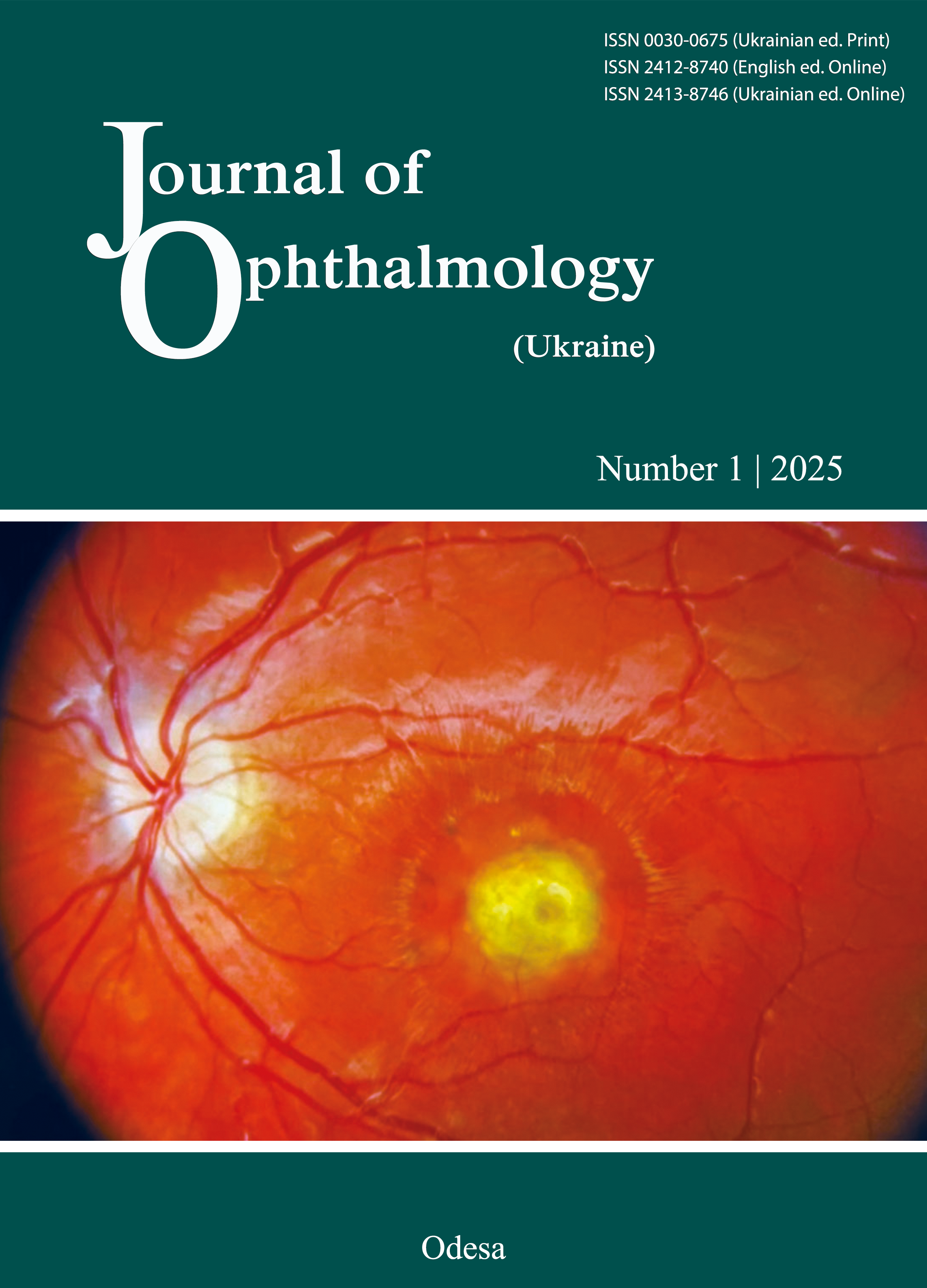
Features of the course of corneal and choroidal inflammation in children infected with SARS-CoV-2
DOI:
https://doi.org/10.31288/oftalmolzh202511721Keywords:
COVID-19, ocular manifestations, uveitis, keratitis, children, ocular inflammatory diseaseAbstract
Background: The consequences of respiratory syndrome coronavirus 2 (SARS-CoV-2) pandemics are still unfolding.
Purpose: To examine the course of corneal and choroid inflammation in children infected with COVID-19 who were treated at the Department of Pediatric Eye Pathology, SI “The Filatov Institute of Eye Diseases and Tissue Therapy of the National Academy of Medical Sciences of Ukraine”, in 2021-2023.
Material and Methods: The study sample included 62 children (94 eyes) with corneal and choroidal inflammation who were treated at the Department of Pediatric Eye Pathology, SI “The Filatov Institute of Eye Diseases and Tissue Therapy of the National Academy of Medical Sciences of Ukraine”, in 2021-2023. SARS-CoV-2 infection was confirmed by laboratory testing in all children. The age of children ranged from 6 months to 17 years (mean age, 10.67 ± 2.95 years). Enzyme-linked immunosorbent assay (ELISA) was used to detect SARS-CoV-2- immunoglobulin (Ig)M antibodies in children suspected for past COVID-19 infection.
Results: The detection of immunoglobulin IgM antibodies indicated a recent past SARS-CoV-2 infection, whereas positive COVID-IgG indicated a prior past infection. Uveitis was the most common ocular finding (48/62 children (77.4%), 78 eyes); 77% of uveitis cases were bilateral. Acute uveitis, acute exacerbation of chronic uveitis and sequelae of chronic uveitis were observed in 33.3%, 53.9%, and 12.8%, respectively, of study eyes. Panuveitis with severe complications (seclusio and occlusio papillae, uveal cataract, secondary glaucoma, retinal detachment, subretinal neovascular membranes, vitreous hemorrhage, vitreous fibrosis and phthisis bulbi) was diagnosed in 57.7% of study eyes, mostly in eyes with bilateral uveitis. Post-COVID-19 keratitis was found in 14 children (16 eyes; 12.4%) and was unilateral in 85.7% and acute in 62.5% of cases.
Conclusion: Past SARS-CoV-2 infection may cause an acute ocular inflammatory disease or recurrent chronic inflammatory process in children. Uveitis with lesions in all uveal tract compartments and severe complications was the most common finding. Family doctors should be encouraged to refer all children with a past COVID-19 infection to an ophthalmologist. Pediatric ophthalmologists should be aware of possible post-COVID-19 ocular complications.
References
Sen M, Honavar SG, Sharma N, Sachdev MS. COVID-19 and Eye: A Review of Ophthalmic Manifestations of COVID-19. Indian J Ophthalmol 2021;69: 488-509. https://doi.org/10.4103/ijo.IJO_297_21
Wu P, Duan F, Luo C. Characteristics of ocular findings of patients with coronavirus disease 2019 (COVID-19) in Hubei Province, China. JAMA Ophthalmol. 2020 May 1; 138(5):575-8. https://doi.org/10.1001/jamaophthalmol.2020.1291
Lu C-W, Liu X-F, Jia Z-F. 2019-nCoV transmission through the ocular surface must not be ignored. Lancet 2020; 395:e39. https://doi.org/10.1016/S0140-6736(20)30313-5
Bezditko PA. [Eye manifestations of COVID-19. Step one]. Arkhiv oftalmologii Ukrainy. 2021; 9(1):30-38. https://doi.org/10.22141/2309-8147.9.1.2021.229522
Dockery DM, Rowe SG, Murphy MA, Krzystolik MG. The ocular manifestations and transmission of COVID-19: recommendations for prevention. J Emerg Med. 2020 Jul; 59(1):137-140. https://doi.org/10.1016/j.jemermed.2020.04.060
Siedlecki J, Brantl V, Schworm B, et al. COVID-19: ophthalmological aspects of the SARS-CoV 2 global pandemic. COVID-19: ophthalmologische Aspekte der globalen SARS-CoV-2-Pandemie. Klin Monatsbl Augenheilkd. 2020; 237(5):675-80. https://doi.org/10.1055/a-1164-9381
Ludvigsson JF. Systematic review of COVID-19 in children shows milder cases and a better prognosis than adults. Acta Paediatrica (Oslo, Norway). 2020; 109:1088-95 https://doi.org/10.1111/apa.15270
Zimmermann P and Curtis N. Coronavirus infections in children including COVID-19: an overview of the epidemiology, clinical features, diagnosis, treatment and prevention options in children. Pediatr Infect Dis J. 2020; 39:355 https://doi.org/10.1097/INF.0000000000002660
Li B, Zhang S, Zhang R, et al. Epidemiological and clinical characteristics of COVID-19 in children: a systematic review and meta-analysis. Front Pediatr. 2020 Nov 2:8:591132. https://doi.org/10.3389/fped.2020.591132
Kawasaki T. Kawasaki disease. Proc Jpn Acad Ser B Phys Biol Sci. 2006;82:59-71. https://doi.org/10.2183/pjab.82.59
Chiotos K, Bassiri H, Behrens EM, et al. Multisystem inflammatory syndrome in children during the COVID-19 pandemic: a case series. J Pediatric Infect Dis. 2020 Jul 13; 9(3):393-8. https://doi.org/10.1093/jpids/piaa069
Mechel E, Trinh M, Kodsi S, et al. Ophthalmia neonatorum as the presenting sign of SARS-CoV-2. J AAPOS. 2021 Aug; 25(4):230-231. https://doi.org/10.1016/j.jaapos.2021.03.001
Pérez-Chimal LG, Cuevas GG, Di-Luciano A, et al. Ophthalmic manifestations associated with SARS-CoV-2 in newborn infants: a preliminary report. J AAPOS. 2021; 25: 102-4. https://doi.org/10.1016/j.jaapos.2020.11.007
Kaur K, Muralikrishnan J, Hussaindeen JR, Deori N, Gurnani B. Impact of Covid-19 on Pediatric Ophthalmology Care: Lessons Learned. Review. Pediatric Health Med Ther. 2023; 14:309-21. https://doi.org/10.2147/PHMT.S395349
Salvetat ML, Salati C, Busatto P, Zeppieri M. The impact of COVID-19 related national lockdown on ophthalmic emergency in Italy: a multicenter study. Eur J Ophthalmol. 2022;32(3):1782-1794. https://doi.org/10.1177/11206721211028046
Iriqat S, Yousef Q, Ereqat S. Clinical profile of COVID-19 patients presenting with uveitis - a short case series. Int Med Case Rep J. 2021; 14:421-7. https://doi.org/10.2147/IMCRJ.S312461
Land P, Shah V, Lovell D J, Miraldi V. Panuveitis and optic neuropathy following SARS-COV-2 in the absence of multisystem inflammatory syndrome in a child. Am J Ophthalmol Case Rep. 2023 Jun 29; 32:101876. https://doi.org/10.1016/j.ajoc.2023.101876
Willcox MD, Walsh K, Nichols JJ, et al. The ocular surface, coronaviruses and COVID-19. Clin Exp Optom. 2020; 103: 418-4. https://doi.org/10.1111/cxo.13088
Li W, Sui J, Huang IC. The S proteins of human coronavirus NL63 and severe acute respiratory syndrome coronavirus bind overlapping regions of ACE2. Virology. 2007; 367(2):367-74. https://doi.org/10.1016/j.virol.2007.04.035
Shah KK, Venkatramani D and Majumder PD. A case series of presumed fungal endogenous endophthalmitis in post COVID-19 patients. Indian J Ophthalmol 2021; 69: 1322-5. https://doi.org/10.4103/ijo.IJO_3755_20
Fernández Alcalde C, Granados Fernández M, Nieves Moreno M, et al. COVID-19 ocular findings in children: a case series. World J Pediatr. 2021 Feb 22;17(3):329-34. https://doi.org/10.1007/s12519-021-00418-z
Öztürk C, Yüce Sezen A, Savaş Ş en Z, et al. Bilateral acute anterior uveitis and corneal punctate epitheliopathy in children diagnosed with multisystem inflammatory syndrome secondary to COVID-19. Ocul Immunol Inflamm. 2021; 29:700-4. https://doi.org/10.1080/09273948.2021.1909070
Chung JEREW, Engin Ö, Wolfs TFW, et al. Anterior uveitis in paediatric inflammatory multisystem syndrome temporally associated with SARS-CoV-2. Lancet. 2021; 397:e10. https://doi.org/10.1016/S0140-6736(21)00579-1
Bettach E, Zadok D, Weill Y, et al. Bilateral anterior uveitis as a part of a multisystem inflammatory syndrome secondary to COVID-19 infection. J Med Virol 2021; 93: 139-40 https://doi.org/10.1002/jmv.26229
Kumar D, Rostad CA, Jaggi P, et al. Distinguishing immune activation and inflammatory signatures of multisystem inflammatory syndrome in children (MIS-C) versus hemophagocytic lymphohistiocytosis (HLH). J Allergy Clin Immunol. 2022 May;149(5):1592-606.e16. https://doi.org/10.1016/j.jaci.2022.02.028
Arruti N. Acute bilateral anterior uveitis in paediatric inflammatory multisystem syndrome temporally associated with COVID-19. Int J Ophthalmol. 2022;15(8):1410-2. https://doi.org/10.18240/ijo.2022.08.28
Downloads
Published
How to Cite
Issue
Section
License
Copyright (c) 2025 Bobrova N.F., Sorochynska T.A., Tronina S.A., Romanova T.V., Dembovetska G.M., Sukhodoieva O.O., Shylyk A.V., Dovhan O.D., Vdovichenko K.S., Romanchuk O.M.

This work is licensed under a Creative Commons Attribution 4.0 International License.
This work is licensed under a Creative Commons Attribution 4.0 International (CC BY 4.0) that allows users to read, download, copy, distribute, print, search, or link to the full texts of the articles, or use them for any other lawful purpose, without asking prior permission from the publisher or the author as long as they cite the source.
COPYRIGHT NOTICE
Authors who publish in this journal agree to the following terms:
- Authors hold copyright immediately after publication of their works and retain publishing rights without any restrictions.
- The copyright commencement date complies the publication date of the issue, where the article is included in.
DEPOSIT POLICY
- Authors are permitted and encouraged to post their work online (e.g., in institutional repositories or on their website) during the editorial process, as it can lead to productive exchanges, as well as earlier and greater citation of published work.
- Authors are able to enter into separate, additional contractual arrangements for the non-exclusive distribution of the journal's published version of the work with an acknowledgement of its initial publication in this journal.
- Post-print (post-refereeing manuscript version) and publisher's PDF-version self-archiving is allowed.
- Archiving the pre-print (pre-refereeing manuscript version) not allowed.
.png)